


NM000380: ieeg dataset, 17 subjects#
iEEG during direct electrical stimulation of orbitofrontal and other sites (Rao, Sellers et al., 2018): continuous-stimulation and single-pulse recordings
Access recordings and metadata through EEGDash.
Citation: Vikram Rao, Kristin Sellers, Deanna Wallace, Morgan Lee, Maryam Bijanzadeh, Omid Sani, Yuxiao Yang, Maryam Shanechi, Heather Dawes, Edward Chang (2018). iEEG during direct electrical stimulation of orbitofrontal and other sites (Rao, Sellers et al., 2018): continuous-stimulation and single-pulse recordings. 10.82901/nemar.nm000380
Modality: ieeg Subjects: 17 Recordings: 45 License: CC-BY-4.0 Source: nemar
Metadata: Complete (100%)
17-participant iEEG dataset — iEEG during direct electrical stimulation of orbitofrontal and other sites (Rao, Sellers et al., 2018): continuous-stimulation and single-pulse recordings.
Quickstart#
Install
pip install eegdash
Access the data
from eegdash.dataset import NM000380

dataset = NM000380(cache_dir="./data")
# Get the raw object of the first recording
raw = dataset.datasets[0].raw
print(raw.info)
Filter by subject
dataset = NM000380(cache_dir="./data", subject="01")
Advanced query
dataset = NM000380(
    cache_dir="./data",
    query={"subject": {"$in": ["01", "02"]}},
)
Iterate recordings
for rec in dataset:
    print(rec.subject, rec.raw.info['sfreq'])
If you use this dataset in your research, please cite the original authors.
BibTeX
@dataset{nm000380,
  title = {iEEG during direct electrical stimulation of orbitofrontal and other sites (Rao, Sellers et al., 2018): continuous-stimulation and single-pulse recordings},
  author = {Vikram Rao and Kristin Sellers and Deanna Wallace and Morgan Lee and Maryam Bijanzadeh and Omid Sani and Yuxiao Yang and Maryam Shanechi and Heather Dawes and Edward Chang},
  doi = {10.82901/nemar.nm000380},
  url = {https://doi.org/10.82901/nemar.nm000380},
}
About This Dataset#
Intracranial EEG (ECoG grids/strips and depth electrodes) from adults undergoing inpatient monitoring for seizure
localization, recorded immediately before, during and after continuous 100 Hz direct electrical stimulation of the lateral orbitofrontal cortex and other sites (task-stim), and during single-pulse electrical stimulation (task-sps). 17 participants (release codes EC##).
The study (Rao et al., 2018) found that unilateral stimulation of the lateral OFC acutely improved mood state in
subjects with moderate-to-severe baseline depression symptoms and suppressed low-frequency OFC power.
iEEG, direct electrical stimulation and mood (Rao, Sellers et al., 2018)
Overview
Source
Dryad: Vikram Rao, Kristin Sellers, Deanna Wallace, Morgan Lee, Maryam Bijanzadeh, Omid Sani, Yuxiao Yang, Maryam Shanechi, Heather Dawes, Edward Chang. iEEG, Direct Electrical Stimulation, and Assessment of Mood in Human Subjects.
View full README
iEEG, direct electrical stimulation and mood (Rao, Sellers et al., 2018)
Overview
Source
Dryad: Vikram Rao, Kristin Sellers, Deanna Wallace, Morgan Lee, Maryam Bijanzadeh, Omid Sani, Yuxiao Yang, Maryam Shanechi, Heather Dawes, Edward Chang. iEEG, Direct Electrical Stimulation, and Assessment of Mood in Human Subjects. doi:10.7272/Q6VD6WM2 (version 2, published 2018-10-05).
License: the Dryad record states CC0-1.0 (
https://spdx.org/licenses/CC0-1.0.html); the Dryad-community Zenodo replica of the same DOI (record 5076490) states CC-BY-4.0. Following the most restrictive of the stated licenses, this dataset is distributed under CC-BY-4.0.Article: Rao et al. (2018) Current Biology, doi:10.1016/j.cub.2018.10.026.
Files were acquired from the Zenodo replica; every file matched the Dryad sha-256 digest.
Participants
Paper cohort (STAR Methods): 25 subjects (14 males, ages 20-60) with drug-resistant epilepsy undergoing intracranial EEG for seizure localization, studied by the University of California, San Francisco group of the senior author (EC codes are assigned to all intracranial patients in the senior author’s clinical practice, hence non-consecutive). Electrode implantation was guided solely by clinical indications; electrodes typically sampled OFC, amygdala, hippocampus, insula and cingulate cortex. Subdural grid, strip and depth electrodes (AdTech, Racine, WI, or Integra, Plainsboro, NJ); localization by co-registering pre-operative 3 T T1 MRI with post-operative CT (SPM12) and FreeSurfer pial reconstructions. Baseline mood trait: Beck Depression Inventory II before implantation (PHQ-9 in two subjects), binned into minimal-mild and moderate-severe.
This dataset: the 17 subjects whose stimulation/SPS recordings are in the Dryad release (10 male, 7 female; ages 20-45, mean 33.5; 13 with grids/strips/depths, 4 with stereo-EEG; 8 left, 7 right, 2 bilateral coverage; 10 with trait depression).
participants.tsv: age, sex, hemisphere, electrode coverage, electrode type, seizure onset zone, surgical pathology, BDI, trait depression, number and version of Immediate Mood Scaler (IMS) data points are copied from Table S1 of the paper’s supplement, which is keyed by the same EC identifiers as the release (direct mapping; consistent with the release electrode types, e.g. depth-only files for the 4 stereo-EEG subjects, and with the Table S3 list of subjects with single-pulse stimulation: EC137, EC139, EC150, EC153, EC155). BDI and trait values also match the release fileBehavior/KS_TraitScores.matin Table S1 order. Handedness is not reported (n/a). Recording dates/years are not given.
Task and stimulation (from the paper)
Mood state was assessed with the tablet-based Immediate Mood Scaler (IMS) several times a day for several days before stimulation. Stimulation took place after clinical seizure data collection and before electrode explantation, typically after anticonvulsants were restarted.
task-stim: bipolar stimulation of adjacent electrodes with the Nicolet Cortical Stimulator (Natus Medical), biphasic constant-current trains, 1 or 6 mA, 100 Hz, 100 us pulse width, 100-200 s; a sham (0 mA) block at the start of each session, OFC and non-OFC sites in varying order. Subjects were blind to condition. About 30 s after onset they gave a verbal mood report (open-ended questions, 30 s), then completed the IMS on a tablet (1-2 min); stimulation stopped after the IMS and the next block started 3-5 min later.
task-sps: single-pulse stimulation, 10 mA, 1 Hz, 500 us pulse width, 20 s, in some subjects before and after continuous OFC stimulation.
Acquisition
Natus clinical EEG system, Tucker-Davis Technologies PZ5M-512 and RZ2, or Alpha Omega NeuroOmega; sampling rates 1-25 kHz (paper). The recording reference and ground are not stated in the paper or the release.
Line noise 60 Hz (the paper’s analysis notch-filtered 60 Hz and harmonics).
Preprocessing
None in this dataset (raw data). The paper’s analysis pipeline (artifact removal with spline interpolation, 2-250 Hz band-pass, common average reference, notch filters, downsampling to 512 Hz) was NOT applied here.
Files
task-stim: one run per release fileEC##_rawDuringContinuous_<amplitude>_<site>.mat(Natus clinical system, acq-natus),EC##_B<block>_rawData.mat/EC175_1mA_rawData.mat(Tucker-Davis Technologies, acq-tdt) orEC##_NO_*.mat(NeuroOmega, acq-neuroomega). Stimulation amplitude and site are taken from the file name and written toElectricalStimulationParameters; per the release README, files without a site in the name were recorded with lateral OFC stimulation. The original file name is incode/conversion_report.json.task-sps:EC##_rawData_SPS.matsingle-pulse stimulation recordings.channels.tsv: names, long names, electrode type and region from the release (selectAnatomy / Anatomy cells). Types: grid/strip -> ECOG, depth -> SEEG, EKG -> ECG, unstated -> MISC.electrodes.tsv: elecmatrix + anatomy from the subject’s clinical_elecs_all.mat / TDT_elecs_all.mat (space-Other, mm, patient space of the authors’ reconstruction); n/a coordinates where the release has no electrode file.
Conversion notes
Samples are float64 in the release and are stored as float32 here (BrainVision); the maximum absolute rounding error per file is listed in
code/conversion_report.json(relative error ~6e-8). The float64 originals are insourcedata/dryad-q6vd6wm2/. No filtering, resampling, re-referencing or channel removal.Units are not stated in the release. Natus and NeuroOmega values are stored as µV (magnitudes of tens to thousands; 34843.74 is a recurring saturation value in Natus files); TDT values (|x| < 0.2) are stored as V.
Stimulation and pulse times are not annotated in the release.
Files not converted: - iEEG_DuringStimulation_EC155.zip::iEEG_DuringStimulation_EC155/EC155_NO_1mA.mat: not readable as MATLAB v7.3/HDF5 (OSError(‘Unable to synchronously open file (file signature not found)’))
The release’s
iEEG_NaturalBehavior.zip(OFC ECoG segments around mood self-reports) is NOT included: itsECoG.timefields contain absolute recording dates and times. Behaviour tables (Behavior.zip: mood scores, BDI/trait scores, speech rate) and the authors’ MATLAB scripts are in sourcedata.Age, sex and handedness are not in the release (n/a). Age and sex (and other Table S1 fields) were added from the paper’s supplement on 2026-10-07; handedness remains n/a.
Known caveats
See “Conversion notes” above (float64 -> float32 storage, units not stated in the release, unannotated stimulation/pulse times, one unreadable file, natural-behavior ECoG not included).
The paper states continuous-stimulation amplitudes of 1 or 6 mA; three release files are labelled 3 mA (EC105 run-03, EC153 run-01 and run-04 in this dataset).
EC105 is a monolingual Spanish speaker (no IMS; verbal report through an interpreter). Word valence scores were excluded for EC99, EC150 and EC152 in the paper (> 50 point change).
How to load
from mne_bids import BIDSPath, read_raw_bids
bp = BIDSPath(root=".", subject="EC105", task="stim", acquisition="natus", run="01", datatype="ieeg",
              suffix="ieeg", extension=".vhdr")

raw = read_raw_bids(bp)
Citation
Rao VR, Sellers KK, Wallace DL, Lee MB, Bijanzadeh M, Sani OG, Yang Y, Shanechi MM, Dawes HE, Chang EF (2018). Direct Electrical Stimulation of Lateral Orbitofrontal Cortex Acutely Improves Mood in Individuals with Symptoms of Depression. Current Biology 28(24):3893-3902.e4. doi:10.1016/j.cub.2018.10.026. Data: doi:10.7272/Q6VD6WM2.
Provenance of the metadata
Release
Data_README.docxandBehavior.zip(sourcedata), and the article (STAR Methods, Acknowledgments) and its Supplemental Document S1 (Tables S1 and S3), read on cell.com (Open Archive) on 2026-10-07.
Ethics approval
Verbatim from Rao VR, Sellers KK, Wallace DL, Lee MB, Bijanzadeh M, Sani OG, Yang Y, Shanechi MM, Dawes HE, Chang EF (2018). Direct Electrical Stimulation of Lateral Orbitofrontal Cortex Acutely Improves Mood in Individuals with Symptoms of Depression. Current Biology 28(24):3893-3902.e4. https://doi.org/10.1016/j.cub.2018.10.026, STAR Methods, “Experimental Model and Subject Details”:
The experimental protocol was approved by the Committee for Human Research at the University of California, San Francisco. Written informed consent was obtained from all subjects.
NEMAR Metadata#
[](https://doi.org/10.82901/nemar.nm000380) # iEEG, direct electrical stimulation and mood (Rao, Sellers et al., 2018) ## Overview Intracranial EEG (ECoG grids/strips and depth electrodes) from adults undergoing inpatient monitoring for seizure localization, recorded immediately before, during and after continuous 100 Hz direct electrical stimulation of the lateral orbitofrontal cortex and other sites (task-stim), and during single-pulse electrical stimulation (task-sps). 17 participants (release codes EC##). The study (Rao et al., 2018) found that unilateral stimulation of the lateral OFC acutely improved mood state in subjects with moderate-to-severe baseline depression symptoms and suppressed low-frequency OFC power. ## Source - Dryad: Vikram Rao, Kristin Sellers, Deanna Wallace, Morgan Lee, Maryam Bijanzadeh, Omid Sani, Yuxiao Yang, Maryam Shanechi, Heather Dawes, Edward Chang. iEEG, Direct Electrical Stimulation, and Assessment of Mood in Human Subjects.
doi:10.7272/Q6VD6WM2 (version 2, published 2018-10-05).
License: the Dryad record states CC0-1.0 (https://spdx.org/licenses/CC0-1.0.html); the Dryad-community Zenodo replica of the same DOI (record 5076490) states CC-BY-4.0. Following the most restrictive of the stated licenses, this dataset is distributed under CC-BY-4.0.
Article: Rao et al. (2018) Current Biology, doi:10.1016/j.cub.2018.10.026.
Files were acquired from the Zenodo replica; every file matched the Dryad sha-256 digest.
## Participants - Paper cohort (STAR Methods): 25 subjects (14 males, ages 20-60) with drug-resistant epilepsy undergoing intracranial
EEG for seizure localization, studied by the University of California, San Francisco group of the senior author (EC codes are assigned to all intracranial patients in the senior author’s clinical practice, hence non-consecutive). Electrode implantation was guided solely by clinical indications; electrodes typically sampled OFC, amygdala, hippocampus, insula and cingulate cortex. Subdural grid, strip and depth electrodes (AdTech, Racine, WI, or Integra, Plainsboro, NJ); localization by co-registering pre-operative 3 T T1 MRI with post-operative CT (SPM12) and FreeSurfer pial reconstructions. Baseline mood trait: Beck Depression Inventory II before implantation (PHQ-9 in two subjects), binned into minimal-mild and moderate-severe.
This dataset: the 17 subjects whose stimulation/SPS recordings are in the Dryad release (10 male, 7 female; ages 20-45, mean 33.5; 13 with grids/strips/depths, 4 with stereo-EEG; 8 left, 7 right, 2 bilateral coverage; 10 with trait depression).
participants.tsv: age, sex, hemisphere, electrode coverage, electrode type, seizure onset zone, surgical pathology, BDI, trait depression, number and version of Immediate Mood Scaler (IMS) data points are copied from Table S1 of the paper’s supplement, which is keyed by the same EC identifiers as the release (direct mapping; consistent with the release electrode types, e.g. depth-only files for the 4 stereo-EEG subjects, and with the Table S3 list of subjects with single-pulse stimulation: EC137, EC139, EC150, EC153, EC155). BDI and trait values also match the release file Behavior/KS_TraitScores.mat in Table S1 order. Handedness is not reported (n/a). Recording dates/years are not given.
## Task and stimulation (from the paper) - Mood state was assessed with the tablet-based Immediate Mood Scaler (IMS) several times a day for several days
before stimulation. Stimulation took place after clinical seizure data collection and before electrode explantation, typically after anticonvulsants were restarted.
task-stim: bipolar stimulation of adjacent electrodes with the Nicolet Cortical Stimulator (Natus Medical), biphasic constant-current trains, 1 or 6 mA, 100 Hz, 100 us pulse width, 100-200 s; a sham (0 mA) block at the start of each session, OFC and non-OFC sites in varying order. Subjects were blind to condition. About 30 s after onset they gave a verbal mood report (open-ended questions, 30 s), then completed the IMS on a tablet (1-2 min); stimulation stopped after the IMS and the next block started 3-5 min later.
task-sps: single-pulse stimulation, 10 mA, 1 Hz, 500 us pulse width, 20 s, in some subjects before and after continuous OFC stimulation.
## Acquisition - Natus clinical EEG system, Tucker-Davis Technologies PZ5M-512 and RZ2, or Alpha Omega NeuroOmega; sampling rates
1-25 kHz (paper). The recording reference and ground are not stated in the paper or the release.
Line noise 60 Hz (the paper’s analysis notch-filtered 60 Hz and harmonics).
## Preprocessing - None in this dataset (raw data). The paper’s analysis pipeline (artifact removal with spline interpolation,
2-250 Hz band-pass, common average reference, notch filters, downsampling to 512 Hz) was NOT applied here.
## Files - task-stim: one run per release file EC##_rawDuringContinuous_<amplitude>_<site>.mat (Natus clinical system,
acq-natus), EC##_B<block>_rawData.mat / EC175_1mA_rawData.mat (Tucker-Davis Technologies, acq-tdt) or EC##_NO_*.mat (NeuroOmega, acq-neuroomega). Stimulation amplitude and site are taken from the file name and written to ElectricalStimulationParameters; per the release README, files without a site in the name were recorded with lateral OFC stimulation. The original file name is in code/conversion_report.json.
task-sps: EC##_rawData_SPS.mat single-pulse stimulation recordings.
channels.tsv: names, long names, electrode type and region from the release (selectAnatomy / Anatomy cells). Types: grid/strip -> ECOG, depth -> SEEG, EKG -> ECG, unstated -> MISC.
electrodes.tsv: elecmatrix + anatomy from the subject’s clinical_elecs_all.mat / TDT_elecs_all.mat (space-Other, mm, patient space of the authors’ reconstruction); n/a coordinates where the release has no electrode file.
## Conversion notes - Samples are float64 in the release and are stored as float32 here (BrainVision); the maximum absolute rounding
error per file is listed in code/conversion_report.json (relative error ~6e-8). The float64 originals are in sourcedata/dryad-q6vd6wm2/. No filtering, resampling, re-referencing or channel removal.
Units are not stated in the release. Natus and NeuroOmega values are stored as µV (magnitudes of tens to thousands; 34843.74 is a recurring saturation value in Natus files); TDT values (|x| < 0.2) are stored as V.
Stimulation and pulse times are not annotated in the release.
Files not converted: - iEEG_DuringStimulation_EC155.zip::iEEG_DuringStimulation_EC155/EC155_NO_1mA.mat: not readable as MATLAB v7.3/HDF5 (OSError(‘Unable to synchronously open file (file signature not found)’))
The release’s iEEG_NaturalBehavior.zip (OFC ECoG segments around mood self-reports) is NOT included: its ECoG.time fields contain absolute recording dates and times. Behaviour tables (Behavior.zip: mood scores, BDI/trait scores, speech rate) and the authors’ MATLAB scripts are in sourcedata.
Age, sex and handedness are not in the release (n/a). Age and sex (and other Table S1 fields) were added from the paper’s supplement on 2026-10-07; handedness remains n/a.
## Known caveats - See “Conversion notes” above (float64 -> float32 storage, units not stated in the release, unannotated
stimulation/pulse times, one unreadable file, natural-behavior ECoG not included).
The paper states continuous-stimulation amplitudes of 1 or 6 mA; three release files are labelled 3 mA (EC105 run-03, EC153 run-01 and run-04 in this dataset).
EC105 is a monolingual Spanish speaker (no IMS; verbal report through an interpreter). Word valence scores were excluded for EC99, EC150 and EC152 in the paper (> 50 point change).
## How to load ```python from mne_bids import BIDSPath, read_raw_bids bp = BIDSPath(root=”.”, subject=”EC105”, task=”stim”, acquisition=”natus”, run=”01”, datatype=”ieeg”,
suffix=”ieeg”, extension=”.vhdr”)
raw = read_raw_bids(bp) ``` ## Citation Rao VR, Sellers KK, Wallace DL, Lee MB, Bijanzadeh M, Sani OG, Yang Y, Shanechi MM, Dawes HE, Chang EF (2018). Direct Electrical Stimulation of Lateral Orbitofrontal Cortex Acutely Improves Mood in Individuals with Symptoms of Depression. Current Biology 28(24):3893-3902.e4. doi:10.1016/j.cub.2018.10.026. Data: doi:10.7272/Q6VD6WM2. ## Provenance of the metadata - Release Data_README.docx and Behavior.zip (sourcedata), and the article (STAR Methods, Acknowledgments) and its
Supplemental Document S1 (Tables S1 and S3), read on cell.com (Open Archive) on 2026-10-07.
## Ethics approval Verbatim from Rao VR, Sellers KK, Wallace DL, Lee MB, Bijanzadeh M, Sani OG, Yang Y, Shanechi MM, Dawes HE, Chang EF (2018). Direct Electrical Stimulation of Lateral Orbitofrontal Cortex Acutely Improves Mood in Individuals with Symptoms of Depression. Current Biology 28(24):3893-3902.e4. https://doi.org/10.1016/j.cub.2018.10.026, STAR Methods, “Experimental Model and Subject Details”: > The experimental protocol was approved by the Committee for Human Research at the University of California, San Francisco. Written informed consent was obtained from all subjects.
License: CC-BY-4.0
Authors:
Vikram Rao
Kristin Sellers
Deanna Wallace
Morgan Lee
Maryam Bijanzadeh
… and 5 more
Versions:
Version |
DOI |
Released |
|---|---|---|
|
Cohort#
Dataset Statistics#
Age distribution by gender (n=17, range 20–45 yr, mean 33.5 yr)
Sex composition
Channel counts (ch)
Sampling frequencies (Hz)
Total recording duration: 3 h 40 min
Signal · Electrodes & live trace#
Live trace viewer — sub-EC153 · task-stim · run-04
Showing one representative recording out of
17 subjects and 45 recordings in this dataset.
Browse the full set on OpenNeuro;
drop any other _ieeg.{set,edf,bdf,vhdr} file onto the
viewer (or pass ?ieeg=<url>) to inspect it.
Electrode layout — iEEG · 136 sensors — 136 channels
NEMAR Processing Statistics#
The plots below are generated by NEMAR’s automated EEG pipeline. The histogram shows pipeline success for data cleaning and ICA decomposition, the percentage of data frames and EEG channels retained after artefact removal, line noise per channel (RMS, dB), and the age/gender distribution of participants.

HED event descriptors word cloud

Manifest#
File Explorer#
Browse the BIDS file structure of this dataset. Records are fetched on demand from the EEGDash catalog the first time you open the explorer.
Full dataset metadata table
Dataset ID |
|
Title |
iEEG during direct electrical stimulation of orbitofrontal and other sites (Rao, Sellers et al., 2018): continuous-stimulation and single-pulse recordings |
Author (year) |
— |
Canonical |
— |
Importable as |
|
Year |
2018 |
Authors |
Vikram Rao, Kristin Sellers, Deanna Wallace, Morgan Lee, Maryam Bijanzadeh, Omid Sani, Yuxiao Yang, Maryam Shanechi, Heather Dawes, Edward Chang |
License |
CC-BY-4.0 |
Citation / DOI |
|
Source links |
OpenNeuro | NeMAR | Source URL |
Copy-paste BibTeX
@dataset{nm000380,
  title = {iEEG during direct electrical stimulation of orbitofrontal and other sites (Rao, Sellers et al., 2018): continuous-stimulation and single-pulse recordings},
  author = {Vikram Rao and Kristin Sellers and Deanna Wallace and Morgan Lee and Maryam Bijanzadeh and Omid Sani and Yuxiao Yang and Maryam Shanechi and Heather Dawes and Edward Chang},
  doi = {10.82901/nemar.nm000380},
  url = {https://doi.org/10.82901/nemar.nm000380},
}
API Reference#
eegdash.datasetEEGDashDataset- class eegdash.dataset.NM000380(cache_dir: str, query: dict | None = None, s3_bucket: str | None = None, **kwargs)[source]#
iEEG during direct electrical stimulation of orbitofrontal and other sites (Rao, Sellers et al., 2018): continuous-stimulation and single-pulse recordings
- Study:
nm000380(NeMAR)- Author (year):
—
- Canonical:
—
Also importable as:
NM000380.Modality:
ieeg; Subject type:Unknown. Subjects: 17; recordings: 45; tasks: 2.- Parameters:
cache_dir (str | Path) – Directory where data are cached locally.
query (dict | None) – Additional MongoDB-style filters to AND with the dataset selection. Must not contain the key
dataset.s3_bucket (str | None) – Base S3 bucket used to locate the data.
**kwargs (dict) – Additional keyword arguments forwarded to
EEGDashDataset.
- data_dir#
Local dataset cache directory (
cache_dir / dataset_id).- Type:
Path
Notes
Each item is a recording; recording-level metadata are available via
dataset.description.querysupports MongoDB-style filters on fields inALLOWED_QUERY_FIELDSand is combined with the dataset filter. Dataset-specific caveats are not provided in the summary metadata.References
OpenNeuro dataset: https://openneuro.org/datasets/nm000380 NeMAR dataset: https://nemar.org/dataexplorer/detail?dataset_id=nm000380 DOI: https://doi.org/10.82901/nemar.nm000380
Examples
>>> from eegdash.dataset import NM000380
>>> dataset = NM000380(cache_dir="./data")
>>> recording = dataset[0]
>>> raw = recording.load()
- __init__(cache_dir: str, query: dict | None = None, s3_bucket: str | None = None, **kwargs)[source]#
- save(path: str, overwrite: bool = False, offset: int = 0)[source]#
Save datasets to files by creating one subdirectory for each dataset:
path/
    0/
        0-raw.fif | 0-epo.fif
        description.json
        raw_preproc_kwargs.json (if raws were preprocessed)
        window_kwargs.json (if this is a windowed dataset)
        window_preproc_kwargs.json  (if windows were preprocessed)
        target_name.json (if target_name is not None and dataset is raw)
    1/
        1-raw.fif | 1-epo.fif
        description.json
        raw_preproc_kwargs.json (if raws were preprocessed)
        window_kwargs.json (if this is a windowed dataset)
        window_preproc_kwargs.json  (if windows were preprocessed)
        target_name.json (if target_name is not None and dataset is raw)
- Parameters:
path (str) –
- Directory in which subdirectories are created to store
-raw.fif | -epo.fif and .json files to.
overwrite (bool) – Whether to delete old subdirectories that will be saved to in this call.
offset (int) – If provided, the integer is added to the id of the dataset in the concat. This is useful in the setting of very large datasets, where one dataset has to be processed and saved at a time to account for its original position.
BaseDataset from braindecode — windowed via create_windows_from_events.braindecodeDataLoader; supports parallel workers and on-the-fly augmentations.pytorch Find datasets with the EEGDash APIQuery the catalogue, filter by task or modality, list candidates.
 Load one EEG recordingResolve a single record to an MNE Raw with channels and events.
 EEG recording to PyTorch DataLoaderWrap braindecode windows in a DataLoader for model training.
 Preprocess EEG and create windowsFilter, resample, epoch — and persist the windowed dataset.
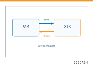 Save and reload prepared dataCache a windowed dataset to disk and reattach it without recompute.
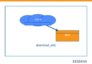 Download a dataset locallyPrefetch BIDS files to a local cache and validate the layout.
Swap any load_dataset(...) call for nm000380 to reproduce the tutorial on this dataset.
Citation
Vikram Rao, Kristin Sellers, Deanna Wallace, Morgan Lee, Maryam Bijanzadeh, … (2018). iEEG during direct electrical stimulation of orbitofrontal and other sites (Rao, Sellers et al., 2018): continuous-stimulation and single-pulse recordings. 10.82901/nemar.nm000380
Provenance
¹Contributed to nemar in BIDS format.
²Curated & ingested by the EEGDash catalog; see CITATION.cff for canonical reference.
³Persistent identifier: 10.82901/nemar.nm000380.
See Also#
eegdash.dataset.EEGDashDataseteegdash.dataset