


NM000353: ieeg dataset, 1 subjects#
Bonn intracranial EEG segments, sets C, D and E (Andrzejak et al. 2001)
Access recordings and metadata through EEGDash.
Citation: Ralph G. Andrzejak, Klaus Lehnertz, Florian Mormann, Christoph Rieke, Peter David, Christian E. Elger (2001). Bonn intracranial EEG segments, sets C, D and E (Andrzejak et al. 2001). 10.82901/nemar.nm000353
Modality: ieeg Subjects: 1 Recordings: 300 License: CC-BY-NC-4.0 Source: nemar
Metadata: Complete (100%)
1-participant iEEG dataset — Bonn intracranial EEG segments, sets C, D and E (Andrzejak et al. 2001).
Quickstart#
Install
pip install eegdash
Access the data
from eegdash.dataset import NM000353

dataset = NM000353(cache_dir="./data")
# Get the raw object of the first recording
raw = dataset.datasets[0].raw
print(raw.info)
Filter by subject
dataset = NM000353(cache_dir="./data", subject="01")
Advanced query
dataset = NM000353(
    cache_dir="./data",
    query={"subject": {"$in": ["01", "02"]}},
)
Iterate recordings
for rec in dataset:
    print(rec.subject, rec.raw.info['sfreq'])
If you use this dataset in your research, please cite the original authors.
BibTeX
@dataset{nm000353,
  title = {Bonn intracranial EEG segments, sets C, D and E (Andrzejak et al. 2001)},
  author = {Ralph G. Andrzejak and Klaus Lehnertz and Florian Mormann and Christoph Rieke and Peter David and Christian E. Elger},
  doi = {10.82901/nemar.nm000353},
  url = {https://doi.org/10.82901/nemar.nm000353},
}
About This Dataset#
Single-channel intracranial EEG segments from five epilepsy patients, from the data set of:
Andrzejak RG, Lehnertz K, Mormann F, Rieke C, David P, Elger CE (2001). Indications of nonlinear deterministic and finite-dimensional structures in time series of brain electrical activity: Dependence on recording region and brain state. Phys. Rev. E 64, 061907. doi:10.1103/PhysRevE.64.061907
Bonn intracranial EEG segments, sets C, D and E (iEEG-BIDS)
Overview
Source dataset: doi:10.34810/data490, hdl:10230/42894 (Repositori Digital de la UPF; mirror of the UPF NTSA download page). The authors ask: “The correct citation is Phys. Rev. E, 64, 061907. No page numbers should be given.”
View full README
Bonn intracranial EEG segments, sets C, D and E (iEEG-BIDS)
Overview
Source dataset: doi:10.34810/data490, hdl:10230/42894 (Repositori Digital de la UPF; mirror of the UPF NTSA download page). The authors ask: “The correct citation is Phys. Rev. E, 64, 061907. No page numbers should be given.” This BIDS dataset is a lossless re-packaging of the intracranial sets only. Every sample is identical to the published text files; nothing was filtered, resampled, re-referenced or rescaled during conversion.
Which sets are included, and why
The source has five sets (A-E) of 100 segments each:
| Set | Source file | Recording | In this dataset |
|---|---|---|---|
| A | `Z.zip` | surface (scalp) EEG, five healthy volunteers, awake, eyes open | **no**: scalp EEG from healthy volunteers, not intracranial |
| B | `O.zip` | surface (scalp) EEG, five healthy volunteers, awake, eyes closed | **no**: scalp EEG from healthy volunteers, not intracranial |
| C | `N.zip` | intracranial, seizure-free interval, hippocampal formation of the opposite hemisphere | yes (`task-interictal_acq-setC`) |
| D | `F.zip` | intracranial, seizure-free interval, within the epileptogenic zone | yes (`task-interictal_acq-setD`) |
| E | `S.zip` | intracranial, seizure activity, all recording sites exhibiting ictal activity | yes (`task-ictal_acq-setE`) |
This is an intracranial EEG (iEEG) release. Sets A and B are scalp recordings from healthy volunteers, so they are not redistributed here. They remain available from the source record.
Cohort and recording
| | |
|---|---|
| Patients | 5 epilepsy patients from the presurgical-diagnosis EEG archive of the Department of Epileptology, University of Bonn; all achieved complete seizure control after resection of one hippocampal formation (paper, Sec. II A) |
| Implant | depth electrodes implanted symmetrically into both hippocampal formations; strip electrodes on the lateral and basal neocortex (paper, Fig. 2) |
| Sets C and D | hippocampal depth-electrode contacts (C: opposite hemisphere; D: within the epileptogenic zone), seizure-free interval |
| Set E | contacts of any depicted electrode (depth or strip) exhibiting ictal activity |
| Amplifier | "the same 128-channel amplifier system" for all sets; manufacturer and model not reported |
| Digitization | 12-bit analog-to-digital conversion, 173.61 Hz |
| Reference | average common reference, omitting electrodes containing pathological activity |
| Age, sex, per-patient details | not reported by the source; segments cannot be attributed to patients |
Recordings (paper, Sec. II A)
“Sets C, D, and E originated from our EEG archive of presurgical diagnosis. For the present study EEGs from five patients were selected, all of whom had achieved complete seizure control after resection of one of the hippocampal formations, which was therefore correctly diagnosed to be the epileptogenic zone.”
Depth electrodes were implanted symmetrically into the hippocampal formations. Segments of sets C and D were taken from all contacts of the respective depth electrode. Strip electrodes were implanted onto the lateral and basal regions of the neocortex. Segments of set E were taken from contacts of all depicted electrodes (paper, Fig. 2).
Segments (23.6 s each) were selected and cut out of continuous multichannel EEG recordings after visual inspection for artifacts, e.g. muscle activity or eye movements. They also had to satisfy a weak-stationarity criterion (paper, Sec. II B).
“All EEG signals were recorded with the same 128-channel amplifier system, using an average common reference [omitting electrodes containing pathological activity (C, D, and E) …]. After 12 bit analog-to-digital conversion, the data were written continuously onto the disk of a data acquisition computer system at a sampling rate of 173.61 Hz. Band-pass filter settings were 0.53-40 Hz (12 dB/oct.).”
Funding: Deutsche Forschungsgemeinschaft. The paper reports no ethics statement.
Task / paradigm and timing
There was no task. Each run is one 23.6 s segment cut by the authors out of a continuous clinical recording:
task-interictal (sets C and D, seizure-free interval) or task-ictal (set E, seizure activity). Segments are
not time-locked to any stimulus or to seizure onset, and their position in the original recording is not
provided. Each ..._events.tsv has one event (onset 0, duration of the run) whose trial_type names the set
(levels described in task-*_events.json).
Files
Content and verified properties
| | |
|---|---|
| Segments | 300 (100 per set C, D, E) |
| Channels per run | 1 (`x`) |
| Samples per segment | **4097** in every file (the paper and the download page say 4096) |
| Sampling rate | 173.61 Hz (duration 4097 / 173.61 = 23.599 s) |
| Values | integers, -1885 to 2047 |
| Patients | 5 (pooled; segment-to-patient mapping not provided) |
The values are integers in the range of a signed 12-bit converter (-2048 to 2047). Three segments reach 2047,
the converter maximum, in 72 samples in total (set D: 1 file; set E: 2 files). These samples are probably
clipped. Per-file counts are in sub-pooled_scans.tsv (n_samples_at_2047).
BIDS layout and source-to-BIDS mapping
The source states: “the signals included in these sets are randomized with regard to the recording contact and
the patient or volunteer. Accordingly, the information which signal corresponds to which recording contact or
patient or volunteer is not available.” All runs therefore sit under one pseudo-subject, sub-pooled. This is
not one person: it pools anonymous segments from five patients. Do not treat segments as independent
subjects.
| Source | BIDS |
|---|---|
| `N.zip` / `N<NNN>.TXT` (set C) | `sub-pooled/ieeg/sub-pooled_task-interictal_acq-setC_run-<NNN>_ieeg.{vhdr,vmrk,eeg}` |
| `F.zip` / `F<NNN>.txt` (set D) | `sub-pooled/ieeg/sub-pooled_task-interictal_acq-setD_run-<NNN>_ieeg.{vhdr,vmrk,eeg}` |
| `S.zip` / `S<NNN>.txt` (set E) | `sub-pooled/ieeg/sub-pooled_task-ictal_acq-setE_run-<NNN>_ieeg.{vhdr,vmrk,eeg}` |
| the single column of each file | channel `x` |
| `N.zip`, `F.zip`, `S.zip`, repository metadata | byte-identical copies in `sourcedata/upf-repositori-10230-42894/` |
| `Z.zip`, `O.zip` (sets A, B, scalp) | not included (see above) |
The run number equals the number in the source file name (001-100). task-interictal / task-ictal label the
brain state; there was no task. sub-pooled_scans.tsv lists for every run the set, source file and its
SHA-256, value range, clipping counts, recording region and brain state. Sidecars are shared by
inheritance:
- sub-pooled_task-interictal_ieeg.json and sub-pooled_task-ictal_ieeg.json
- sub-pooled_task-interictal_channels.tsv: type SEEG, because sets C and D come from depth-electrode
contacts in the hippocampal formation.
sub-pooled_task-ictal_channels.tsv: typeOTHER, because a set E segment may come from a depth or a strip contact and the source does not say which.one
..._acq-setX_events.tsvper set, with a single event spanning the run.
Electrode positions are not available, so there is no electrodes.tsv.
Data format and exactness
The source integers are stored as BrainVision INT_16 (multiplexed, resolution 1). A separate round-trip check
re-read all 300 files and compared every sample with the source text (see the campaign ledger).
Preprocessing already applied by the source
Segment selection: segments were “selected and cut out of continuous multichannel EEG recordings after visual inspection for artifacts, e.g., due to muscle activity or eye movements” and had to fulfil a weak-stationarity criterion (paper, Sec. II A and II B 2).
Segment boundaries (paper, Sec. II B 1, paraphrased): to avoid spurious spectral components from discontinuities, 4396-sample stretches were first cut out of the recordings; within each, the start of the final 4096-sample segment was chosen so that the amplitude difference between the last and first samples was within the range of differences between consecutive samples, and the slopes at the end and start had the same sign.
Filtering: see below.
Filtering: the paper and the download page disagree
Paper: “Band-pass filter settings were 0.53-40 Hz (12 dB/oct.).”
UPF download page (captured 2026-10-06): “The time series you can download here are not filtered. The application of a low-pass filter of 40 Hz, as described in the manuscript, is regarded as the first step of analysis and therefore not carried out for the downloadable time series.”
Both statements are reproduced here; the BIDS sidecars therefore record SoftwareFilters and
HardwareFilters as n/a and quote both in FilterNotes. No filtering was done during conversion.
Known caveats
sub-pooledis a pseudo-subject that pools five patients; see “BIDS layout and source-to-BIDS mapping”.Every file has 4097 samples although the paper and the download page say 4096; see “Content and verified properties”.
Three segments reach the converter maximum (probable clipping); see “Content and verified properties”.
The physical unit of the integer values is not documented; see “Units”.
The paper and the download page disagree on filtering; see “Filtering”.
Set E channels are typed
OTHERbecause a segment may come from a depth or a strip contact.
Units
The source does not state the physical scale of the integer values (for example, microvolts per unit). The
paper’s Fig. 3 caption says intracranial amplitudes are “around some 100 µV” and seizure activity “can exceed
1000 µV”, which is consistent with roughly 1 µV per unit. That is not a documented calibration. Channel units
are therefore n/a (in channels.tsv and in the BrainVision header), and the values are the source integers
unchanged.
Privacy
The published files contain only integer samples (no headers, names, dates or identifiers). The source randomized segments across patients and contacts. Converted headers contain no dates or identifiers.
How to load
The signal files are stored with git-annex on NEMAR; fetch them first (for example git annex get sub-pooled
or the NEMAR download tools). Then, with MNE-BIDS:
from mne_bids import BIDSPath, read_raw_bids
bids_path = BIDSPath(root="nm000353", subject="pooled", task="ictal",
                     acquisition="setE", run="001", datatype="ieeg")

raw = read_raw_bids(bids_path)
The channel unit is n/a (see “Units”), so MNE may warn about it. Compare raw.get_data() with the
min/max columns of sub-pooled_scans.tsv to check whether your reader applied any scaling to the source
integers.
License and terms of use
The repository record lists two rights statements (dc.rights), quoted verbatim: 1. “Licensed under a Creative Commons License (CC-BY) 4.0” (https://creativecommons.org/licenses/by/4.0/) 2. “The source codes, data and results on these sites are free of charge for research and education purposes
only. Any commercial or military use is prohibited. All resources are provided without any expressed or implied warranty. In no event the authors of the article or any of their host institutions are liable for any damages arising from the use of the software, data or results.”
The UPF download page carries the same Legal Agreement. To respect the research-and-education-only condition,
this BIDS release uses the closest standard license, CC-BY-NC-4.0 (attribution, non-commercial). No
standard license expresses the additional prohibition of military use, and it still applies: by using
these data you agree to use them for research and education only, and not for commercial or military
purposes. See LICENSE.
How to cite
Cite Andrzejak et al. (2001), Phys. Rev. E 64, 061907 (doi:10.1103/PhysRevE.64.061907) and the dataset doi:10.34810/data490. Also cite this BIDS release by its NEMAR identifier.
Conversion provenance
Converted 2026-10-06 on SDSC Voyager (Kubernetes jobs) by the iEEG-NEMAR campaign (lane G) with
laneG_convert.py and laneG_finalize.py. Source files were downloaded through the repository’s DSpace REST
API, and every bitstream’s MD5 matched the repository checksum. See sourcedata/provenance.json. Paper
details were taken from the published version deposited at hdl:10230/43637 (repository full-text extraction)
and from the UPF NTSA download page.
Additional details in this README (affiliations, acknowledgments, segment-boundary procedure) were read on
2026-10-06 from the repository text extraction (Andrzejak_PhysRevE2001.pdf.txt) of the published article
deposited at hdl:10230/43637.
NEMAR Metadata#
[](https://doi.org/10.82901/nemar.nm000353) # Bonn intracranial EEG segments, sets C, D and E (iEEG-BIDS) ## Overview Single-channel intracranial EEG segments from five epilepsy patients, from the data set of: > Andrzejak RG, Lehnertz K, Mormann F, Rieke C, David P, Elger CE (2001). Indications of nonlinear > deterministic and finite-dimensional structures in time series of brain electrical activity: Dependence on > recording region and brain state. Phys. Rev. E 64, 061907. > doi:[10.1103/PhysRevE.64.061907](https://doi.org/10.1103/PhysRevE.64.061907) Source dataset: doi:[10.34810/data490](https://doi.org/10.34810/data490), hdl:[10230/42894](http://hdl.handle.net/10230/42894) (Repositori Digital de la UPF; mirror of the UPF NTSA download page). The authors ask: “The correct citation is Phys. Rev. E, 64, 061907. No page numbers should be given.” This BIDS dataset is a lossless re-packaging of the intracranial sets only. Every sample is identical to the published text files; nothing was filtered, resampled, re-referenced or rescaled during conversion. ## Which sets are included, and why The source has five sets (A-E) of 100 segments each: | Set | Source file | Recording | In this dataset | |---|—|---|—| | A | Z.zip | surface (scalp) EEG, five healthy volunteers, awake, eyes open | no: scalp EEG from healthy volunteers, not intracranial | | B | O.zip | surface (scalp) EEG, five healthy volunteers, awake, eyes closed | no: scalp EEG from healthy volunteers, not intracranial | | C | N.zip | intracranial, seizure-free interval, hippocampal formation of the opposite hemisphere | yes (task-interictal_acq-setC) | | D | F.zip | intracranial, seizure-free interval, within the epileptogenic zone | yes (task-interictal_acq-setD) | | E | S.zip | intracranial, seizure activity, all recording sites exhibiting ictal activity | yes (task-ictal_acq-setE) | This is an intracranial EEG (iEEG) release. Sets A and B are scalp recordings from healthy volunteers, so they are not redistributed here. They remain available from the source record. ## Cohort and recording | | | |---|—| | Patients | 5 epilepsy patients from the presurgical-diagnosis EEG archive of the Department of Epileptology, University of Bonn; all achieved complete seizure control after resection of one hippocampal formation (paper, Sec. II A) | | Implant | depth electrodes implanted symmetrically into both hippocampal formations; strip electrodes on the lateral and basal neocortex (paper, Fig. 2) | | Sets C and D | hippocampal depth-electrode contacts (C: opposite hemisphere; D: within the epileptogenic zone), seizure-free interval | | Set E | contacts of any depicted electrode (depth or strip) exhibiting ictal activity | | Amplifier | “the same 128-channel amplifier system” for all sets; manufacturer and model not reported | | Digitization | 12-bit analog-to-digital conversion, 173.61 Hz | | Reference | average common reference, omitting electrodes containing pathological activity | | Age, sex, per-patient details | not reported by the source; segments cannot be attributed to patients | ### Recordings (paper, Sec. II A) - “Sets C, D, and E originated from our EEG archive of presurgical diagnosis. For the present study EEGs from
five patients were selected, all of whom had achieved complete seizure control after resection of one of the hippocampal formations, which was therefore correctly diagnosed to be the epileptogenic zone.”
Depth electrodes were implanted symmetrically into the hippocampal formations. Segments of sets C and D were taken from all contacts of the respective depth electrode. Strip electrodes were implanted onto the lateral and basal regions of the neocortex. Segments of set E were taken from contacts of all depicted electrodes (paper, Fig. 2).
Segments (23.6 s each) were selected and cut out of continuous multichannel EEG recordings after visual inspection for artifacts, e.g. muscle activity or eye movements. They also had to satisfy a weak-stationarity criterion (paper, Sec. II B).
“All EEG signals were recorded with the same 128-channel amplifier system, using an average common reference [omitting electrodes containing pathological activity (C, D, and E) …]. After 12 bit analog-to-digital conversion, the data were written continuously onto the disk of a data acquisition computer system at a sampling rate of 173.61 Hz. Band-pass filter settings were 0.53-40 Hz (12 dB/oct.).”
Funding: Deutsche Forschungsgemeinschaft. The paper reports no ethics statement.
## Task / paradigm and timing There was no task. Each run is one 23.6 s segment cut by the authors out of a continuous clinical recording: task-interictal (sets C and D, seizure-free interval) or task-ictal (set E, seizure activity). Segments are not time-locked to any stimulus or to seizure onset, and their position in the original recording is not provided. Each …_events.tsv has one event (onset 0, duration of the run) whose trial_type names the set (levels described in task-*_events.json). ## Files ### Content and verified properties | | | |---|—| | Segments | 300 (100 per set C, D, E) | | Channels per run | 1 (x) | | Samples per segment | 4097 in every file (the paper and the download page say 4096) | | Sampling rate | 173.61 Hz (duration 4097 / 173.61 = 23.599 s) | | Values | integers, -1885 to 2047 | | Patients | 5 (pooled; segment-to-patient mapping not provided) | The values are integers in the range of a signed 12-bit converter (-2048 to 2047). Three segments reach 2047, the converter maximum, in 72 samples in total (set D: 1 file; set E: 2 files). These samples are probably clipped. Per-file counts are in sub-pooled_scans.tsv (n_samples_at_2047). ### BIDS layout and source-to-BIDS mapping The source states: “the signals included in these sets are randomized with regard to the recording contact and the patient or volunteer. Accordingly, the information which signal corresponds to which recording contact or patient or volunteer is not available.” All runs therefore sit under one pseudo-subject, sub-pooled. This is not one person: it pools anonymous segments from five patients. Do not treat segments as independent subjects. | Source | BIDS | |---|—| | N.zip / N<NNN>.TXT (set C) | sub-pooled/ieeg/sub-pooled_task-interictal_acq-setC_run-<NNN>_ieeg.{vhdr,vmrk,eeg} | | F.zip / F<NNN>.txt (set D) | sub-pooled/ieeg/sub-pooled_task-interictal_acq-setD_run-<NNN>_ieeg.{vhdr,vmrk,eeg} | | S.zip / S<NNN>.txt (set E) | sub-pooled/ieeg/sub-pooled_task-ictal_acq-setE_run-<NNN>_ieeg.{vhdr,vmrk,eeg} | | the single column of each file | channel x | | N.zip, F.zip, S.zip, repository metadata | byte-identical copies in sourcedata/upf-repositori-10230-42894/ | | Z.zip, O.zip (sets A, B, scalp) | not included (see above) | The run number equals the number in the source file name (001-100). task-interictal / task-ictal label the brain state; there was no task. sub-pooled_scans.tsv lists for every run the set, source file and its SHA-256, value range, clipping counts, recording region and brain state. Sidecars are shared by inheritance: - sub-pooled_task-interictal_ieeg.json and sub-pooled_task-ictal_ieeg.json - sub-pooled_task-interictal_channels.tsv: type SEEG, because sets C and D come from depth-electrode
contacts in the hippocampal formation.
sub-pooled_task-ictal_channels.tsv: type OTHER, because a set E segment may come from a depth or a strip contact and the source does not say which.
one …_acq-setX_events.tsv per set, with a single event spanning the run.
Electrode positions are not available, so there is no electrodes.tsv. ### Data format and exactness The source integers are stored as BrainVision INT_16 (multiplexed, resolution 1). A separate round-trip check re-read all 300 files and compared every sample with the source text (see the campaign ledger). ## Preprocessing already applied by the source - Segment selection: segments were “selected and cut out of continuous multichannel EEG recordings after visual
inspection for artifacts, e.g., due to muscle activity or eye movements” and had to fulfil a weak-stationarity criterion (paper, Sec. II A and II B 2).
Segment boundaries (paper, Sec. II B 1, paraphrased): to avoid spurious spectral components from discontinuities, 4396-sample stretches were first cut out of the recordings; within each, the start of the final 4096-sample segment was chosen so that the amplitude difference between the last and first samples was within the range of differences between consecutive samples, and the slopes at the end and start had the same sign.
Filtering: see below.
### Filtering: the paper and the download page disagree - Paper: “Band-pass filter settings were 0.53-40 Hz (12 dB/oct.).” - UPF download page (captured 2026-10-06): “The time series you can download here are not filtered. The
application of a low-pass filter of 40 Hz, as described in the manuscript, is regarded as the first step of analysis and therefore not carried out for the downloadable time series.”
Both statements are reproduced here; the BIDS sidecars therefore record SoftwareFilters and HardwareFilters as n/a and quote both in FilterNotes. No filtering was done during conversion. ## Known caveats - sub-pooled is a pseudo-subject that pools five patients; see “BIDS layout and source-to-BIDS mapping”. - Every file has 4097 samples although the paper and the download page say 4096; see “Content and verified properties”. - Three segments reach the converter maximum (probable clipping); see “Content and verified properties”. - The physical unit of the integer values is not documented; see “Units”. - The paper and the download page disagree on filtering; see “Filtering”. - Set E channels are typed OTHER because a segment may come from a depth or a strip contact. ### Units The source does not state the physical scale of the integer values (for example, microvolts per unit). The paper’s Fig. 3 caption says intracranial amplitudes are “around some 100 µV” and seizure activity “can exceed 1000 µV”, which is consistent with roughly 1 µV per unit. That is not a documented calibration. Channel units are therefore n/a (in channels.tsv and in the BrainVision header), and the values are the source integers unchanged. ## Privacy The published files contain only integer samples (no headers, names, dates or identifiers). The source randomized segments across patients and contacts. Converted headers contain no dates or identifiers. ## How to load The signal files are stored with git-annex on NEMAR; fetch them first (for example git annex get sub-pooled or the NEMAR download tools). Then, with MNE-BIDS: ```python from mne_bids import BIDSPath, read_raw_bids bids_path = BIDSPath(root=”nm000353”, subject=”pooled”, task=”ictal”,
acquisition=”setE”, run=”001”, datatype=”ieeg”)
raw = read_raw_bids(bids_path) ``` The channel unit is n/a (see “Units”), so MNE may warn about it. Compare raw.get_data() with the min/max columns of sub-pooled_scans.tsv to check whether your reader applied any scaling to the source integers. ## License and terms of use The repository record lists two rights statements (dc.rights), quoted verbatim: 1. “Licensed under a Creative Commons License (CC-BY) 4.0” (https://creativecommons.org/licenses/by/4.0/) 2. “The source codes, data and results on these sites are free of charge for research and education purposes
only. Any commercial or military use is prohibited. All resources are provided without any expressed or implied warranty. In no event the authors of the article or any of their host institutions are liable for any damages arising from the use of the software, data or results.”
The UPF download page carries the same Legal Agreement. To respect the research-and-education-only condition, this BIDS release uses the closest standard license, CC-BY-NC-4.0 (attribution, non-commercial). No standard license expresses the additional prohibition of military use, and it still applies: by using these data you agree to use them for research and education only, and not for commercial or military purposes. See LICENSE. ## How to cite Cite Andrzejak et al. (2001), Phys. Rev. E 64, 061907 (doi:10.1103/PhysRevE.64.061907) and the dataset doi:10.34810/data490. Also cite this BIDS release by its NEMAR identifier. ## Conversion provenance Converted 2026-10-06 on SDSC Voyager (Kubernetes jobs) by the iEEG-NEMAR campaign (lane G) with laneG_convert.py and laneG_finalize.py. Source files were downloaded through the repository’s DSpace REST API, and every bitstream’s MD5 matched the repository checksum. See sourcedata/provenance.json. Paper details were taken from the published version deposited at hdl:10230/43637 (repository full-text extraction) and from the UPF NTSA download page. Additional details in this README (affiliations, acknowledgments, segment-boundary procedure) were read on 2026-10-06 from the repository text extraction (Andrzejak_PhysRevE2001.pdf.txt) of the published article deposited at hdl:[10230/43637](http://hdl.handle.net/10230/43637).
License: CC-BY-NC-4.0
Authors:
Ralph G. Andrzejak
Klaus Lehnertz
Florian Mormann
Christoph Rieke
Peter David
… and 1 more
Versions:
Version |
DOI |
Released |
|---|---|---|
|
Cohort#
Dataset Statistics#
Channel counts: 1 ch (n=300 recordings)
Sampling frequencies: 173.61 Hz (n=300 recordings)
Total recording duration: 1 h 57 min
Signal · Electrodes & live trace#
Live trace viewer — sub-pooled · task-ictal · run-032
Showing one representative recording out of
1 subjects and 300 recordings in this dataset.
Browse the full set on OpenNeuro;
drop any other _ieeg.{set,edf,bdf,vhdr} file onto the
viewer (or pass ?ieeg=<url>) to inspect it.
No scalp electrode layout is currently indexed for this dataset. Once the eegdash montage registry ingests it, the interactive viewer will appear here automatically.
NEMAR Processing Statistics#
The plots below are generated by NEMAR’s automated EEG pipeline. The histogram shows pipeline success for data cleaning and ICA decomposition, the percentage of data frames and EEG channels retained after artefact removal, line noise per channel (RMS, dB), and the age/gender distribution of participants.

HED event descriptors word cloud

Manifest#
File Explorer#
Browse the BIDS file structure of this dataset. Records are fetched on demand from the EEGDash catalog the first time you open the explorer.
Full dataset metadata table
Dataset ID |
|
Title |
Bonn intracranial EEG segments, sets C, D and E (Andrzejak et al. 2001) |
Author (year) |
— |
Canonical |
— |
Importable as |
|
Year |
2001 |
Authors |
Ralph G. Andrzejak, Klaus Lehnertz, Florian Mormann, Christoph Rieke, Peter David, Christian E. Elger |
License |
CC-BY-NC-4.0 |
Citation / DOI |
|
Source links |
OpenNeuro | NeMAR | Source URL |
Copy-paste BibTeX
@dataset{nm000353,
  title = {Bonn intracranial EEG segments, sets C, D and E (Andrzejak et al. 2001)},
  author = {Ralph G. Andrzejak and Klaus Lehnertz and Florian Mormann and Christoph Rieke and Peter David and Christian E. Elger},
  doi = {10.82901/nemar.nm000353},
  url = {https://doi.org/10.82901/nemar.nm000353},
}
API Reference#
eegdash.datasetEEGDashDataset- class eegdash.dataset.NM000353(cache_dir: str, query: dict | None = None, s3_bucket: str | None = None, **kwargs)[source]#
Bonn intracranial EEG segments, sets C, D and E (Andrzejak et al. 2001)
- Study:
nm000353(NeMAR)- Author (year):
—
- Canonical:
—
Also importable as:
NM000353.Modality:
ieeg; Subject type:Unknown. Subjects: 1; recordings: 300; tasks: 2.- Parameters:
cache_dir (str | Path) – Directory where data are cached locally.
query (dict | None) – Additional MongoDB-style filters to AND with the dataset selection. Must not contain the key
dataset.s3_bucket (str | None) – Base S3 bucket used to locate the data.
**kwargs (dict) – Additional keyword arguments forwarded to
EEGDashDataset.
- data_dir#
Local dataset cache directory (
cache_dir / dataset_id).- Type:
Path
Notes
Each item is a recording; recording-level metadata are available via
dataset.description.querysupports MongoDB-style filters on fields inALLOWED_QUERY_FIELDSand is combined with the dataset filter. Dataset-specific caveats are not provided in the summary metadata.References
OpenNeuro dataset: https://openneuro.org/datasets/nm000353 NeMAR dataset: https://nemar.org/dataexplorer/detail?dataset_id=nm000353 DOI: https://doi.org/10.82901/nemar.nm000353
Examples
>>> from eegdash.dataset import NM000353
>>> dataset = NM000353(cache_dir="./data")
>>> recording = dataset[0]
>>> raw = recording.load()
- __init__(cache_dir: str, query: dict | None = None, s3_bucket: str | None = None, **kwargs)[source]#
- save(path: str, overwrite: bool = False, offset: int = 0)[source]#
Save datasets to files by creating one subdirectory for each dataset:
path/
    0/
        0-raw.fif | 0-epo.fif
        description.json
        raw_preproc_kwargs.json (if raws were preprocessed)
        window_kwargs.json (if this is a windowed dataset)
        window_preproc_kwargs.json  (if windows were preprocessed)
        target_name.json (if target_name is not None and dataset is raw)
    1/
        1-raw.fif | 1-epo.fif
        description.json
        raw_preproc_kwargs.json (if raws were preprocessed)
        window_kwargs.json (if this is a windowed dataset)
        window_preproc_kwargs.json  (if windows were preprocessed)
        target_name.json (if target_name is not None and dataset is raw)
- Parameters:
path (str) –
- Directory in which subdirectories are created to store
-raw.fif | -epo.fif and .json files to.
overwrite (bool) – Whether to delete old subdirectories that will be saved to in this call.
offset (int) – If provided, the integer is added to the id of the dataset in the concat. This is useful in the setting of very large datasets, where one dataset has to be processed and saved at a time to account for its original position.
BaseDataset from braindecode — windowed via create_windows_from_events.braindecodeDataLoader; supports parallel workers and on-the-fly augmentations.pytorch Find datasets with the EEGDash APIQuery the catalogue, filter by task or modality, list candidates.
 Load one EEG recordingResolve a single record to an MNE Raw with channels and events.
 EEG recording to PyTorch DataLoaderWrap braindecode windows in a DataLoader for model training.
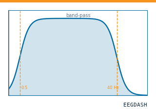 Preprocess EEG and create windowsFilter, resample, epoch — and persist the windowed dataset.
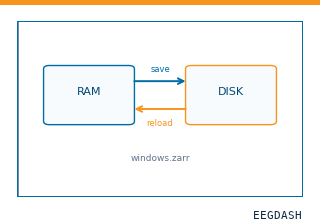 Save and reload prepared dataCache a windowed dataset to disk and reattach it without recompute.
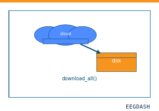 Download a dataset locallyPrefetch BIDS files to a local cache and validate the layout.
Swap any load_dataset(...) call for nm000353 to reproduce the tutorial on this dataset.
Citation
Ralph G. Andrzejak, Klaus Lehnertz, Florian Mormann, Christoph Rieke, Peter David, … (2001). Bonn intracranial EEG segments, sets C, D and E (Andrzejak et al. 2001). 10.82901/nemar.nm000353
Provenance
¹Contributed to nemar in BIDS format.
²Curated & ingested by the EEGDash catalog; see CITATION.cff for canonical reference.
³Persistent identifier: 10.82901/nemar.nm000353.
See Also#
eegdash.dataset.EEGDashDataseteegdash.dataset