


NM000396: ieeg dataset, 13 subjects#
Developmental ECoG: resting-state electrocorticography from 13 epilepsy patients aged 3-33 years (Miles, Weaver, Webb, Ojemann 2025)
Access recordings and metadata through EEGDash.
Citation: Jesse T. Miles, Kurt E. Weaver, Sara Jane Webb, Jeffrey G. Ojemann (2026). Developmental ECoG: resting-state electrocorticography from 13 epilepsy patients aged 3-33 years (Miles, Weaver, Webb, Ojemann 2025). 10.82901/nemar.nm000396
Modality: ieeg Subjects: 13 Recordings: 13 License: CC-BY-NC-4.0 Source: nemar
Metadata: Complete (100%)
13-participant iEEG dataset — Developmental ECoG: resting-state electrocorticography from 13 epilepsy patients aged 3-33 years (Miles, Weaver, Webb, Ojemann 2025).
Quickstart#
Install
pip install eegdash
Access the data
from eegdash.dataset import NM000396

dataset = NM000396(cache_dir="./data")
# Get the raw object of the first recording
raw = dataset.datasets[0].raw
print(raw.info)
Filter by subject
dataset = NM000396(cache_dir="./data", subject="01")
Advanced query
dataset = NM000396(
    cache_dir="./data",
    query={"subject": {"$in": ["01", "02"]}},
)
Iterate recordings
for rec in dataset:
    print(rec.subject, rec.raw.info['sfreq'])
If you use this dataset in your research, please cite the original authors.
BibTeX
@dataset{nm000396,
  title = {Developmental ECoG: resting-state electrocorticography from 13 epilepsy patients aged 3-33 years (Miles, Weaver, Webb, Ojemann 2025)},
  author = {Jesse T. Miles and Kurt E. Weaver and Sara Jane Webb and Jeffrey G. Ojemann},
  doi = {10.82901/nemar.nm000396},
  url = {https://doi.org/10.82901/nemar.nm000396},
}
About This Dataset#
Resting-state intracranial recordings (subdural grids and strips; depth electrodes in some participants) from **13
patients with epilepsy aged 3.4-33.9 years** (6 male, 7 female) undergoing intracranial monitoring at Seattle Children’s Hospital and Harborview Medical Center (Seattle, WA, USA). Re-packaged in iEEG-BIDS from the authors’ public release (Zenodo record 16954184, “Developmental ECoG”, CC-BY-NC-4.0).
Reference article: Miles JT, Weaver KE, Webb SJ, Ojemann JG. *Developmental relationships between the human alpha
rhythm and intrinsic neural timescales are dependent on neural hierarchy.* Journal of Neurophysiology 135:143-152 (2026). https://doi.org/10.1152/jn.00435.2025 (preprint: https://doi.org/10.1101/2025.08.21.671637).
Developmental ECoG (Miles, Weaver, Webb, Ojemann 2025): resting-state electrocorticography, ages 3-33
Participants
participants.tsv gives age, sex, implanted electrodes, coverage and epileptiform zones for each participant from
Table 1 of the paper. The release orders participants by age (sub01 = P1, 3.42 years … sub13 = P13, 33.92 years);
the ages in the release montage files match Table 1 (checked by the converter). release_id is the pseudonymous
code of the release folder.
View full README
Developmental ECoG (Miles, Weaver, Webb, Ojemann 2025): resting-state electrocorticography, ages 3-33
Participants
participants.tsv gives age, sex, implanted electrodes, coverage and epileptiform zones for each participant from
Table 1 of the paper. The release orders participants by age (sub01 = P1, 3.42 years … sub13 = P13, 33.92 years);
the ages in the release montage files match Table 1 (checked by the converter). release_id is the pseudonymous
code of the release folder.
Task
task-rest: dedicated resting-state period. Participants were asked to lie quietly, upright, for at least three
minutes before other research experiments (paper, Methods). There are no events.
Recording
Sampling rates differ by site and system (512-4800 Hz; release channel tables: 1200 Hz for 9 participants, 4800,
1525.878906, 1220.703125, 1000 and 512 Hz for the others). Filter settings and notch ranges are copied from the
release channel tables into channels.tsv. Power line frequency: 60 Hz.
- Units. The release channel tables give the ECoG units as unscaled for 12 of the 13 participants: the values
are as exported from the acquisition system and the release does not give a scale factor to volts.
channels.tsvtherefore listsn/aunits for these channels, and the BrainVision header saysunscaled. Amplitudes are not comparable across participants. sub-02’s values are in microvolts.
sub-02 also has the depth electrodes (
A'…``S’, named as in the ROSA planning file), one ECG channel (``EKG-Ref) and one unlabelled channel (C154-Ref, type MISC). These are not in the release channel table. Their types are set from the channel names, and the units are assumed to be the same microvolts as the other channels of that export. The release Sample/Time columns show that the excerpt starts at sample 4234240 (8270 s) of the clinical file (seeRecordingDescriptionin the sidecar). The release’s per-contact anatomical labels for the depth electrodes (d419f2_Anatomical_Labels.txt) are kept insourcedata/.sub-08: channels 49-64 are marked
bad(release note: “49-64 are empty channels”; 1-32 are a left parietal half grid and 33-48 a left interhemispheric strip).analysis_regioninchannels.tsvmarks the 12 channels the paper analysed (two per gyrus: middle frontal, precentral, postcentral, supramarginal, superior temporal and middle temporal), from the release montage files.Electrode coordinates are not part of the release.
electrodes.tsvlists the ECoG/sEEG electrode names withn/acoordinates (spaceOther).
What was converted, and how
Each RestingState_<id>_iEEG.csv (samples × channels) became one BrainVision run (sub-XX_task-rest_ieeg.vhdr,
IEEE float32). The file values are the CSV values rounded to float32, and the round-trip check compares every run
with the CSV. No filtering, resampling, re-referencing or channel removal was applied. The paper’s processing
(resampling to 1024 Hz, bipolar re-referencing, drift and line-noise filtering, z-scoring) is not applied.
sourcedata/zenodo-16954184/ holds the original release zip byte for byte (CSV tables, channel tables, montage files,
the sub02 anatomical labels and the sub08 note) and the Zenodo record metadata.
License
CC-BY-NC-4.0
Additional metadata and localisation (added 2026-10-08)
Compiled after the upload from the article, its supplement and the source deposit (each statement names its source). Text and sidecar metadata only; no data file was changed.
Recording system. Sampled “at a minimum of 512 Hz and maximum of 4800 Hz (depending on acquisition system and site)” (preprint Methods “Signal processing”); per-subject rates in the deposit channels.txt: 1200 Hz (9 subjects), 512, 4800, 1525.88, 1220.70 and 1000 Hz. channels.txt lists low_cutoff 0, high_cutoff 500, notch 58-62, units “unscaled” (deposit, e.g. sub06 channels.txt). Amplifier make: n/a (not stated).
Reference scheme. The deposit CSVs hold the recorded channels; the paper analysed bipolar pairs of two adjacent contacts within the same gyrus (preprint Methods). channels.txt status_description reads “keep for referencing”. Original recording reference: n/a.
Electrode types. Subdural grids (8x8, 4x8) and strips, some patients also with depth electrodes (preprint Table 1). Deposit note sub08_a9952e/note.txt: “1-32 are parietal half grid (left); 33-48 are intrahemispheric strip (left); 49-64 are empty channels”.
Localisation method. “ECoG contact locations were identified by clinical MRI reconstructions aligned to CT scans. Electrode orientations (and, by extension, channel numbering) were cross-referenced to a combination of intra-operative placement photos, surgical notes, and clinical monitoring notes.” “Gyri were identified based on automated registration of Harvard-Oxford atlas labels to patient clinical neuroimaging using the Localizing Electrodes GUI and manual inspection of co-registered CT and T1-weighted MR images.” (preprint Methods). The deposit montage.csv per subject gives the gyrus of each analysed contact (empty ch = gyrus not analysed for that subject). No coordinates or imaging are deposited. For sub02 the deposit also contains d419f2_Anatomical_Labels.txt (ROSA-planned depth electrodes A’-S’, 116 contacts, atlas labels “not independently verified”); those depth channels are not part of the deposited resting-state recording.
These gyrus labels are now in the anat_label column of each *_electrodes.tsv (n/a for contacts that montage.csv does not list).
NEMAR Metadata#
[](https://doi.org/10.82901/nemar.nm000396) # Developmental ECoG (Miles, Weaver, Webb, Ojemann 2025): resting-state electrocorticography, ages 3-33 Resting-state intracranial recordings (subdural grids and strips; depth electrodes in some participants) from 13 patients with epilepsy aged 3.4-33.9 years (6 male, 7 female) undergoing intracranial monitoring at Seattle Children’s Hospital and Harborview Medical Center (Seattle, WA, USA). Re-packaged in iEEG-BIDS from the authors’ public release (Zenodo record [16954184](https://doi.org/10.5281/zenodo.16954184), “Developmental ECoG”, CC-BY-NC-4.0). Reference article: Miles JT, Weaver KE, Webb SJ, Ojemann JG. Developmental relationships between the human alpha rhythm and intrinsic neural timescales are dependent on neural hierarchy. Journal of Neurophysiology 135:143-152 (2026). https://doi.org/10.1152/jn.00435.2025 (preprint: https://doi.org/10.1101/2025.08.21.671637). ## Participants participants.tsv gives age, sex, implanted electrodes, coverage and epileptiform zones for each participant from Table 1 of the paper. The release orders participants by age (sub01 = P1, 3.42 years … sub13 = P13, 33.92 years); the ages in the release montage files match Table 1 (checked by the converter). release_id is the pseudonymous code of the release folder. ## Task task-rest: dedicated resting-state period. Participants were asked to lie quietly, upright, for at least three minutes before other research experiments (paper, Methods). There are no events. ## Recording Sampling rates differ by site and system (512-4800 Hz; release channel tables: 1200 Hz for 9 participants, 4800, 1525.878906, 1220.703125, 1000 and 512 Hz for the others). Filter settings and notch ranges are copied from the release channel tables into channels.tsv. Power line frequency: 60 Hz. - Units. The release channel tables give the ECoG units as unscaled for 12 of the 13 participants: the values
are as exported from the acquisition system and the release does not give a scale factor to volts. channels.tsv therefore lists n/a units for these channels, and the BrainVision header says unscaled. Amplitudes are not comparable across participants. sub-02’s values are in microvolts.
sub-02 also has the depth electrodes (A’…`S’, named as in the ROSA planning file), one ECG channel (`EKG-Ref) and one unlabelled channel (C154-Ref, type MISC). These are not in the release channel table. Their types are set from the channel names, and the units are assumed to be the same microvolts as the other channels of that export. The release Sample/Time columns show that the excerpt starts at sample 4234240 (8270 s) of the clinical file (see RecordingDescription in the sidecar). The release’s per-contact anatomical labels for the depth electrodes (d419f2_Anatomical_Labels.txt) are kept in sourcedata/.
sub-08: channels 49-64 are marked bad (release note: “49-64 are empty channels”; 1-32 are a left parietal half grid and 33-48 a left interhemispheric strip).
analysis_region in channels.tsv marks the 12 channels the paper analysed (two per gyrus: middle frontal, precentral, postcentral, supramarginal, superior temporal and middle temporal), from the release montage files.
Electrode coordinates are not part of the release. electrodes.tsv lists the ECoG/sEEG electrode names with n/a coordinates (space Other).
## What was converted, and how Each RestingState_<id>_iEEG.csv (samples × channels) became one BrainVision run (sub-XX_task-rest_ieeg.vhdr, IEEE float32). The file values are the CSV values rounded to float32, and the round-trip check compares every run with the CSV. No filtering, resampling, re-referencing or channel removal was applied. The paper’s processing (resampling to 1024 Hz, bipolar re-referencing, drift and line-noise filtering, z-scoring) is not applied. sourcedata/zenodo-16954184/ holds the original release zip byte for byte (CSV tables, channel tables, montage files, the sub02 anatomical labels and the sub08 note) and the Zenodo record metadata. ## License CC-BY-NC-4.0 ## Additional metadata and localisation (added 2026-10-08) Compiled after the upload from the article, its supplement and the source deposit (each statement names its source). Text and sidecar metadata only; no data file was changed. Recording system. Sampled “at a minimum of 512 Hz and maximum of 4800 Hz (depending on acquisition system and site)” (preprint Methods “Signal processing”); per-subject rates in the deposit channels.txt: 1200 Hz (9 subjects), 512, 4800, 1525.88, 1220.70 and 1000 Hz. channels.txt lists low_cutoff 0, high_cutoff 500, notch 58-62, units “unscaled” (deposit, e.g. sub06 channels.txt). Amplifier make: n/a (not stated). Reference scheme. The deposit CSVs hold the recorded channels; the paper analysed bipolar pairs of two adjacent contacts within the same gyrus (preprint Methods). channels.txt status_description reads “keep for referencing”. Original recording reference: n/a. Electrode types. Subdural grids (8x8, 4x8) and strips, some patients also with depth electrodes (preprint Table 1). Deposit note sub08_a9952e/note.txt: “1-32 are parietal half grid (left); 33-48 are intrahemispheric strip (left); 49-64 are empty channels”. Localisation method. “ECoG contact locations were identified by clinical MRI reconstructions aligned to CT scans. Electrode orientations (and, by extension, channel numbering) were cross-referenced to a combination of intra-operative placement photos, surgical notes, and clinical monitoring notes.” “Gyri were identified based on automated registration of Harvard-Oxford atlas labels to patient clinical neuroimaging using the Localizing Electrodes GUI and manual inspection of co-registered CT and T1-weighted MR images.” (preprint Methods). The deposit montage.csv per subject gives the gyrus of each analysed contact (empty ch = gyrus not analysed for that subject). No coordinates or imaging are deposited. For sub02 the deposit also contains d419f2_Anatomical_Labels.txt (ROSA-planned depth electrodes A’-S’, 116 contacts, atlas labels “not independently verified”); those depth channels are not part of the deposited resting-state recording. These gyrus labels are now in the anat_label column of each *_electrodes.tsv (n/a for contacts that montage.csv does not list).
License: CC-BY-NC-4.0
Authors:
Jesse T. Miles
Kurt E. Weaver
Sara Jane Webb
Jeffrey G. Ojemann
Versions:
Version |
DOI |
Released |
|---|---|---|
|
Cohort#
Dataset Statistics#
Age distribution by gender (n=13, range 3–34 yr, mean 14.8 yr)
Sex composition
Channel counts (ch)
Sampling frequencies (Hz)
Total recording duration: 1 h 47 min
Signal · Electrodes & live trace#
Live trace viewer — sub-01 · task-rest
Showing one representative recording out of
13 subjects and 13 recordings in this dataset.
Browse the full set on OpenNeuro;
drop any other _ieeg.{set,edf,bdf,vhdr} file onto the
viewer (or pass ?ieeg=<url>) to inspect it.
No scalp electrode layout is currently indexed for this dataset. Once the eegdash montage registry ingests it, the interactive viewer will appear here automatically.
NEMAR Processing Statistics#
The plots below are generated by NEMAR’s automated EEG pipeline. The histogram shows pipeline success for data cleaning and ICA decomposition, the percentage of data frames and EEG channels retained after artefact removal, line noise per channel (RMS, dB), and the age/gender distribution of participants.

HED event descriptors word cloud

Manifest#
File Explorer#
Browse the BIDS file structure of this dataset. Records are fetched on demand from the EEGDash catalog the first time you open the explorer.
Full dataset metadata table
Dataset ID |
|
Title |
Developmental ECoG: resting-state electrocorticography from 13 epilepsy patients aged 3-33 years (Miles, Weaver, Webb, Ojemann 2025) |
Author (year) |
— |
Canonical |
— |
Importable as |
|
Year |
2026 |
Authors |
Jesse T. Miles, Kurt E. Weaver, Sara Jane Webb, Jeffrey G. Ojemann |
License |
CC-BY-NC-4.0 |
Citation / DOI |
|
Source links |
OpenNeuro | NeMAR | Source URL |
Copy-paste BibTeX
@dataset{nm000396,
  title = {Developmental ECoG: resting-state electrocorticography from 13 epilepsy patients aged 3-33 years (Miles, Weaver, Webb, Ojemann 2025)},
  author = {Jesse T. Miles and Kurt E. Weaver and Sara Jane Webb and Jeffrey G. Ojemann},
  doi = {10.82901/nemar.nm000396},
  url = {https://doi.org/10.82901/nemar.nm000396},
}
API Reference#
eegdash.datasetEEGDashDataset- class eegdash.dataset.NM000396(cache_dir: str, query: dict | None = None, s3_bucket: str | None = None, **kwargs)[source]#
Developmental ECoG: resting-state electrocorticography from 13 epilepsy patients aged 3-33 years (Miles, Weaver, Webb, Ojemann 2025)
- Study:
nm000396(NeMAR)- Author (year):
—
- Canonical:
—
Also importable as:
NM000396.Modality:
ieeg; Subject type:Unknown. Subjects: 13; recordings: 13; tasks: 1.- Parameters:
cache_dir (str | Path) – Directory where data are cached locally.
query (dict | None) – Additional MongoDB-style filters to AND with the dataset selection. Must not contain the key
dataset.s3_bucket (str | None) – Base S3 bucket used to locate the data.
**kwargs (dict) – Additional keyword arguments forwarded to
EEGDashDataset.
- data_dir#
Local dataset cache directory (
cache_dir / dataset_id).- Type:
Path
Notes
Each item is a recording; recording-level metadata are available via
dataset.description.querysupports MongoDB-style filters on fields inALLOWED_QUERY_FIELDSand is combined with the dataset filter. Dataset-specific caveats are not provided in the summary metadata.References
OpenNeuro dataset: https://openneuro.org/datasets/nm000396 NeMAR dataset: https://nemar.org/dataexplorer/detail?dataset_id=nm000396 DOI: https://doi.org/10.82901/nemar.nm000396
Examples
>>> from eegdash.dataset import NM000396
>>> dataset = NM000396(cache_dir="./data")
>>> recording = dataset[0]
>>> raw = recording.load()
- __init__(cache_dir: str, query: dict | None = None, s3_bucket: str | None = None, **kwargs)[source]#
- save(path: str, overwrite: bool = False, offset: int = 0)[source]#
Save datasets to files by creating one subdirectory for each dataset:
path/
    0/
        0-raw.fif | 0-epo.fif
        description.json
        raw_preproc_kwargs.json (if raws were preprocessed)
        window_kwargs.json (if this is a windowed dataset)
        window_preproc_kwargs.json  (if windows were preprocessed)
        target_name.json (if target_name is not None and dataset is raw)
    1/
        1-raw.fif | 1-epo.fif
        description.json
        raw_preproc_kwargs.json (if raws were preprocessed)
        window_kwargs.json (if this is a windowed dataset)
        window_preproc_kwargs.json  (if windows were preprocessed)
        target_name.json (if target_name is not None and dataset is raw)
- Parameters:
path (str) –
- Directory in which subdirectories are created to store
-raw.fif | -epo.fif and .json files to.
overwrite (bool) – Whether to delete old subdirectories that will be saved to in this call.
offset (int) – If provided, the integer is added to the id of the dataset in the concat. This is useful in the setting of very large datasets, where one dataset has to be processed and saved at a time to account for its original position.
BaseDataset from braindecode — windowed via create_windows_from_events.braindecodeDataLoader; supports parallel workers and on-the-fly augmentations.pytorch Find datasets with the EEGDash APIQuery the catalogue, filter by task or modality, list candidates.
 Load one EEG recordingResolve a single record to an MNE Raw with channels and events.
 EEG recording to PyTorch DataLoaderWrap braindecode windows in a DataLoader for model training.
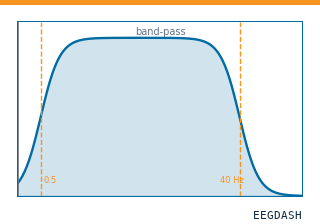 Preprocess EEG and create windowsFilter, resample, epoch — and persist the windowed dataset.
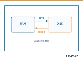 Save and reload prepared dataCache a windowed dataset to disk and reattach it without recompute.
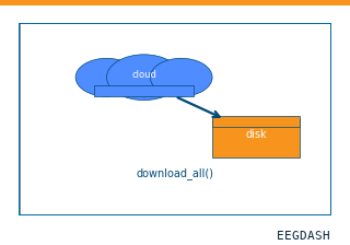 Download a dataset locallyPrefetch BIDS files to a local cache and validate the layout.
Swap any load_dataset(...) call for nm000396 to reproduce the tutorial on this dataset.
Citation
Jesse T. Miles, Kurt E. Weaver, Sara Jane Webb, Jeffrey G. Ojemann (2026). Developmental ECoG: resting-state electrocorticography from 13 epilepsy patients aged 3-33 years (Miles, Weaver, Webb, Ojemann 2025). 10.82901/nemar.nm000396
Provenance
¹Contributed to nemar in BIDS format.
²Curated & ingested by the EEGDash catalog; see CITATION.cff for canonical reference.
³Persistent identifier: 10.82901/nemar.nm000396.
See Also#
eegdash.dataset.EEGDashDataseteegdash.dataset