


NM000356: ieeg dataset, 29 subjects#
Bellier et al. 2023 Pink Floyd music listening ECoG: authors’ high-frequency activity (HFA, 70-150 Hz) for 29 patients
Access recordings and metadata through EEGDash.
Citation: Ludovic Bellier, Anaïs Llorens, Déborah Marciano, Aysegul Gunduz, Gerwin Schalk, Peter Brunner, Robert T. Knight (2023). Bellier et al. 2023 Pink Floyd music listening ECoG: authors’ high-frequency activity (HFA, 70-150 Hz) for 29 patients. 10.82901/nemar.nm000356
Modality: ieeg Subjects: 29 Recordings: 29 License: CC-BY-4.0 Source: nemar
Metadata: Complete (100%)
29-participant iEEG dataset — Bellier et al. 2023 Pink Floyd music listening ECoG: authors' high-frequency activity (HFA, 70-150 Hz) for 29 patients.
Quickstart#
Install
pip install eegdash
Access the data
from eegdash.dataset import NM000356

dataset = NM000356(cache_dir="./data")
# Get the raw object of the first recording
raw = dataset.datasets[0].raw
print(raw.info)
Filter by subject
dataset = NM000356(cache_dir="./data", subject="01")
Advanced query
dataset = NM000356(
    cache_dir="./data",
    query={"subject": {"$in": ["01", "02"]}},
)
Iterate recordings
for rec in dataset:
    print(rec.subject, rec.raw.info['sfreq'])
If you use this dataset in your research, please cite the original authors.
BibTeX
@dataset{nm000356,
  title = {Bellier et al. 2023 Pink Floyd music listening ECoG: authors' high-frequency activity (HFA, 70-150 Hz) for 29 patients},
  author = {Ludovic Bellier and Anaïs Llorens and Déborah Marciano and Aysegul Gunduz and Gerwin Schalk and Peter Brunner and Robert T. Knight},
  doi = {10.82901/nemar.nm000356},
  url = {https://doi.org/10.82901/nemar.nm000356},
}
About This Dataset#
**These are not raw recordings.**This dataset is a BIDS packaging of the*preprocessed* neural data released with
Bellier et al. (2023), “Music can be reconstructed from human auditory cortex activity using nonlinear decoding models”,
PLoS Biology 21(8): e3002176. For each of 29 patients the release holds the authors’ high-frequency activity (HFA,
70-150 Hz) estimate at 100 Hz, aligned to the 190.72 s song, plus the electrode coordinates in MNI space. The raw ECoG
was not released. dataset_description.json therefore declares DatasetType: derivative, with GeneratedBy and
SourceDatasets pointing at the authors’ Zenodo record (doi:10.5281/zenodo.7876019, CC-BY-4.0).
covering at least part of the superior temporal gyrus. Recorded at Albany Medical Center at 1200 Hz with g.USBamp
and BCI2000. Approved by Albany Medical College IRB #2061 and UC Berkeley CPHS #2010-01-520; written informed consent. Per-patient age and sex are not in the release.
participants.tsvgives the implantation laterality stored by the authors.
Bellier et al. 2023: ECoG high-frequency activity while listening to a Pink Floyd song (29 patients)
Overview
Task: passive listening to Another Brick in the Wall, Part 1*(Pink Floyd,*The Wall, 1979). The song was delivered through in-ear headphones at 50-60 dB SL.
View full README
Bellier et al. 2023: ECoG high-frequency activity while listening to a Pink Floyd song (29 patients)
Overview
Task: passive listening to Another Brick in the Wall, Part 1*(Pink Floyd,*The Wall, 1979). The song was delivered through in-ear headphones at 50-60 dB SL.
Further cohort and acquisition details (Bellier et al. 2023, Methods; PMC10427021)
29 patients with pharmacoresistant epilepsy: 15 females; age range 16 to 60 (mean 33.4, SD 12.7); 23 right-handed; full-scale IQ range 74 to 122 (mean 96.6, SD 13.1); all with self-declared normal hearing. Electrode location “was solely guided by clinical concern”; patients were included if their implantation map covered at least partially the superior temporal gyrus.
Implant: grids or strips of platinum-iridium electrodes (Ad-Tech Medical, Oak Creek, WI), centre-to-centre distance 10 mm (21 patients), 6 mm (4), 4 mm (3) or 3 mm (1; the paper describes P29 as the patient with high-density 3-mm coverage). 28 unilateral cases (18 left, 10 right) and 1 bilateral case; 2,668 electrodes in total (range 36 to 250, mean 92 per patient).
Acquisition: “ECoG activity was recorded at a sampling rate of 1,200 Hz using g.USBamp biosignal acquisition devices (g.tec, Graz, Austria) and BCI2000”. Hardware filter settings and the acquisition reference are not reported.
Eight patients had more than one recording of the task; the authors kept the cleanest one (“containing the least epileptic activity or noisy electrodes”).
Task / paradigm and timing
Patients passively listened to Another Brick in the Wall, Part 1*(Pink Floyd,*The Wall, Harvest Records/Columbia Records, 1979) and “were instructed to listen attentively to the music, without focusing on any special detail”. The stimulus (190.72 s) was digitized at 44.1 kHz and delivered through in-ear monitor headphones (bandwidth 12 Hz to 23.5 kHz, 20 dB isolation from surrounding noise) at a comfortable level adjusted for each patient (50 to 60 dB SL). Time 0 of every file is song onset; the HFA and the authors’ auditory spectrograms share the same 100 Hz time base (19072 samples).
Files
sub-PXX/ieeg/sub-PXX_task-musiclistening_ieeg.vhdr/.vmrk/.eeg: BrainVision, IEEE float32, 100 Hz, 19072 samples. Each file has one channel per electrode, with the authors’ labels “1”..”n”. Time 0 is song onset...._channels.tsv: electrodes the authors flagged as noisy or epileptic (dataInfo.idxNoisyElecs,idxEpilepticElecs) are markedstatus = bad. The paper excluded these from its analyses; their HFA is kept as released.author_reference_electrodemarksdataInfo.idxRefElec. Column i of the authors’ecogmatrix is matched to the i-th label of the coordinate file; the counts agree for all 29 patients (2668 electrodes in total, as in the paper). The stored flags mark 98 noisy and 151 epileptic electrodes. The paper reports removing 106 noisy and 183 epileptic electrodes. The flags are transcribed as stored, without reconciling the counts...._events.tsv: onesongrow (onset 0, duration 190.72 s). It also has oneoutlier_samplesrow per run of consecutive samples that the authors’artifactsmatrix marks for a channel. These rows reconstruct that matrix exactly.sub-PXX_space-Other_electrodes.tsv+_coordsystem.json: the authors’ MNI coordinates in mm and their anatomical labels. The BIDS coordinate system isOtherbecause the release does not say which MNI template variant was used. The P2 coordinate structure has nounitfield. Like all the others, it is taken as mm (its value range matches).sourcedata/zenodo-7876019/: the authors’ 58 HFA and coordinate.matfiles, byte-identical, with SHA-256 insourcedata/sourcedata_provenance.json.
Preprocessing already applied by the source
How the HFA was computed (authors, Methods “Preprocessing - ECoG data”)
The authors first notch-filtered 60 Hz and its harmonics up to 300 Hz, then high-pass filtered at 1 Hz. They band-passed the signal into 20-Hz sub-bands from 70-90 to 130-150 Hz in 5-Hz steps and applied a median-based common average reference to each sub-band. For some patients the reference was computed per splitter box. They took the Hilbert envelope of each sub-band and robust-scaled it (median removed, divided by the 10th-90th percentile range).
The sub-bands were then averaged, the 10-s pads removed, and the result downsampled to 100 Hz. Samples above 7 SD were
tagged as outliers. The HFA values are dimensionless, so units is n/a.
FieldTrip (version from May 11, 2021) and the authors’ own scripts were used for these steps (paper, Methods).
Before extracting HFA the authors visually inspected the raw signals and identified noisy and epileptic electrodes
with a neurologist; these are the status = bad channels (see Files).
Known caveats
Not raw data: dimensionless HFA envelopes at 100 Hz only (see Overview and the HFA section).
Bad-channel flag counts in the release (98 noisy, 151 epileptic) differ from the paper (106, 183); see Files.
The MNI template variant is not stated, so the coordinate system is
Other; see Files.The stimulus audio and spectrograms are not redistributed; see “Stimulus: not included”.
Precision
The source stores float64 and BrainVision float32. The relative rounding error is below 1e-7; the maximum per subject
is in the conversion report. For bit-exact values, use the .mat files in sourcedata/.
Stimulus: not included
The Zenodo record also contains thewall1.wav (the song) and its 32- and 128-bin auditory spectrograms
(thewall1_stim32.mat, thewall1_stim128.mat). The record’s CC-BY-4.0 licence does not establish a right to
redistribute a commercial recording, so these three files are not part of this package. To reproduce the
stimulus-based analyses, download them from https://zenodo.org/records/7876019. Alternatively, use a commercial copy of
the song and compute the auditory spectrogram with the NSL toolbox as described in the paper, using the authors’ code
at ludovicbellier/PF_HFAdecoding. The spectrograms are sampled at 100 Hz and have the same 19072
samples as the HFA.
How to load
The signal files are stored with git-annex on NEMAR; fetch them first (for example git annex get sub-P01).
Then, with MNE-BIDS:
from mne_bids import BIDSPath, read_raw_bids
bids_path = BIDSPath(root="nm000356", subject="P01", task="musiclistening",
                     datatype="ieeg")

raw = read_raw_bids(bids_path)       # 100 Hz HFA, one channel per electrode
The HFA is dimensionless (channel units n/a), so ignore any unit scaling your reader reports and use the values
as stored. Channels with status = bad were excluded by the authors; rows with trial_type = outlier_samples in
_events.tsv mark the samples the authors tagged as outliers per channel.
Licence and citation
CC-BY-4.0 (Zenodo record licence). Please cite Bellier et al. (2023), doi:10.1371/journal.pbio.3002176, and the data record doi:10.5281/zenodo.7876019.
Source and provenance
Data record: Zenodo doi:10.5281/zenodo.7876019 (published 2023-04-28, CC-BY-4.0).
Article: Bellier et al. (2023) PLoS Biol 21(8): e3002176, doi:10.1371/journal.pbio.3002176 (PMC10427021). Cohort, acquisition, task, funding and acknowledgment details were read from the PMC full text on 2026-10-06.
Packaging:
laneD_convert.py(iEEG-NEMAR campaign); seeGeneratedByindataset_description.jsonand SHA-256 checksums insourcedata/sourcedata_provenance.json.
NEMAR Metadata#
[](https://doi.org/10.82901/nemar.nm000356) # Bellier et al. 2023: ECoG high-frequency activity while listening to a Pink Floyd song (29 patients) ## Overview These are not raw recordings. This dataset is a BIDS packaging of the preprocessed neural data released with Bellier et al. (2023), “Music can be reconstructed from human auditory cortex activity using nonlinear decoding models”, PLoS Biology 21(8): e3002176. For each of 29 patients the release holds the authors’ high-frequency activity (HFA, 70-150 Hz) estimate at 100 Hz, aligned to the 190.72 s song, plus the electrode coordinates in MNI space. The raw ECoG was not released. dataset_description.json therefore declares DatasetType: derivative, with GeneratedBy and SourceDatasets pointing at the authors’ Zenodo record (doi:10.5281/zenodo.7876019, CC-BY-4.0). ## Cohort and recording ### Participants, recording and task (from the paper) - 29 patients with pharmacoresistant epilepsy (15 female, age 16-60), with clinical ECoG grids or strips (Ad-Tech)
covering at least part of the superior temporal gyrus. Recorded at Albany Medical Center at 1200 Hz with g.USBamp and BCI2000. Approved by Albany Medical College IRB #2061 and UC Berkeley CPHS #2010-01-520; written informed consent. Per-patient age and sex are not in the release. participants.tsv gives the implantation laterality stored by the authors.
Task: passive listening to Another Brick in the Wall, Part 1 (Pink Floyd, The Wall, 1979). The song was delivered through in-ear headphones at 50-60 dB SL.
### Further cohort and acquisition details (Bellier et al. 2023, Methods; PMC10427021) - 29 patients with pharmacoresistant epilepsy: 15 females; age range 16 to 60 (mean 33.4, SD 12.7); 23
right-handed; full-scale IQ range 74 to 122 (mean 96.6, SD 13.1); all with self-declared normal hearing. Electrode location “was solely guided by clinical concern”; patients were included if their implantation map covered at least partially the superior temporal gyrus.
Implant: grids or strips of platinum-iridium electrodes (Ad-Tech Medical, Oak Creek, WI), centre-to-centre distance 10 mm (21 patients), 6 mm (4), 4 mm (3) or 3 mm (1; the paper describes P29 as the patient with high-density 3-mm coverage). 28 unilateral cases (18 left, 10 right) and 1 bilateral case; 2,668 electrodes in total (range 36 to 250, mean 92 per patient).
Acquisition: “ECoG activity was recorded at a sampling rate of 1,200 Hz using g.USBamp biosignal acquisition devices (g.tec, Graz, Austria) and BCI2000”. Hardware filter settings and the acquisition reference are not reported.
Eight patients had more than one recording of the task; the authors kept the cleanest one (“containing the least epileptic activity or noisy electrodes”).
## Task / paradigm and timing Patients passively listened to Another Brick in the Wall, Part 1 (Pink Floyd, The Wall, Harvest Records/Columbia Records, 1979) and “were instructed to listen attentively to the music, without focusing on any special detail”. The stimulus (190.72 s) was digitized at 44.1 kHz and delivered through in-ear monitor headphones (bandwidth 12 Hz to 23.5 kHz, 20 dB isolation from surrounding noise) at a comfortable level adjusted for each patient (50 to 60 dB SL). Time 0 of every file is song onset; the HFA and the authors’ auditory spectrograms share the same 100 Hz time base (19072 samples). ## Files - sub-PXX/ieeg/sub-PXX_task-musiclistening_ieeg.vhdr/.vmrk/.eeg: BrainVision, IEEE float32, 100 Hz, 19072 samples.
Each file has one channel per electrode, with the authors’ labels “1”..”n”. Time 0 is song onset.
…_channels.tsv: electrodes the authors flagged as noisy or epileptic (dataInfo.idxNoisyElecs, idxEpilepticElecs) are marked status = bad. The paper excluded these from its analyses; their HFA is kept as released. author_reference_electrode marks dataInfo.idxRefElec. Column i of the authors’ ecog matrix is matched to the i-th label of the coordinate file; the counts agree for all 29 patients (2668 electrodes in total, as in the paper). The stored flags mark 98 noisy and 151 epileptic electrodes. The paper reports removing 106 noisy and 183 epileptic electrodes. The flags are transcribed as stored, without reconciling the counts.
…_events.tsv: one song row (onset 0, duration 190.72 s). It also has one outlier_samples row per run of consecutive samples that the authors’ artifacts matrix marks for a channel. These rows reconstruct that matrix exactly.
sub-PXX_space-Other_electrodes.tsv + _coordsystem.json: the authors’ MNI coordinates in mm and their anatomical labels. The BIDS coordinate system is Other because the release does not say which MNI template variant was used. The P2 coordinate structure has no unit field. Like all the others, it is taken as mm (its value range matches).
sourcedata/zenodo-7876019/: the authors’ 58 HFA and coordinate .mat files, byte-identical, with SHA-256 in sourcedata/sourcedata_provenance.json.
## Preprocessing already applied by the source ### How the HFA was computed (authors, Methods “Preprocessing - ECoG data”) The authors first notch-filtered 60 Hz and its harmonics up to 300 Hz, then high-pass filtered at 1 Hz. They band-passed the signal into 20-Hz sub-bands from 70-90 to 130-150 Hz in 5-Hz steps and applied a median-based common average reference to each sub-band. For some patients the reference was computed per splitter box. They took the Hilbert envelope of each sub-band and robust-scaled it (median removed, divided by the 10th-90th percentile range). The sub-bands were then averaged, the 10-s pads removed, and the result downsampled to 100 Hz. Samples above 7 SD were tagged as outliers. The HFA values are dimensionless, so units is n/a. FieldTrip (version from May 11, 2021) and the authors’ own scripts were used for these steps (paper, Methods). Before extracting HFA the authors visually inspected the raw signals and identified noisy and epileptic electrodes with a neurologist; these are the status = bad channels (see Files). ## Known caveats - Not raw data: dimensionless HFA envelopes at 100 Hz only (see Overview and the HFA section). - Bad-channel flag counts in the release (98 noisy, 151 epileptic) differ from the paper (106, 183); see Files. - The MNI template variant is not stated, so the coordinate system is Other; see Files. - The stimulus audio and spectrograms are not redistributed; see “Stimulus: not included”. ### Precision The source stores float64 and BrainVision float32. The relative rounding error is below 1e-7; the maximum per subject is in the conversion report. For bit-exact values, use the .mat files in sourcedata/. ### Stimulus: not included The Zenodo record also contains thewall1.wav (the song) and its 32- and 128-bin auditory spectrograms (thewall1_stim32.mat, thewall1_stim128.mat). The record’s CC-BY-4.0 licence does not establish a right to redistribute a commercial recording, so these three files are not part of this package. To reproduce the stimulus-based analyses, download them from https://zenodo.org/records/7876019. Alternatively, use a commercial copy of the song and compute the auditory spectrogram with the NSL toolbox as described in the paper, using the authors’ code at ludovicbellier/PF_HFAdecoding. The spectrograms are sampled at 100 Hz and have the same 19072 samples as the HFA. ## How to load The signal files are stored with git-annex on NEMAR; fetch them first (for example git annex get sub-P01). Then, with MNE-BIDS: ```python from mne_bids import BIDSPath, read_raw_bids bids_path = BIDSPath(root=”nm000356”, subject=”P01”, task=”musiclistening”,
datatype=”ieeg”)
raw = read_raw_bids(bids_path) # 100 Hz HFA, one channel per electrode ``` The HFA is dimensionless (channel units n/a), so ignore any unit scaling your reader reports and use the values as stored. Channels with status = bad were excluded by the authors; rows with trial_type = outlier_samples in _events.tsv mark the samples the authors tagged as outliers per channel. ## Licence and citation CC-BY-4.0 (Zenodo record licence). Please cite Bellier et al. (2023), doi:10.1371/journal.pbio.3002176, and the data record doi:10.5281/zenodo.7876019. ## Source and provenance - Data record: Zenodo doi:10.5281/zenodo.7876019 (published 2023-04-28, CC-BY-4.0). - Article: Bellier et al. (2023) PLoS Biol 21(8): e3002176, doi:10.1371/journal.pbio.3002176 (PMC10427021).
Cohort, acquisition, task, funding and acknowledgment details were read from the PMC full text on 2026-10-06.
Packaging: laneD_convert.py (iEEG-NEMAR campaign); see GeneratedBy in dataset_description.json and SHA-256 checksums in sourcedata/sourcedata_provenance.json.
License: CC-BY-4.0
Authors:
Ludovic Bellier
Anaïs Llorens
Déborah Marciano
Aysegul Gunduz
Gerwin Schalk
… and 2 more
Versions:
Version |
DOI |
Released |
|---|---|---|
|
Cohort#
Dataset Statistics#
Channel counts (ch)
Sampling frequencies: 100.0 Hz (n=29 recordings)
Total recording duration: 1 h 32 min
Signal · Electrodes & live trace#
Live trace viewer — sub-P10 · task-musiclistening
Showing one representative recording out of
29 subjects and 29 recordings in this dataset.
Browse the full set on OpenNeuro;
drop any other _ieeg.{set,edf,bdf,vhdr} file onto the
viewer (or pass ?ieeg=<url>) to inspect it.
Electrode layout — iEEG · 76 sensors — 76 channels
NEMAR Processing Statistics#
The plots below are generated by NEMAR’s automated EEG pipeline. The histogram shows pipeline success for data cleaning and ICA decomposition, the percentage of data frames and EEG channels retained after artefact removal, line noise per channel (RMS, dB), and the age/gender distribution of participants.

HED event descriptors word cloud

Manifest#
File Explorer#
Browse the BIDS file structure of this dataset. Records are fetched on demand from the EEGDash catalog the first time you open the explorer.
Full dataset metadata table
Dataset ID |
|
Title |
Bellier et al. 2023 Pink Floyd music listening ECoG: authors’ high-frequency activity (HFA, 70-150 Hz) for 29 patients |
Author (year) |
— |
Canonical |
— |
Importable as |
|
Year |
2023 |
Authors |
Ludovic Bellier, Anaïs Llorens, Déborah Marciano, Aysegul Gunduz, Gerwin Schalk, Peter Brunner, Robert T. Knight |
License |
CC-BY-4.0 |
Citation / DOI |
|
Source links |
OpenNeuro | NeMAR | Source URL |
Copy-paste BibTeX
@dataset{nm000356,
  title = {Bellier et al. 2023 Pink Floyd music listening ECoG: authors' high-frequency activity (HFA, 70-150 Hz) for 29 patients},
  author = {Ludovic Bellier and Anaïs Llorens and Déborah Marciano and Aysegul Gunduz and Gerwin Schalk and Peter Brunner and Robert T. Knight},
  doi = {10.82901/nemar.nm000356},
  url = {https://doi.org/10.82901/nemar.nm000356},
}
API Reference#
eegdash.datasetEEGDashDataset- class eegdash.dataset.NM000356(cache_dir: str, query: dict | None = None, s3_bucket: str | None = None, **kwargs)[source]#
Bellier et al. 2023 Pink Floyd music listening ECoG: authors’ high-frequency activity (HFA, 70-150 Hz) for 29 patients
- Study:
nm000356(NeMAR)- Author (year):
—
- Canonical:
—
Also importable as:
NM000356.Modality:
ieeg; Subject type:Unknown. Subjects: 29; recordings: 29; tasks: 1.- Parameters:
cache_dir (str | Path) – Directory where data are cached locally.
query (dict | None) – Additional MongoDB-style filters to AND with the dataset selection. Must not contain the key
dataset.s3_bucket (str | None) – Base S3 bucket used to locate the data.
**kwargs (dict) – Additional keyword arguments forwarded to
EEGDashDataset.
- data_dir#
Local dataset cache directory (
cache_dir / dataset_id).- Type:
Path
Notes
Each item is a recording; recording-level metadata are available via
dataset.description.querysupports MongoDB-style filters on fields inALLOWED_QUERY_FIELDSand is combined with the dataset filter. Dataset-specific caveats are not provided in the summary metadata.References
OpenNeuro dataset: https://openneuro.org/datasets/nm000356 NeMAR dataset: https://nemar.org/dataexplorer/detail?dataset_id=nm000356 DOI: https://doi.org/10.82901/nemar.nm000356
Examples
>>> from eegdash.dataset import NM000356
>>> dataset = NM000356(cache_dir="./data")
>>> recording = dataset[0]
>>> raw = recording.load()
- __init__(cache_dir: str, query: dict | None = None, s3_bucket: str | None = None, **kwargs)[source]#
- save(path: str, overwrite: bool = False, offset: int = 0)[source]#
Save datasets to files by creating one subdirectory for each dataset:
path/
    0/
        0-raw.fif | 0-epo.fif
        description.json
        raw_preproc_kwargs.json (if raws were preprocessed)
        window_kwargs.json (if this is a windowed dataset)
        window_preproc_kwargs.json  (if windows were preprocessed)
        target_name.json (if target_name is not None and dataset is raw)
    1/
        1-raw.fif | 1-epo.fif
        description.json
        raw_preproc_kwargs.json (if raws were preprocessed)
        window_kwargs.json (if this is a windowed dataset)
        window_preproc_kwargs.json  (if windows were preprocessed)
        target_name.json (if target_name is not None and dataset is raw)
- Parameters:
path (str) –
- Directory in which subdirectories are created to store
-raw.fif | -epo.fif and .json files to.
overwrite (bool) – Whether to delete old subdirectories that will be saved to in this call.
offset (int) – If provided, the integer is added to the id of the dataset in the concat. This is useful in the setting of very large datasets, where one dataset has to be processed and saved at a time to account for its original position.
BaseDataset from braindecode — windowed via create_windows_from_events.braindecodeDataLoader; supports parallel workers and on-the-fly augmentations.pytorch Find datasets with the EEGDash APIQuery the catalogue, filter by task or modality, list candidates.
 Load one EEG recordingResolve a single record to an MNE Raw with channels and events.
 EEG recording to PyTorch DataLoaderWrap braindecode windows in a DataLoader for model training.
 Preprocess EEG and create windowsFilter, resample, epoch — and persist the windowed dataset.
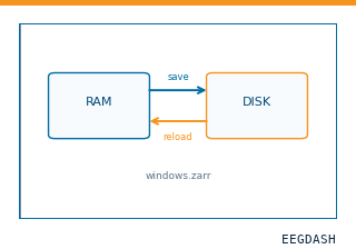 Save and reload prepared dataCache a windowed dataset to disk and reattach it without recompute.
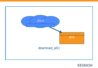 Download a dataset locallyPrefetch BIDS files to a local cache and validate the layout.
Swap any load_dataset(...) call for nm000356 to reproduce the tutorial on this dataset.
Citation
Ludovic Bellier, Anaïs Llorens, Déborah Marciano, Aysegul Gunduz, Gerwin Schalk, … (2023). Bellier et al. 2023 Pink Floyd music listening ECoG: authors' high-frequency activity (HFA, 70-150 Hz) for 29 patients. 10.82901/nemar.nm000356
Provenance
¹Contributed to nemar in BIDS format.
²Curated & ingested by the EEGDash catalog; see CITATION.cff for canonical reference.
³Persistent identifier: 10.82901/nemar.nm000356.
See Also#
eegdash.dataset.EEGDashDataseteegdash.dataset