


NM000414: ieeg dataset, 3 subjects#
Intracranial EEG Epilepsy - Studies 3, 5 and 6 (Mayo Clinic; IEEG.org)
Access recordings and metadata through EEGDash.
Citation: Jenaye Johnson, Gregory Worrell, Brian Litt (2018). Intracranial EEG Epilepsy - Studies 3, 5 and 6 (Mayo Clinic; IEEG.org). 10.82901/nemar.nm000414
Modality: ieeg Subjects: 3 Recordings: 41 License: CC0-1.0 Source: nemar
Metadata: Complete (100%)
3-participant iEEG dataset — Intracranial EEG Epilepsy - Studies 3, 5 and 6 (Mayo Clinic; IEEG.org).
Quickstart#
Install
pip install eegdash
Access the data
from eegdash.dataset import NM000414

dataset = NM000414(cache_dir="./data")
# Get the raw object of the first recording
raw = dataset.datasets[0].raw
print(raw.info)
Filter by subject
dataset = NM000414(cache_dir="./data", subject="01")
Advanced query
dataset = NM000414(
    cache_dir="./data",
    query={"subject": {"$in": ["01", "02"]}},
)
Iterate recordings
for rec in dataset:
    print(rec.subject, rec.raw.info['sfreq'])
If you use this dataset in your research, please cite the original authors.
BibTeX
@dataset{nm000414,
  title = {Intracranial EEG Epilepsy - Studies 3, 5 and 6 (Mayo Clinic; IEEG.org)},
  author = {Jenaye Johnson and Gregory Worrell and Brian Litt},
  doi = {10.82901/nemar.nm000414},
  url = {https://doi.org/10.82901/nemar.nm000414},
}
About This Dataset#
Long-term clinical intracranial EEG (subdural grids/strips and depth electrodes) from three adults/adolescents with drug-resistant
epilepsy recorded at Mayo Clinic, Rochester, MN, with expert seizure annotations. Each Pennsieve record (“Intracranial EEG Epilepsy - Study 3/5/6”, published 2019-07-12, licence “Creative Commons Zero 1.0 Universal”, creator Jenaye Johnson, contributors Gregory Worrell and Brian Litt) holds one patient, originally shared on the IEEG.org portal. Record description: “Data for a patient with epilepsy obtained for clinical treatment and collected for research, consisting of a large intracranial EEG dataset … with multiple seizures.”
with resolution = MEF voltage conversion factor (0.2658 µV per count). Acquisition system “XLTek EEG 128”; hardware filter
settings from the MEF headers (0.1 Hz high-pass; low-pass field 500 Hz as stored). One run per continuous MEF segment: the MEF files flag recording discontinuities, and each run starts at one.
sub-*_scans.tsvgives each run’s start offset in seconds from the start of the recording (calendar times are not released; see Privacy).
Intracranial EEG Epilepsy - Studies 3, 5 and 6 (Mayo Clinic, IEEG.org)
Overview
_channels.tsv: channel type from the record readme’s electrode description: depth electrodes SEEG (Study 3: RTD; Study 5: AD, PD), grids and strips ECOG._electrodes.tsv: names only (no coordinates are released; an electrode diagram is in sourcedatapackages/Cartoon Map.pdf).
View full README
Intracranial EEG Epilepsy - Studies 3, 5 and 6 (Mayo Clinic, IEEG.org)
Overview
_channels.tsv: channel type from the record readme’s electrode description: depth electrodes SEEG (Study 3: RTD; Study 5: AD, PD), grids and strips ECOG._electrodes.tsv: names only (no coordinates are released; an electrode diagram is in sourcedatapackages/Cartoon Map.pdf)._events.tsv: every annotation of the IEEG.org layer (packages/EEG/annotations.xml: type, layer, annotator, channels), placed with the MEF block timestamps. Timing note: within a run the block timestamps deviate from the nominal 500 Hz clock (per run: sub-study3 run-01: max |block timestamp - nominal| = 0.794 s over 5.96 h; sub-study3 run-02: max |block timestamp - nominal| = 0.777 s over 15.67 h; sub-study3 run-03: max |block timestamp - nominal| = 1.673 s over 6.47 h; sub-study3 run-04: max |block timestamp - nominal| = 0.257 s over 1.59 h; sub-study3 run-05: max |block timestamp - nominal| = 1.072 s over 24.03 h; sub-study3 run-06: max |block timestamp - nominal| = 0.884 s over 17.63 h; sub-study3 run-07: max |block timestamp - nominal| = 0.162 s over 3.22 h; sub-study3 run-08: max |block timestamp - nominal| = 1.097 s over 2.55 h; sub-study3 run-09: max |block timestamp - nominal| = 0.625 s over 24.17 h; sub-study3 run-10: max |block timestamp - nominal| = 0.762 s over 24.61 h; sub-study3 run-11: max |block timestamp - nominal| = 0.000 s over 0.01 h; sub-study3 run-12: max |block timestamp - nominal| = 0.316 s over 2.58 h; sub-study3 run-13: max |block timestamp - nominal| = 1.098 s over 13.68 h; sub-study3 run-14: max |block timestamp - nominal| = 0.969 s over 7.27 h; sub-study3 run-15: max |block timestamp - nominal| = 0.608 s over 4.78 h; sub-study3 run-16: max |block timestamp - nominal| = 0.049 s over 0.53 h; sub-study5 run-01: max |block timestamp - nominal| = 0.002 s over 0.01 h; sub-study5 run-02: max |block timestamp - nominal| = 0.084 s over 0.24 h; sub-study5 run-03: max |block timestamp - nominal| = 0.990 s over 2.22 h; sub-study5 run-04: max |block timestamp - nominal| = 1.014 s over 3.31 h; sub-study5 run-05: max |block timestamp - nominal| = 1.853 s over 9.02 h; sub-study5 run-06: max |block timestamp - nominal| = 4.295 s over 24.07 h; sub-study5 run-07: max |block timestamp - nominal| = 1.677 s over 8.61 h; sub-study6 run-01: max |block timestamp - nominal| = 0.380 s over 1.00 h; sub-study6 run-02: max |block timestamp - nominal| = 0.000 s over 0.03 h; sub-study6 run-03: max |block timestamp - nominal| = 0.132 s over 11.87 h; sub-study6 run-04: max |block timestamp - nominal| = 0.114 s over 4.93 h; sub-study6 run-05: max |block timestamp - nominal| = 0.298 s over 14.96 h; sub-study6 run-06: max |block timestamp - nominal| = 0.202 s over 4.16 h; sub-study6 run-07: max |block timestamp - nominal| = 0.354 s over 24.86 h; sub-study6 run-08: max |block timestamp - nominal| = 0.382 s over 22.94 h; sub-study6 run-09: max |block timestamp - nominal| = 0.546 s over 23.77 h; sub-study6 run-10: max |block timestamp - nominal| = 0.004 s over 0.02 h; sub-study6 run-11: max |block timestamp - nominal| = 0.018 s over 0.73 h; sub-study6 run-12: max |block timestamp - nominal| = 0.396 s over 24.10 h; sub-study6 run-13: max |block timestamp - nominal| = 0.874 s over 4.55 h; sub-study6 run-14: max |block timestamp - nominal| = 0.018 s over 0.21 h; sub-study6 run-15: max |block timestamp - nominal| = 0.094 s over 0.63 h; sub-study6 run-16: max |block timestamp - nominal| = 2.400 s over 14.55 h; sub-study6 run-17: max |block timestamp - nominal| = 0.301 s over 23.72 h; sub-study6 run-18: max |block timestamp - nominal| = 3.393 s over 17.49 h); onsets follow the timestamps. All annotations fall inside a run.participants.tsv: age, sex, handedness and age at onset from the record readmes; seizure type, ILAE outcome and resection fields fromgraph/patient.csv.sourcedata/pennsieve-<id>-v1/: the complete records: the MEF 2.0 files byte-identical (packages/EEG/<Study> IEEG/*.mef),packages/EEG/annotations.xml,packages/Cartoon Map.pdf, the Pennsieve graph CSVs (patient, EEG study, seizures, imaging studies, MR/CT details, clinical reports), readme, metadata.json, banner and the Pennsieve file listing with checksums.date_scrub.tsvlists the date edits (see Privacy).graph/seizures.csvgives seizure clock times on portal-shifted dates; they do not line up one-to-one with the annotation offsets, so events are taken from annotations.xml.
Record readmes (verbatim)
sub-study3 (Study 3)
The patient is a right-handed, 21-year old male who was admitted to the epilepsy monitoring unit for **intracranial monitoring**. The age at onset was 13 years old.
These data are from a 6 x 8 grid that was placed over the right frontal region, two 4 x 6 grids placed over the right temporal lobe and right parietal region, respectively, a 4-contact strip wrapped beneath the right anterior temporal lobe, a 4-contact strip wrapped beneath the right posterior temporal lobe, and a 4-contact depth electrode inserted in the anterior temporal region. This is an abnormal computer-assisted prolonged intracranial EEG monitoring session due to the presence of frequent independent right frontal, right parietal and right temporal epileptiform discharges. During the monitoring session, the patient had a total of **eleven seizures, including nine clinical seizures and two seizures which were subclinical**. The first six seizures showed a diffuse ictal onset over the right frontal and right temporal grids. The last five seizures showed either a focal ictal onset simultaneously from the right anterior temporal strip and the posterior superior region of the right frontal grid, or focal onset from these two foci independently. These findings could be consistent with a localization-related epilepsy with seizure onset in the right frontotemporal neocortex.
The patient underwent surgery of reopening the right forntotemporoparietal craniotomy and removing the subdural grid electrodes, then a **right anterior-superior frontocortical resection, a right temporal lobectomy, and a right amygdalohippocampectomy**. Pathology samples of leptomeningeal and focal parenchymal lymphohistiocytic infiltrate consistent with grid/electrode placement. Mild subpial and subcortical gliosis. The patient also underwent surgery for a right mesial temporal lobe resection. Pathology samples of mesial temporal structures with focal parenchymal macrophage aggregate consistent with electrode placement. **Mild subpial and subcortical gliosis**.
sub-study5 (Study 5)
This patient is a right-handed, 16-year old male who was admitted to the epilepsy monitoring unit for **intracranial monitoring**. The age of onset was 1 year old.
These data are from an 8-contact straight strip inserted into the left frontal region parallel to the superior temporal gyrus, an 8x8 grid inserted over the left posterior temporo-occipital region, a 4-contact straight strip inserted over the left anterior temporal pole, a 4-contact straight strip inserted over the left inferior subtemporal region, posterior to the anterior temporal strip, a 4-contact orthogonal depth electrode inserted between the anterior temporal strip and inferior subtemporal strip, targeting the hippocampus, and another 4-contact orthogonal depth electrode targeting the hippocampus. This is an abnormal computer-assisted prolonged intracranial EEG monitoring session, lasting approximately 2 days, due to the presence of frequent independent left posterior temporo-occipital, left mesial temporal and left anterior temporal epileptiform discharges. During the monitoring session, the patient had **four complex partial seizures**. All of these seizures began in the posterior contacts of the left temporo-occipital grid. The findings would be consistent with a localization-related epilepsy with seizure onset in the left occipital neocortex.
The patient underwent a surgery for reopening of left tempooccipital craniotomy, removal of subdural grids and depth electrodes, and left occipital pole resection**. Pathology samples for cortex and underlying white matter with changes consistent with remote destructive injury. The cortex shows multiple areas of severe neuronal loss and gliosis associated with atrophy and focal dystrophic calcification. **Calcifications are present focally in the meninges and vascular walls**. An immunohistochemical stain was performed on paraffin-embedded tissue using antibodies to GFAP. GFAP highlights disruption of the normal cortical architecture, reactive gliosis and subpial gliosis. An immunohistochemical stain for NeuN highlights neurons. GROSS DESCRIPTION: Received fresh labeled “left occipital brain lobe” is a 7.51 gram, 5.2 x 2.8 x 0.8 cm portion of brain. The specimen is sent in formalin to the neuropathology working group for sectioning. **46-month post-resection ILAE 1 outcome score
sub-study6 (Study 6)
The patient is a right-handed, 23-year-old female who was admitted to the epilepsy monitoring unit for intracranial monitoring. Age of onset was 9 years old.
These data are from _three 8-contact strips_ placed over the left frontopolar cortex, left temporalcortex, and left parietal cortex, a _4-contact strip_ inserted in the left frontal interhemispheric region, _a 15-contact hybrid strip_ placed over the left central interhemispheric region, _three 8-contact strips_ placed over the right frontopolar cortex, right temporal cortex, and right parietal cortex, _a 4-contact strip_ placed over the right central interhemispheric region, and _a 15-contact hybrid strip_ inserted in the right frontal interhemispheric region. This is an abnormal computer-assisted prolonged intracranial EEG monitoring session due to the presence of frequent independent left frontal, left parietal, left temporal, right central interhemispheric, and right parietal epileptiform discharges. During the monitoring session, the patient had **forty-two typical complex partial seizures**. The seizure onset was diffuse and bilateral and therefore non-localized. These findings are consistent with partial epilepsy, with indeterminate seizure onset.
The patient _did not_ undergo resective surgery.
Privacy
MEF headers: subject name fields “none”/”Anonymized”, subject id placeholder “9-999-000”, recording start 2000-01-01
00:00:00 UTC (the IEEG.org de-identification epoch, not a calendar date); data unencrypted. The MEF header CRCs no longer match
(consistent with the portal’s header anonymisation); all data-block CRCs are valid. Dates in the text sources (clinical
report, imaging and seizure dates, annotation creation times) are written with day 01 in the sourcedata copies
(date_scrub.tsv); the unedited originals are not part of this deposit. The clinical reports are narrative text without
names or record numbers (scan in the NEMAR conversion log).
Ethics approval
The Pennsieve records and the IEEG.org release give no ethics approval statement beyond the record description (“obtained for clinical treatment and collected for research”). No statement has been added.
Licence
CC0-1.0 (Creative Commons Zero 1.0 Universal), as released.
Conversion
MEF 2.0 was decoded with mef_lib_2_1 (github.com/benbrinkmann/mef_lib_2_1, Apache-2.0); note that the library’s built-in block CRC check reads the output buffer, so the CRCs were validated on the compressed bytes instead. Every written sample was compared with the decoded MEF sample (exact equality).
Additional metadata and localisation (added 2026-10-08)
Compiled after the upload from the article, its supplement and the source deposit (each statement names its source). Text and sidecar metadata only; no data file was changed. Recording system. 500 Hz sampling, 88 / 108 / 88 channels (graph/eeg_study.csv of 13 / 14 / 15), one MEF file per channel. Amplifier, filters: n/a. Reference scheme. Study 5 cartoon map marks a GND/REF strip (Cartoon Map.pdf, Pennsieve 13); otherwise not stated. Electrode types. Study 5: subdural grid (8x8), strips and two 4-contact depths targeting the hippocampus; Study 3: 6x8 and two 4x6 grids, two 4-contact strips, one 4-contact depth; Study 6: strips only, bilateral, including 15-contact hybrid interhemispheric strips (readme.md; eeg_study.csv type_of_electrodes; Cartoon Map.pdf). Localisation method. Only schematic placement: readme.md text and a cartoon map (Cartoon Map.pdf) naming each electrode on a lateral brain drawing. Post-implant CT and pre-implant MRI acquisitions are listed in graph/imaging_study.csv / ct_details.csv / mr_details.csv (metadata only; no image files in the listings), so no contact coordinates exist in the deposits.
Regions per participant (as published; no coordinates exist)
| participant | region (as stated) | hemisphere | contacts | source |
|---|---|---|---|---|
| sub-study5 = Study023 (Pennsieve 13, Study 5) | an 8x8 grid inserted over the left posterior temporo-occipital region | L | 64 | https://discover.pennsieve.io/datasets/13/version/1 readme.md + packages/Cartoon Map.pdf, readme.md paragraph 2 (placement) |
| sub-study5 = Study023 (Pennsieve 13, Study 5) | an 8-contact straight strip inserted into the left frontal region parallel to the superior temporal gyrus | L | 8 | https://discover.pennsieve.io/datasets/13/version/1 readme.md + packages/Cartoon Map.pdf, readme.md paragraph 2 (placement) |
| sub-study5 = Study023 (Pennsieve 13, Study 5) | a 4-contact straight strip inserted over the left anterior temporal pole | L | 4 | https://discover.pennsieve.io/datasets/13/version/1 readme.md + packages/Cartoon Map.pdf, readme.md paragraph 2 (placement) |
| sub-study5 = Study023 (Pennsieve 13, Study 5) | a 4-contact straight strip inserted over the left inferior subtemporal region, posterior to the anterior temporal strip | L | 4 | https://discover.pennsieve.io/datasets/13/version/1 readme.md + packages/Cartoon Map.pdf, readme.md paragraph 2 (placement) |
| sub-study5 = Study023 (Pennsieve 13, Study 5) | a 4-contact orthogonal depth electrode inserted between the anterior temporal strip and inferior subtemporal strip, targeting the hippocampus | L | 4 | https://discover.pennsieve.io/datasets/13/version/1 readme.md + packages/Cartoon Map.pdf, readme.md paragraph 2 (placement) |
| sub-study5 = Study023 (Pennsieve 13, Study 5) | another 4-contact orthogonal depth electrode targeting the hippocampus | L | 4 | https://discover.pennsieve.io/datasets/13/version/1 readme.md + packages/Cartoon Map.pdf, readme.md paragraph 2 (placement) |
| sub-study3 = Study021 (Pennsieve 14, Study 3) | a 6 x 8 grid that was placed over the right frontal region | R | 48 | https://discover.pennsieve.io/datasets/14/version/1 readme.md + packages/Cartoon Map.pdf, readme.md paragraph 2 (placement) |
| sub-study3 = Study021 (Pennsieve 14, Study 3) | two 4 x 6 grids placed over the right temporal lobe and right parietal region, respectively (temporal grid) | R | 24 | https://discover.pennsieve.io/datasets/14/version/1 readme.md + packages/Cartoon Map.pdf, readme.md paragraph 2 (placement) |
| sub-study3 = Study021 (Pennsieve 14, Study 3) | two 4 x 6 grids placed over the right temporal lobe and right parietal region, respectively (parietal grid) | R | 24 | https://discover.pennsieve.io/datasets/14/version/1 readme.md + packages/Cartoon Map.pdf, readme.md paragraph 2 (placement) |
| sub-study3 = Study021 (Pennsieve 14, Study 3) | a 4-contact strip wrapped beneath the right anterior temporal lobe | R | 4 | https://discover.pennsieve.io/datasets/14/version/1 readme.md + packages/Cartoon Map.pdf, readme.md paragraph 2 (placement) |
| sub-study3 = Study021 (Pennsieve 14, Study 3) | a 4-contact strip wrapped beneath the right posterior temporal lobe | R | 4 | https://discover.pennsieve.io/datasets/14/version/1 readme.md + packages/Cartoon Map.pdf, readme.md paragraph 2 (placement) |
| sub-study3 = Study021 (Pennsieve 14, Study 3) | a 4-contact depth electrode inserted in the anterior temporal region | R | 4 | https://discover.pennsieve.io/datasets/14/version/1 readme.md + packages/Cartoon Map.pdf, readme.md paragraph 2 (placement) |
| sub-study6 = Study024 (Pennsieve 15, Study 6) | 8-contact strip placed over the left frontopolar cortex | L | 8 | https://discover.pennsieve.io/datasets/15/version/1 readme.md + packages/Cartoon Map.pdf, readme.md paragraph 2 (placement) |
| sub-study6 = Study024 (Pennsieve 15, Study 6) | 8-contact strip placed over the left temporal cortex | L | 8 | https://discover.pennsieve.io/datasets/15/version/1 readme.md + packages/Cartoon Map.pdf, readme.md paragraph 2 (placement) |
| sub-study6 = Study024 (Pennsieve 15, Study 6) | 8-contact strip placed over the left parietal cortex | L | 8 | https://discover.pennsieve.io/datasets/15/version/1 readme.md + packages/Cartoon Map.pdf, readme.md paragraph 2 (placement) |
| sub-study6 = Study024 (Pennsieve 15, Study 6) | a 4-contact strip inserted in the left frontal interhemispheric region | L | 4 | https://discover.pennsieve.io/datasets/15/version/1 readme.md + packages/Cartoon Map.pdf, readme.md paragraph 2 (placement) |
| sub-study6 = Study024 (Pennsieve 15, Study 6) | a 15-contact hybrid strip placed over the left central interhemispheric region | L | 16 | https://discover.pennsieve.io/datasets/15/version/1 readme.md + packages/Cartoon Map.pdf, readme.md paragraph 2 (placement) |
| sub-study6 = Study024 (Pennsieve 15, Study 6) | 8-contact strip placed over the right frontopolar cortex | R | 8 | https://discover.pennsieve.io/datasets/15/version/1 readme.md + packages/Cartoon Map.pdf, readme.md paragraph 2 (placement) |
| sub-study6 = Study024 (Pennsieve 15, Study 6) | 8-contact strip placed over the right temporal cortex | R | 8 | https://discover.pennsieve.io/datasets/15/version/1 readme.md + packages/Cartoon Map.pdf, readme.md paragraph 2 (placement) |
| sub-study6 = Study024 (Pennsieve 15, Study 6) | 8-contact strip placed over the right parietal cortex | R | 8 | https://discover.pennsieve.io/datasets/15/version/1 readme.md + packages/Cartoon Map.pdf, readme.md paragraph 2 (placement) |
| sub-study6 = Study024 (Pennsieve 15, Study 6) | a 4-contact strip placed over the right central interhemispheric region | R | 4 | https://discover.pennsieve.io/datasets/15/version/1 readme.md + packages/Cartoon Map.pdf, readme.md paragraph 2 (placement) |
| sub-study6 = Study024 (Pennsieve 15, Study 6) | a 15-contact hybrid strip inserted in the right frontal interhemispheric region | R | 16 | https://discover.pennsieve.io/datasets/15/version/1 readme.md + packages/Cartoon Map.pdf, readme.md paragraph 2 (placement) |
Ethics
The source records (Pennsieve Discover datasets 13, 14 and 15 = IEEG.org Studies 021, 023 and 024; contributors Brian Litt and Gregory Worrell) say only that the data were “obtained for clinical treatment and collected for research”; they contain no ethics approval statement.
Research intracranial EEG from the Mayo Clinic laboratory of Gregory Worrell is described as collected under a Mayo Clinic IRB-approved wide-bandwidth electrophysiology study in Cimbalnik et al. (2018), Methods, “Patients” (verbatim):
Each patient consented to this Mayo Institutional Review Board approved study investigating wide bandwidth electrophysiology.
Cimbalnik J, Brinkmann B, Kremen V, Jurak P, Berry B, Van Gompel J, Stead M, Worrell G (2018). Physiological and pathological high frequency oscillations in focal epilepsy. Annals of Clinical and Translational Neurology 5(9):1062-1076. https://doi.org/10.1002/acn3.618 (PMC6144446).
Scope of this statement: neither the Pennsieve/IEEG.org records nor the paper link these three patients to the paper’s 112-patient cohort, and the paper does not mention IEEG.org. The statement documents the Mayo Clinic IRB-approved research protocol of the contributing laboratory; it is not a per-patient approval confirmed by the source records.
NEMAR Metadata#
[](https://doi.org/10.82901/nemar.nm000414) # Intracranial EEG Epilepsy - Studies 3, 5 and 6 (Mayo Clinic, IEEG.org) ## Overview Long-term clinical intracranial EEG (subdural grids/strips and depth electrodes) from three adults/adolescents with drug-resistant epilepsy recorded at Mayo Clinic, Rochester, MN, with expert seizure annotations. Each Pennsieve record (“Intracranial EEG Epilepsy - Study 3/5/6”, published 2019-07-12, licence “Creative Commons Zero 1.0 Universal”, creator Jenaye Johnson, contributors Gregory Worrell and Brian Litt) holds one patient, originally shared on the IEEG.org portal. Record description: “Data for a patient with epilepsy obtained for clinical treatment and collected for research, consisting of a large intracranial EEG dataset … with multiple seizures.” | participant | record | IEEG.org study | channels | runs | hours | annotations | |---|—|---|—|---|—|---| | sub-study3 | Study 3 (Pennsieve 14, doi:10.26275/psj7-wggf) | Study 021 | 108 (104 ECOG, 4 SEEG) | 16 | 154.7 | 13 (13 inside runs) | | sub-study5 | Study 5 (Pennsieve 13, doi:10.26275/hqyv-oagj) | Study 023 | 88 (80 ECOG, 8 SEEG) | 7 | 47.5 | 4 (4 inside runs) | | sub-study6 | Study 6 (Pennsieve 15, doi:10.26275/pfxu-irqf) | Study 024 | 88 (88 ECOG, 0 SEEG) | 18 | 194.5 | 19 (19 inside runs) | ## Data - sub-*/ieeg/*_task-clinical_run-NN_ieeg.vhdr/.eeg/.vmrk: BrainVision, INT_32, 500 Hz, the MEF integer samples unchanged
with resolution = MEF voltage conversion factor (0.2658 µV per count). Acquisition system “XLTek EEG 128”; hardware filter settings from the MEF headers (0.1 Hz high-pass; low-pass field 500 Hz as stored). One run per continuous MEF segment: the MEF files flag recording discontinuities, and each run starts at one. sub-*_scans.tsv gives each run’s start offset in seconds from the start of the recording (calendar times are not released; see Privacy).
_channels.tsv: channel type from the record readme’s electrode description: depth electrodes SEEG (Study 3: RTD; Study 5: AD, PD), grids and strips ECOG. _electrodes.tsv: names only (no coordinates are released; an electrode diagram is in sourcedata packages/Cartoon Map.pdf).
_events.tsv: every annotation of the IEEG.org layer (packages/EEG/annotations.xml: type, layer, annotator, channels), placed with the MEF block timestamps. Timing note: within a run the block timestamps deviate from the nominal 500 Hz clock (per run: sub-study3 run-01: max |block timestamp - nominal| = 0.794 s over 5.96 h; sub-study3 run-02: max |block timestamp - nominal| = 0.777 s over 15.67 h; sub-study3 run-03: max |block timestamp - nominal| = 1.673 s over 6.47 h; sub-study3 run-04: max |block timestamp - nominal| = 0.257 s over 1.59 h; sub-study3 run-05: max |block timestamp - nominal| = 1.072 s over 24.03 h; sub-study3 run-06: max |block timestamp - nominal| = 0.884 s over 17.63 h; sub-study3 run-07: max |block timestamp - nominal| = 0.162 s over 3.22 h; sub-study3 run-08: max |block timestamp - nominal| = 1.097 s over 2.55 h; sub-study3 run-09: max |block timestamp - nominal| = 0.625 s over 24.17 h; sub-study3 run-10: max |block timestamp - nominal| = 0.762 s over 24.61 h; sub-study3 run-11: max |block timestamp - nominal| = 0.000 s over 0.01 h; sub-study3 run-12: max |block timestamp - nominal| = 0.316 s over 2.58 h; sub-study3 run-13: max |block timestamp - nominal| = 1.098 s over 13.68 h; sub-study3 run-14: max |block timestamp - nominal| = 0.969 s over 7.27 h; sub-study3 run-15: max |block timestamp - nominal| = 0.608 s over 4.78 h; sub-study3 run-16: max |block timestamp - nominal| = 0.049 s over 0.53 h; sub-study5 run-01: max |block timestamp - nominal| = 0.002 s over 0.01 h; sub-study5 run-02: max |block timestamp - nominal| = 0.084 s over 0.24 h; sub-study5 run-03: max |block timestamp - nominal| = 0.990 s over 2.22 h; sub-study5 run-04: max |block timestamp - nominal| = 1.014 s over 3.31 h; sub-study5 run-05: max |block timestamp - nominal| = 1.853 s over 9.02 h; sub-study5 run-06: max |block timestamp - nominal| = 4.295 s over 24.07 h; sub-study5 run-07: max |block timestamp - nominal| = 1.677 s over 8.61 h; sub-study6 run-01: max |block timestamp - nominal| = 0.380 s over 1.00 h; sub-study6 run-02: max |block timestamp - nominal| = 0.000 s over 0.03 h; sub-study6 run-03: max |block timestamp - nominal| = 0.132 s over 11.87 h; sub-study6 run-04: max |block timestamp - nominal| = 0.114 s over 4.93 h; sub-study6 run-05: max |block timestamp - nominal| = 0.298 s over 14.96 h; sub-study6 run-06: max |block timestamp - nominal| = 0.202 s over 4.16 h; sub-study6 run-07: max |block timestamp - nominal| = 0.354 s over 24.86 h; sub-study6 run-08: max |block timestamp - nominal| = 0.382 s over 22.94 h; sub-study6 run-09: max |block timestamp - nominal| = 0.546 s over 23.77 h; sub-study6 run-10: max |block timestamp - nominal| = 0.004 s over 0.02 h; sub-study6 run-11: max |block timestamp - nominal| = 0.018 s over 0.73 h; sub-study6 run-12: max |block timestamp - nominal| = 0.396 s over 24.10 h; sub-study6 run-13: max |block timestamp - nominal| = 0.874 s over 4.55 h; sub-study6 run-14: max |block timestamp - nominal| = 0.018 s over 0.21 h; sub-study6 run-15: max |block timestamp - nominal| = 0.094 s over 0.63 h; sub-study6 run-16: max |block timestamp - nominal| = 2.400 s over 14.55 h; sub-study6 run-17: max |block timestamp - nominal| = 0.301 s over 23.72 h; sub-study6 run-18: max |block timestamp - nominal| = 3.393 s over 17.49 h); onsets follow the timestamps. All annotations fall inside a run.
participants.tsv: age, sex, handedness and age at onset from the record readmes; seizure type, ILAE outcome and resection fields from graph/patient.csv.
sourcedata/pennsieve-<id>-v1/: the complete records: the MEF 2.0 files byte-identical (packages/EEG/<Study> IEEG/*.mef), packages/EEG/annotations.xml, packages/Cartoon Map.pdf, the Pennsieve graph CSVs (patient, EEG study, seizures, imaging studies, MR/CT details, clinical reports), readme, metadata.json, banner and the Pennsieve file listing with checksums. date_scrub.tsv lists the date edits (see Privacy). graph/seizures.csv gives seizure clock times on portal-shifted dates; they do not line up one-to-one with the annotation offsets, so events are taken from annotations.xml.
## Record readmes (verbatim) ### sub-study3 (Study 3) The patient is a right-handed, 21-year old male who was admitted to the epilepsy monitoring unit for intracranial monitoring. The age at onset was 13 years old. These data are from a 6 x 8 grid that was placed over the right frontal region, two 4 x 6 grids placed over the right temporal lobe and right parietal region, respectively, a 4-contact strip wrapped beneath the right anterior temporal lobe, a 4-contact strip wrapped beneath the right posterior temporal lobe, and a 4-contact depth electrode inserted in the anterior temporal region. This is an abnormal computer-assisted prolonged intracranial EEG monitoring session due to the presence of frequent independent right frontal, right parietal and right temporal epileptiform discharges. During the monitoring session, the patient had a total of eleven seizures, including nine clinical seizures and two seizures which were subclinical. The first six seizures showed a diffuse ictal onset over the right frontal and right temporal grids. The last five seizures showed either a focal ictal onset simultaneously from the right anterior temporal strip and the posterior superior region of the right frontal grid, or focal onset from these two foci independently. These findings could be consistent with a localization-related epilepsy with seizure onset in the right frontotemporal neocortex. The patient underwent surgery of reopening the right forntotemporoparietal craniotomy and removing the subdural grid electrodes, then a right anterior-superior frontocortical resection, a right temporal lobectomy, and a right amygdalohippocampectomy. Pathology samples of leptomeningeal and focal parenchymal lymphohistiocytic infiltrate consistent with grid/electrode placement. Mild subpial and subcortical gliosis. The patient also underwent surgery for a right mesial temporal lobe resection. Pathology samples of mesial temporal structures with focal parenchymal macrophage aggregate consistent with electrode placement. Mild subpial and subcortical gliosis. ### sub-study5 (Study 5) This patient is a right-handed, 16-year old male who was admitted to the epilepsy monitoring unit for intracranial monitoring. The age of onset was 1 year old. These data are from an 8-contact straight strip inserted into the left frontal region parallel to the superior temporal gyrus, an 8x8 grid inserted over the left posterior temporo-occipital region, a 4-contact straight strip inserted over the left anterior temporal pole, a 4-contact straight strip inserted over the left inferior subtemporal region, posterior to the anterior temporal strip, a 4-contact orthogonal depth electrode inserted between the anterior temporal strip and inferior subtemporal strip, targeting the hippocampus, and another 4-contact orthogonal depth electrode targeting the hippocampus. This is an abnormal computer-assisted prolonged intracranial EEG monitoring session, lasting approximately 2 days, due to the presence of frequent independent left posterior temporo-occipital, left mesial temporal and left anterior temporal epileptiform discharges. During the monitoring session, the patient had four complex partial seizures. All of these seizures began in the posterior contacts of the left temporo-occipital grid. The findings would be consistent with a localization-related epilepsy with seizure onset in the left occipital neocortex. The patient underwent a surgery for reopening of left tempooccipital craniotomy, removal of subdural grids and depth electrodes, and left occipital pole resection. Pathology samples for cortex and underlying white matter with changes consistent with remote destructive injury. The cortex shows multiple areas of severe neuronal loss and gliosis associated with atrophy and focal dystrophic calcification. Calcifications are present focally in the meninges and vascular walls. An immunohistochemical stain was performed on paraffin-embedded tissue using antibodies to GFAP. GFAP highlights disruption of the normal cortical architecture, reactive gliosis and subpial gliosis. An immunohistochemical stain for NeuN highlights neurons. GROSS DESCRIPTION: Received fresh labeled “left occipital brain lobe” is a 7.51 gram, 5.2 x 2.8 x 0.8 cm portion of brain. The specimen is sent in formalin to the neuropathology working group for sectioning. 46-month post-resection ILAE 1 outcome score ### sub-study6 (Study 6) The patient is a right-handed, 23-year-old female who was admitted to the epilepsy monitoring unit for intracranial monitoring. Age of onset was 9 years old. These data are from _three 8-contact strips_ placed over the left frontopolar cortex, left temporalcortex, and left parietal cortex, a _4-contact strip_ inserted in the left frontal interhemispheric region, _a 15-contact hybrid strip_ placed over the left central interhemispheric region, _three 8-contact strips_ placed over the right frontopolar cortex, right temporal cortex, and right parietal cortex, _a 4-contact strip_ placed over the right central interhemispheric region, and _a 15-contact hybrid strip_ inserted in the right frontal interhemispheric region. This is an abnormal computer-assisted prolonged intracranial EEG monitoring session due to the presence of frequent independent left frontal, left parietal, left temporal, right central interhemispheric, and right parietal epileptiform discharges. During the monitoring session, the patient had forty-two typical complex partial seizures. The seizure onset was diffuse and bilateral and therefore non-localized. These findings are consistent with partial epilepsy, with indeterminate seizure onset. The patient _did not_ undergo resective surgery. ## Privacy MEF headers: subject name fields “none”/”Anonymized”, subject id placeholder “9-999-000”, recording start 2000-01-01 00:00:00 UTC (the IEEG.org de-identification epoch, not a calendar date); data unencrypted. The MEF header CRCs no longer match (consistent with the portal’s header anonymisation); all data-block CRCs are valid. Dates in the text sources (clinical report, imaging and seizure dates, annotation creation times) are written with day 01 in the sourcedata copies (date_scrub.tsv); the unedited originals are not part of this deposit. The clinical reports are narrative text without names or record numbers (scan in the NEMAR conversion log). ## Ethics approval The Pennsieve records and the IEEG.org release give no ethics approval statement beyond the record description (“obtained for clinical treatment and collected for research”). No statement has been added. ## Licence CC0-1.0 (Creative Commons Zero 1.0 Universal), as released. ## Conversion MEF 2.0 was decoded with mef_lib_2_1 (github.com/benbrinkmann/mef_lib_2_1, Apache-2.0); note that the library’s built-in block CRC check reads the output buffer, so the CRCs were validated on the compressed bytes instead. Every written sample was compared with the decoded MEF sample (exact equality). ## Additional metadata and localisation (added 2026-10-08) Compiled after the upload from the article, its supplement and the source deposit (each statement names its source). Text and sidecar metadata only; no data file was changed. Recording system. 500 Hz sampling, 88 / 108 / 88 channels (graph/eeg_study.csv of 13 / 14 / 15), one MEF file per channel. Amplifier, filters: n/a. Reference scheme. Study 5 cartoon map marks a GND/REF strip (Cartoon Map.pdf, Pennsieve 13); otherwise not stated. Electrode types. Study 5: subdural grid (8x8), strips and two 4-contact depths targeting the hippocampus; Study 3: 6x8 and two 4x6 grids, two 4-contact strips, one 4-contact depth; Study 6: strips only, bilateral, including 15-contact hybrid interhemispheric strips (readme.md; eeg_study.csv type_of_electrodes; Cartoon Map.pdf). Localisation method. Only schematic placement: readme.md text and a cartoon map (Cartoon Map.pdf) naming each electrode on a lateral brain drawing. Post-implant CT and pre-implant MRI acquisitions are listed in graph/imaging_study.csv / ct_details.csv / mr_details.csv (metadata only; no image files in the listings), so no contact coordinates exist in the deposits. ### Regions per participant (as published; no coordinates exist) | participant | region (as stated) | hemisphere | contacts | source | |---|—|---|—|---| | sub-study5 = Study023 (Pennsieve 13, Study 5) | an 8x8 grid inserted over the left posterior temporo-occipital region | L | 64 | https://discover.pennsieve.io/datasets/13/version/1 readme.md + packages/Cartoon Map.pdf, readme.md paragraph 2 (placement) | | sub-study5 = Study023 (Pennsieve 13, Study 5) | an 8-contact straight strip inserted into the left frontal region parallel to the superior temporal gyrus | L | 8 | https://discover.pennsieve.io/datasets/13/version/1 readme.md + packages/Cartoon Map.pdf, readme.md paragraph 2 (placement) | | sub-study5 = Study023 (Pennsieve 13, Study 5) | a 4-contact straight strip inserted over the left anterior temporal pole | L | 4 | https://discover.pennsieve.io/datasets/13/version/1 readme.md + packages/Cartoon Map.pdf, readme.md paragraph 2 (placement) | | sub-study5 = Study023 (Pennsieve 13, Study 5) | a 4-contact straight strip inserted over the left inferior subtemporal region, posterior to the anterior temporal strip | L | 4 | https://discover.pennsieve.io/datasets/13/version/1 readme.md + packages/Cartoon Map.pdf, readme.md paragraph 2 (placement) | | sub-study5 = Study023 (Pennsieve 13, Study 5) | a 4-contact orthogonal depth electrode inserted between the anterior temporal strip and inferior subtemporal strip, targeting the hippocampus | L | 4 | https://discover.pennsieve.io/datasets/13/version/1 readme.md + packages/Cartoon Map.pdf, readme.md paragraph 2 (placement) | | sub-study5 = Study023 (Pennsieve 13, Study 5) | another 4-contact orthogonal depth electrode targeting the hippocampus | L | 4 | https://discover.pennsieve.io/datasets/13/version/1 readme.md + packages/Cartoon Map.pdf, readme.md paragraph 2 (placement) | | sub-study3 = Study021 (Pennsieve 14, Study 3) | a 6 x 8 grid that was placed over the right frontal region | R | 48 | https://discover.pennsieve.io/datasets/14/version/1 readme.md + packages/Cartoon Map.pdf, readme.md paragraph 2 (placement) | | sub-study3 = Study021 (Pennsieve 14, Study 3) | two 4 x 6 grids placed over the right temporal lobe and right parietal region, respectively (temporal grid) | R | 24 | https://discover.pennsieve.io/datasets/14/version/1 readme.md + packages/Cartoon Map.pdf, readme.md paragraph 2 (placement) | | sub-study3 = Study021 (Pennsieve 14, Study 3) | two 4 x 6 grids placed over the right temporal lobe and right parietal region, respectively (parietal grid) | R | 24 | https://discover.pennsieve.io/datasets/14/version/1 readme.md + packages/Cartoon Map.pdf, readme.md paragraph 2 (placement) | | sub-study3 = Study021 (Pennsieve 14, Study 3) | a 4-contact strip wrapped beneath the right anterior temporal lobe | R | 4 | https://discover.pennsieve.io/datasets/14/version/1 readme.md + packages/Cartoon Map.pdf, readme.md paragraph 2 (placement) | | sub-study3 = Study021 (Pennsieve 14, Study 3) | a 4-contact strip wrapped beneath the right posterior temporal lobe | R | 4 | https://discover.pennsieve.io/datasets/14/version/1 readme.md + packages/Cartoon Map.pdf, readme.md paragraph 2 (placement) | | sub-study3 = Study021 (Pennsieve 14, Study 3) | a 4-contact depth electrode inserted in the anterior temporal region | R | 4 | https://discover.pennsieve.io/datasets/14/version/1 readme.md + packages/Cartoon Map.pdf, readme.md paragraph 2 (placement) | | sub-study6 = Study024 (Pennsieve 15, Study 6) | 8-contact strip placed over the left frontopolar cortex | L | 8 | https://discover.pennsieve.io/datasets/15/version/1 readme.md + packages/Cartoon Map.pdf, readme.md paragraph 2 (placement) | | sub-study6 = Study024 (Pennsieve 15, Study 6) | 8-contact strip placed over the left temporal cortex | L | 8 | https://discover.pennsieve.io/datasets/15/version/1 readme.md + packages/Cartoon Map.pdf, readme.md paragraph 2 (placement) | | sub-study6 = Study024 (Pennsieve 15, Study 6) | 8-contact strip placed over the left parietal cortex | L | 8 | https://discover.pennsieve.io/datasets/15/version/1 readme.md + packages/Cartoon Map.pdf, readme.md paragraph 2 (placement) | | sub-study6 = Study024 (Pennsieve 15, Study 6) | a 4-contact strip inserted in the left frontal interhemispheric region | L | 4 | https://discover.pennsieve.io/datasets/15/version/1 readme.md + packages/Cartoon Map.pdf, readme.md paragraph 2 (placement) | | sub-study6 = Study024 (Pennsieve 15, Study 6) | a 15-contact hybrid strip placed over the left central interhemispheric region | L | 16 | https://discover.pennsieve.io/datasets/15/version/1 readme.md + packages/Cartoon Map.pdf, readme.md paragraph 2 (placement) | | sub-study6 = Study024 (Pennsieve 15, Study 6) | 8-contact strip placed over the right frontopolar cortex | R | 8 | https://discover.pennsieve.io/datasets/15/version/1 readme.md + packages/Cartoon Map.pdf, readme.md paragraph 2 (placement) | | sub-study6 = Study024 (Pennsieve 15, Study 6) | 8-contact strip placed over the right temporal cortex | R | 8 | https://discover.pennsieve.io/datasets/15/version/1 readme.md + packages/Cartoon Map.pdf, readme.md paragraph 2 (placement) | | sub-study6 = Study024 (Pennsieve 15, Study 6) | 8-contact strip placed over the right parietal cortex | R | 8 | https://discover.pennsieve.io/datasets/15/version/1 readme.md + packages/Cartoon Map.pdf, readme.md paragraph 2 (placement) | | sub-study6 = Study024 (Pennsieve 15, Study 6) | a 4-contact strip placed over the right central interhemispheric region | R | 4 | https://discover.pennsieve.io/datasets/15/version/1 readme.md + packages/Cartoon Map.pdf, readme.md paragraph 2 (placement) | | sub-study6 = Study024 (Pennsieve 15, Study 6) | a 15-contact hybrid strip inserted in the right frontal interhemispheric region | R | 16 | https://discover.pennsieve.io/datasets/15/version/1 readme.md + packages/Cartoon Map.pdf, readme.md paragraph 2 (placement) | ## Ethics The source records (Pennsieve Discover datasets 13, 14 and 15 = IEEG.org Studies 021, 023 and 024; contributors Brian Litt and Gregory Worrell) say only that the data were “obtained for clinical treatment and collected for research”; they contain no ethics approval statement. Research intracranial EEG from the Mayo Clinic laboratory of Gregory Worrell is described as collected under a Mayo Clinic IRB-approved wide-bandwidth electrophysiology study in Cimbalnik et al. (2018), Methods, “Patients” (verbatim): > Each patient consented to this Mayo Institutional Review Board approved study investigating wide bandwidth electrophysiology. Cimbalnik J, Brinkmann B, Kremen V, Jurak P, Berry B, Van Gompel J, Stead M, Worrell G (2018). Physiological and pathological high frequency oscillations in focal epilepsy. Annals of Clinical and Translational Neurology 5(9):1062-1076. https://doi.org/10.1002/acn3.618 (PMC6144446). Scope of this statement: neither the Pennsieve/IEEG.org records nor the paper link these three patients to the paper’s 112-patient cohort, and the paper does not mention IEEG.org. The statement documents the Mayo Clinic IRB-approved research protocol of the contributing laboratory; it is not a per-patient approval confirmed by the source records.
License: CC0-1.0
Authors:
Jenaye Johnson
Gregory Worrell
Brian Litt
Versions:
Version |
DOI |
Released |
|---|---|---|
|
Cohort#
Dataset Statistics#
Age distribution by gender (n=3, range 16–23 yr, mean 20.0 yr)
Sex composition
Channel counts (ch)
Sampling frequencies: 500.0 Hz (n=41 recordings)
Total recording duration: 396 h
Signal · Electrodes & live trace#
Live trace viewer — sub-study3 · task-clinical · run-16
Showing one representative recording out of
3 subjects and 41 recordings in this dataset.
Browse the full set on OpenNeuro;
drop any other _ieeg.{set,edf,bdf,vhdr} file onto the
viewer (or pass ?ieeg=<url>) to inspect it.
No scalp electrode layout is currently indexed for this dataset. Once the eegdash montage registry ingests it, the interactive viewer will appear here automatically.
NEMAR Processing Statistics#
The plots below are generated by NEMAR’s automated EEG pipeline. The histogram shows pipeline success for data cleaning and ICA decomposition, the percentage of data frames and EEG channels retained after artefact removal, line noise per channel (RMS, dB), and the age/gender distribution of participants.

HED event descriptors word cloud

Manifest#
File Explorer#
Browse the BIDS file structure of this dataset. Records are fetched on demand from the EEGDash catalog the first time you open the explorer.
Full dataset metadata table
Dataset ID |
|
Title |
Intracranial EEG Epilepsy - Studies 3, 5 and 6 (Mayo Clinic; IEEG.org) |
Author (year) |
— |
Canonical |
— |
Importable as |
|
Year |
2018 |
Authors |
Jenaye Johnson, Gregory Worrell, Brian Litt |
License |
CC0-1.0 |
Citation / DOI |
|
Source links |
OpenNeuro | NeMAR | Source URL |
Copy-paste BibTeX
@dataset{nm000414,
  title = {Intracranial EEG Epilepsy - Studies 3, 5 and 6 (Mayo Clinic; IEEG.org)},
  author = {Jenaye Johnson and Gregory Worrell and Brian Litt},
  doi = {10.82901/nemar.nm000414},
  url = {https://doi.org/10.82901/nemar.nm000414},
}
API Reference#
eegdash.datasetEEGDashDataset- class eegdash.dataset.NM000414(cache_dir: str, query: dict | None = None, s3_bucket: str | None = None, **kwargs)[source]#
Intracranial EEG Epilepsy - Studies 3, 5 and 6 (Mayo Clinic; IEEG.org)
- Study:
nm000414(NeMAR)- Author (year):
—
- Canonical:
—
Also importable as:
NM000414.Modality:
ieeg; Subject type:Unknown. Subjects: 3; recordings: 41; tasks: 1.- Parameters:
cache_dir (str | Path) – Directory where data are cached locally.
query (dict | None) – Additional MongoDB-style filters to AND with the dataset selection. Must not contain the key
dataset.s3_bucket (str | None) – Base S3 bucket used to locate the data.
**kwargs (dict) – Additional keyword arguments forwarded to
EEGDashDataset.
- data_dir#
Local dataset cache directory (
cache_dir / dataset_id).- Type:
Path
Notes
Each item is a recording; recording-level metadata are available via
dataset.description.querysupports MongoDB-style filters on fields inALLOWED_QUERY_FIELDSand is combined with the dataset filter. Dataset-specific caveats are not provided in the summary metadata.References
OpenNeuro dataset: https://openneuro.org/datasets/nm000414 NeMAR dataset: https://nemar.org/dataexplorer/detail?dataset_id=nm000414 DOI: https://doi.org/10.82901/nemar.nm000414
Examples
>>> from eegdash.dataset import NM000414
>>> dataset = NM000414(cache_dir="./data")
>>> recording = dataset[0]
>>> raw = recording.load()
- __init__(cache_dir: str, query: dict | None = None, s3_bucket: str | None = None, **kwargs)[source]#
- save(path: str, overwrite: bool = False, offset: int = 0)[source]#
Save datasets to files by creating one subdirectory for each dataset:
path/
    0/
        0-raw.fif | 0-epo.fif
        description.json
        raw_preproc_kwargs.json (if raws were preprocessed)
        window_kwargs.json (if this is a windowed dataset)
        window_preproc_kwargs.json  (if windows were preprocessed)
        target_name.json (if target_name is not None and dataset is raw)
    1/
        1-raw.fif | 1-epo.fif
        description.json
        raw_preproc_kwargs.json (if raws were preprocessed)
        window_kwargs.json (if this is a windowed dataset)
        window_preproc_kwargs.json  (if windows were preprocessed)
        target_name.json (if target_name is not None and dataset is raw)
- Parameters:
path (str) –
- Directory in which subdirectories are created to store
-raw.fif | -epo.fif and .json files to.
overwrite (bool) – Whether to delete old subdirectories that will be saved to in this call.
offset (int) – If provided, the integer is added to the id of the dataset in the concat. This is useful in the setting of very large datasets, where one dataset has to be processed and saved at a time to account for its original position.
BaseDataset from braindecode — windowed via create_windows_from_events.braindecodeDataLoader; supports parallel workers and on-the-fly augmentations.pytorch Find datasets with the EEGDash APIQuery the catalogue, filter by task or modality, list candidates.
 Load one EEG recordingResolve a single record to an MNE Raw with channels and events.
 EEG recording to PyTorch DataLoaderWrap braindecode windows in a DataLoader for model training.
 Preprocess EEG and create windowsFilter, resample, epoch — and persist the windowed dataset.
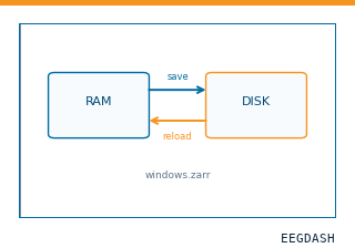 Save and reload prepared dataCache a windowed dataset to disk and reattach it without recompute.
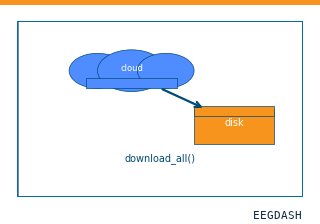 Download a dataset locallyPrefetch BIDS files to a local cache and validate the layout.
Swap any load_dataset(...) call for nm000414 to reproduce the tutorial on this dataset.
Citation
Jenaye Johnson, Gregory Worrell, Brian Litt (2018). Intracranial EEG Epilepsy - Studies 3, 5 and 6 (Mayo Clinic; IEEG.org). 10.82901/nemar.nm000414
Provenance
¹Contributed to nemar in BIDS format.
²Curated & ingested by the EEGDash catalog; see CITATION.cff for canonical reference.
³Persistent identifier: 10.82901/nemar.nm000414.
See Also#
eegdash.dataset.EEGDashDataseteegdash.dataset