


NM000369: ieeg dataset, 3 subjects#
Sensorimotor alpha and beta ECoG during movement imagery (Stolk et al. 2019, shared participants S4-S6)
Access recordings and metadata through EEGDash.
Citation: Arjen Stolk, Loek Brinkman, Mariska J. Vansteensel, Erik Aarnoutse, Frans S.S. Leijten, Chris H. Dijkerman, Robert T. Knight, Floris P. de Lange, Ivan Toni (2019). Sensorimotor alpha and beta ECoG during movement imagery (Stolk et al. 2019, shared participants S4-S6). 10.82901/nemar.nm000369
Modality: ieeg Subjects: 3 Recordings: 3 License: CC-BY-4.0 Source: nemar
Metadata: Complete (100%)
3-participant iEEG dataset — Sensorimotor alpha and beta ECoG during movement imagery (Stolk et al. 2019, shared participants S4-S6).
Quickstart#
Install
pip install eegdash
Access the data
from eegdash.dataset import NM000369

dataset = NM000369(cache_dir="./data")
# Get the raw object of the first recording
raw = dataset.datasets[0].raw
print(raw.info)
Filter by subject
dataset = NM000369(cache_dir="./data", subject="01")
Advanced query
dataset = NM000369(
    cache_dir="./data",
    query={"subject": {"$in": ["01", "02"]}},
)
Iterate recordings
for rec in dataset:
    print(rec.subject, rec.raw.info['sfreq'])
If you use this dataset in your research, please cite the original authors.
BibTeX
@dataset{nm000369,
  title = {Sensorimotor alpha and beta ECoG during movement imagery (Stolk et al. 2019, shared participants S4-S6)},
  author = {Arjen Stolk and Loek Brinkman and Mariska J. Vansteensel and Erik Aarnoutse and Frans S.S. Leijten and Chris H. Dijkerman and Robert T. Knight and Floris P. de Lange and Ivan Toni},
  doi = {10.82901/nemar.nm000369},
  url = {https://doi.org/10.82901/nemar.nm000369},
}
About This Dataset#
Electrocorticography (subdural grids/strips, University Medical Center Utrecht) recorded while epilepsy patients imagined
grasping a cylinder with the left or right hand (movement imagery task, 60 trials per session). The paper analysed 9 of 11 implanted participants; the authors’ OSF record (https://osf.io/z4hfm/) states: “Due to privacy issues, we are not allowed to share the data of all the patients reported in our paper. The data of the patients that have explicitly granted permission to share their data is shared here.” Those are participants S4, S5 and S6, released as trial segments.
Paper: Stolk A, Brinkman L, Vansteensel MJ, Aarnoutse E, Leijten FSS, Dijkerman CH, Knight RT, de Lange FP, Toni I (2019).
Electrocorticographic dissociation of alpha and beta rhythmic activity in the human sensorimotor system. eLife 8:e48065. https://doi.org/10.7554/eLife.48065 . License of the OSF data record: CC-By Attribution 4.0 International.
Sensorimotor alpha and beta ECoG during movement imagery (Stolk et al. 2019) - participants S4, S5, S6
Overview
Participants / cohort (paper, Methods ‘Participants’)
Paper cohort: 11 epilepsy patients (7 males, 14-45 years) implanted subdurally with grid and strip arrays at the University
View full README
Sensorimotor alpha and beta ECoG during movement imagery (Stolk et al. 2019) - participants S4, S5, S6
Overview
Participants / cohort (paper, Methods ‘Participants’)
Paper cohort: 11 epilepsy patients (7 males, 14-45 years) implanted subdurally with grid and strip arrays at the University Medical Center Utrecht (The Netherlands) to localise the seizure onset zone for surgical resection; arrays on the left hemisphere in 8 cases, on the right in 3; 81.3 +/- 11.2 electrodes (mean +/- SEM). All had normal hearing and vision. Two participants were excluded for not adhering to the task instructions (9 analysed behaviourally); two had no upper-limb sensorimotor coverage (7 analysed neurally). No seizures occurred during task administration.
This release: S4 (right hemisphere, 112 electrodes), S5 (left, 120), S6 (left, 64); hemisphere and counts from the released electrode positions/atlas labels. In the paper, S4 and S5 contributed alpha/beta local-maxima electrodes; S6 had limited sensorimotor coverage and only its four stimulation-positive electrodes were used, for temporal dynamics only.
Per-participant age, sex and handedness are not published (n/a in participants.tsv); recording dates/years are not stated.
Task (paper, Methods ‘Movement imagery task’)
Participants lay semi-recumbent in their hospital bed and performed up to three sessions (mean 2 +/- 0.2) of 60 trials (10 min). A black-white cylinder (17.5 x 3.5 cm), tilted in 1 of 15 orientations (24 deg apart, pseudo-random), was shown for 2-5 s (adjusted per participant); participants imagined grasping its middle third with the left or right hand (hand alternating every ten trials, visual cue). A response screen (black and white squares, order pseudo-random) followed, and participants reported whether the thumb ended on the black or white part by pressing the left or right button with the left or right thumb on a button box held with both hands; a fixation cross (3-4 s) followed. A control task (same visual input, judge which side of the cylinder is larger) was completed by 8 of 9 participants; it is not identified in this release.
Acquisition (paper, Methods ‘ECoG acquisition and analysis’)
128-channel Micromed system (Treviso, Italy; 22 bits), analog band-pass 0.15-134.4 Hz, sampled at 512 Hz. Ad-Tech subdural grids and strips (Racine, USA), 10 mm spacing, 2.3 mm exposed diameter. Epochs with overt movements or distracting events were excluded by the authors (6 +/- 2 % of trials). Electrode localisation: post-implantation CT fused with the pre-operative T1 MRI (Philips 3T Achieva; CT Philips Tomoscan SR7000), projection to FreeSurfer surfaces (Stolk et al. 2018, https://doi.org/10.1038/s41596-018-0009-6).
Ethics: Medical Ethical Committee of UMC Utrecht, reference 12-075. The paper’s own analysis then filtered (1-200 Hz), removed line noise and re-referenced to the common average; that processing is NOT applied to the shared segments (see below).
Content, preprocessing and conversion (known caveats)
sub-S4: 112 ECoG channels, 173 epochs of 2305 samples (-1.5 s to 3.000 s around the trial time zero), 512 Hz
sub-S5: 120 ECoG channels, 49 epochs of 2049 samples (-1.5 s to 2.500 s around the trial time zero), 512 Hz
sub-S6: 64 ECoG channels, 180 epochs of 2561 samples (-1.5 s to 3.500 s around the trial time zero), 512 Hz
The source contains only trial segments (FieldTrip
data.trial), not continuous recordings. Each participant’s segments are written back-to-back into one BrainVision file (RecordingType:epoched;events.tsvmarks every epoch, its time-zero sample and the 9 undocumentedtrialinfocolumns verbatim). Do not treat the file as continuous: epoch boundaries are discontinuities.No filtering, re-referencing or resampling was applied. Values are stored losslessly (round trip exact, values in microvolts): sub-S4 BrainVision INT_16 x 0.09765625; sub-S5 BrainVision INT_16 x 0.09765625; sub-S6 no exact integer coding exists (off-grid values, likely resampled by the authors), so the float64 samples are stored losslessly in NWB (acquisition ‘ECoG’). Units: the .mat files do not state the signal unit (FieldTrip
elec.chanunit‘V’ is the toolbox default for sensor definitions). The values are labelled microvolts because the recording system is Micromed and the quantum (0.09765625) is the same as in Micromed data with declared microvolt units; this is an inference, not a source statement.Electrodes: positions from the FieldTrip
elecstructure (mm, coordinate system not named by the source), plus the authors’ electrode table (11 mm atlas look-ups, discard/epileptic/out-of-brain flags) as extra columns. Channels flagged by the authors arestatus=badin channels.tsv (no channel removed).Reference: not reported for the shared segments (n/a).
sourcedata/osf-z4hfm/: the original OSF files, byte-identical:S*_raw_segmented.mat,S*_electable_11mm.xlsxand FreeSurfer pial surfacesS*_lh.pial/S*_rh.pial(cortical meshes, no facial information).Per-participant age/sex are not released (paper: 11 participants, 7 males, 14-45 years): n/a in participants.tsv.
How to load
import mne_bids
bp = mne_bids.BIDSPath(root=".", subject="S4", task="motorimagery", datatype="ieeg")
raw = mne_bids.read_raw_bids(bp)  # sub-S4/S5 BrainVision; sub-S6 is NWB (read with pynwb)
Remember that the files are epoched (RecordingType = epoched): use events.tsv (sample, time_zero_sample) to cut epochs.
Citation
Stolk A et al. (2019) eLife 8:e48065, https://doi.org/10.7554/eLife.48065 ; data: https://osf.io/z4hfm/ .
Provenance
OSF project z4hfm (“Data accompanying Stolk et al. 2019 …”, created 2020-04-21); paper full text PMC6785220. Metadata enriched 2026-10-07 from the paper (participants, task, acquisition, acknowledgements) and from the released electrode tables.
NEMAR Metadata#
[](https://doi.org/10.82901/nemar.nm000369) # Sensorimotor alpha and beta ECoG during movement imagery (Stolk et al. 2019) - participants S4, S5, S6 ## Overview Electrocorticography (subdural grids/strips, University Medical Center Utrecht) recorded while epilepsy patients imagined grasping a cylinder with the left or right hand (movement imagery task, 60 trials per session). The paper analysed 9 of 11 implanted participants; the authors’ OSF record (https://osf.io/z4hfm/) states: “Due to privacy issues, we are not allowed to share the data of all the patients reported in our paper. The data of the patients that have explicitly granted permission to share their data is shared here.” Those are participants S4, S5 and S6, released as trial segments. Paper: Stolk A, Brinkman L, Vansteensel MJ, Aarnoutse E, Leijten FSS, Dijkerman CH, Knight RT, de Lange FP, Toni I (2019). Electrocorticographic dissociation of alpha and beta rhythmic activity in the human sensorimotor system. eLife 8:e48065. https://doi.org/10.7554/eLife.48065 . License of the OSF data record: CC-By Attribution 4.0 International. ## Participants / cohort (paper, Methods ‘Participants’) - Paper cohort: 11 epilepsy patients (7 males, 14-45 years) implanted subdurally with grid and strip arrays at the University
Medical Center Utrecht (The Netherlands) to localise the seizure onset zone for surgical resection; arrays on the left hemisphere in 8 cases, on the right in 3; 81.3 +/- 11.2 electrodes (mean +/- SEM). All had normal hearing and vision. Two participants were excluded for not adhering to the task instructions (9 analysed behaviourally); two had no upper-limb sensorimotor coverage (7 analysed neurally). No seizures occurred during task administration.
This release: S4 (right hemisphere, 112 electrodes), S5 (left, 120), S6 (left, 64); hemisphere and counts from the released electrode positions/atlas labels. In the paper, S4 and S5 contributed alpha/beta local-maxima electrodes; S6 had limited sensorimotor coverage and only its four stimulation-positive electrodes were used, for temporal dynamics only.
Per-participant age, sex and handedness are not published (n/a in participants.tsv); recording dates/years are not stated.
## Task (paper, Methods ‘Movement imagery task’) Participants lay semi-recumbent in their hospital bed and performed up to three sessions (mean 2 +/- 0.2) of 60 trials (10 min). A black-white cylinder (17.5 x 3.5 cm), tilted in 1 of 15 orientations (24 deg apart, pseudo-random), was shown for 2-5 s (adjusted per participant); participants imagined grasping its middle third with the left or right hand (hand alternating every ten trials, visual cue). A response screen (black and white squares, order pseudo-random) followed, and participants reported whether the thumb ended on the black or white part by pressing the left or right button with the left or right thumb on a button box held with both hands; a fixation cross (3-4 s) followed. A control task (same visual input, judge which side of the cylinder is larger) was completed by 8 of 9 participants; it is not identified in this release. ## Acquisition (paper, Methods ‘ECoG acquisition and analysis’) 128-channel Micromed system (Treviso, Italy; 22 bits), analog band-pass 0.15-134.4 Hz, sampled at 512 Hz. Ad-Tech subdural grids and strips (Racine, USA), 10 mm spacing, 2.3 mm exposed diameter. Epochs with overt movements or distracting events were excluded by the authors (6 +/- 2 % of trials). Electrode localisation: post-implantation CT fused with the pre-operative T1 MRI (Philips 3T Achieva; CT Philips Tomoscan SR7000), projection to FreeSurfer surfaces (Stolk et al. 2018, https://doi.org/10.1038/s41596-018-0009-6). Ethics: Medical Ethical Committee of UMC Utrecht, reference 12-075. The paper’s own analysis then filtered (1-200 Hz), removed line noise and re-referenced to the common average; that processing is NOT applied to the shared segments (see below). ## Content, preprocessing and conversion (known caveats) - sub-S4: 112 ECoG channels, 173 epochs of 2305 samples (-1.5 s to 3.000 s around the trial time zero), 512 Hz - sub-S5: 120 ECoG channels, 49 epochs of 2049 samples (-1.5 s to 2.500 s around the trial time zero), 512 Hz - sub-S6: 64 ECoG channels, 180 epochs of 2561 samples (-1.5 s to 3.500 s around the trial time zero), 512 Hz - The source contains only trial segments (FieldTrip data.trial), not continuous recordings. Each participant’s segments are
written back-to-back into one BrainVision file (RecordingType: epoched; events.tsv marks every epoch, its time-zero sample and the 9 undocumented trialinfo columns verbatim). Do not treat the file as continuous: epoch boundaries are discontinuities.
No filtering, re-referencing or resampling was applied. Values are stored losslessly (round trip exact, values in microvolts): sub-S4 BrainVision INT_16 x 0.09765625; sub-S5 BrainVision INT_16 x 0.09765625; sub-S6 no exact integer coding exists (off-grid values, likely resampled by the authors), so the float64 samples are stored losslessly in NWB (acquisition ‘ECoG’). Units: the .mat files do not state the signal unit (FieldTrip elec.chanunit ‘V’ is the toolbox default for sensor definitions). The values are labelled microvolts because the recording system is Micromed and the quantum (0.09765625) is the same as in Micromed data with declared microvolt units; this is an inference, not a source statement.
Electrodes: positions from the FieldTrip elec structure (mm, coordinate system not named by the source), plus the authors’ electrode table (11 mm atlas look-ups, discard/epileptic/out-of-brain flags) as extra columns. Channels flagged by the authors are status=bad in channels.tsv (no channel removed).
Reference: not reported for the shared segments (n/a).
sourcedata/osf-z4hfm/: the original OSF files, byte-identical: S*_raw_segmented.mat, S*_electable_11mm.xlsx and FreeSurfer pial surfaces S*_lh.pial/S*_rh.pial (cortical meshes, no facial information).
Per-participant age/sex are not released (paper: 11 participants, 7 males, 14-45 years): n/a in participants.tsv.
## How to load
`python
import mne_bids
bp = mne_bids.BIDSPath(root=".", subject="S4", task="motorimagery", datatype="ieeg")
raw = mne_bids.read_raw_bids(bp)  # sub-S4/S5 BrainVision; sub-S6 is NWB (read with pynwb)
`
Remember that the files are epoched (RecordingType = epoched): use events.tsv (sample, time_zero_sample) to cut epochs.
## Citation
Stolk A et al. (2019) eLife 8:e48065, https://doi.org/10.7554/eLife.48065 ; data: https://osf.io/z4hfm/ .
## Provenance
OSF project z4hfm (“Data accompanying Stolk et al. 2019 …”, created 2020-04-21); paper full text PMC6785220. Metadata enriched
2026-10-07 from the paper (participants, task, acquisition, acknowledgements) and from the released electrode tables.
License: CC-BY-4.0
Authors:
Arjen Stolk
Loek Brinkman
Mariska J. Vansteensel
Erik Aarnoutse
Frans S.S. Leijten
… and 4 more
Versions:
Version |
DOI |
Released |
|---|---|---|
|
Cohort#
Dataset Statistics#
Channel counts (ch)
Sampling frequencies: 512.0 Hz (n=3 recordings)
Total recording duration: 31 min
Signal · Electrodes & live trace#
Live trace viewer — sub-S5 · task-motorimagery
Showing one representative recording out of
3 subjects and 3 recordings in this dataset.
Browse the full set on OpenNeuro;
drop any other _ieeg.{set,edf,bdf,vhdr} file onto the
viewer (or pass ?ieeg=<url>) to inspect it.
Electrode layout — iEEG · 112 sensors — 112 channels
NEMAR Processing Statistics#
The plots below are generated by NEMAR’s automated EEG pipeline. The histogram shows pipeline success for data cleaning and ICA decomposition, the percentage of data frames and EEG channels retained after artefact removal, line noise per channel (RMS, dB), and the age/gender distribution of participants.

HED event descriptors word cloud

Manifest#
File Explorer#
Browse the BIDS file structure of this dataset. Records are fetched on demand from the EEGDash catalog the first time you open the explorer.
Full dataset metadata table
Dataset ID |
|
Title |
Sensorimotor alpha and beta ECoG during movement imagery (Stolk et al. 2019, shared participants S4-S6) |
Author (year) |
— |
Canonical |
— |
Importable as |
|
Year |
2019 |
Authors |
Arjen Stolk, Loek Brinkman, Mariska J. Vansteensel, Erik Aarnoutse, Frans S.S. Leijten, Chris H. Dijkerman, Robert T. Knight, Floris P. de Lange, Ivan Toni |
License |
CC-BY-4.0 |
Citation / DOI |
|
Source links |
OpenNeuro | NeMAR | Source URL |
Copy-paste BibTeX
@dataset{nm000369,
  title = {Sensorimotor alpha and beta ECoG during movement imagery (Stolk et al. 2019, shared participants S4-S6)},
  author = {Arjen Stolk and Loek Brinkman and Mariska J. Vansteensel and Erik Aarnoutse and Frans S.S. Leijten and Chris H. Dijkerman and Robert T. Knight and Floris P. de Lange and Ivan Toni},
  doi = {10.82901/nemar.nm000369},
  url = {https://doi.org/10.82901/nemar.nm000369},
}
API Reference#
eegdash.datasetEEGDashDataset- class eegdash.dataset.NM000369(cache_dir: str, query: dict | None = None, s3_bucket: str | None = None, **kwargs)[source]#
Sensorimotor alpha and beta ECoG during movement imagery (Stolk et al. 2019, shared participants S4-S6)
- Study:
nm000369(NeMAR)- Author (year):
—
- Canonical:
—
Also importable as:
NM000369.Modality:
ieeg; Subject type:Unknown. Subjects: 3; recordings: 3; tasks: 1.- Parameters:
cache_dir (str | Path) – Directory where data are cached locally.
query (dict | None) – Additional MongoDB-style filters to AND with the dataset selection. Must not contain the key
dataset.s3_bucket (str | None) – Base S3 bucket used to locate the data.
**kwargs (dict) – Additional keyword arguments forwarded to
EEGDashDataset.
- data_dir#
Local dataset cache directory (
cache_dir / dataset_id).- Type:
Path
Notes
Each item is a recording; recording-level metadata are available via
dataset.description.querysupports MongoDB-style filters on fields inALLOWED_QUERY_FIELDSand is combined with the dataset filter. Dataset-specific caveats are not provided in the summary metadata.References
OpenNeuro dataset: https://openneuro.org/datasets/nm000369 NeMAR dataset: https://nemar.org/dataexplorer/detail?dataset_id=nm000369 DOI: https://doi.org/10.82901/nemar.nm000369
Examples
>>> from eegdash.dataset import NM000369
>>> dataset = NM000369(cache_dir="./data")
>>> recording = dataset[0]
>>> raw = recording.load()
- __init__(cache_dir: str, query: dict | None = None, s3_bucket: str | None = None, **kwargs)[source]#
- save(path: str, overwrite: bool = False, offset: int = 0)[source]#
Save datasets to files by creating one subdirectory for each dataset:
path/
    0/
        0-raw.fif | 0-epo.fif
        description.json
        raw_preproc_kwargs.json (if raws were preprocessed)
        window_kwargs.json (if this is a windowed dataset)
        window_preproc_kwargs.json  (if windows were preprocessed)
        target_name.json (if target_name is not None and dataset is raw)
    1/
        1-raw.fif | 1-epo.fif
        description.json
        raw_preproc_kwargs.json (if raws were preprocessed)
        window_kwargs.json (if this is a windowed dataset)
        window_preproc_kwargs.json  (if windows were preprocessed)
        target_name.json (if target_name is not None and dataset is raw)
- Parameters:
path (str) –
- Directory in which subdirectories are created to store
-raw.fif | -epo.fif and .json files to.
overwrite (bool) – Whether to delete old subdirectories that will be saved to in this call.
offset (int) – If provided, the integer is added to the id of the dataset in the concat. This is useful in the setting of very large datasets, where one dataset has to be processed and saved at a time to account for its original position.
BaseDataset from braindecode — windowed via create_windows_from_events.braindecodeDataLoader; supports parallel workers and on-the-fly augmentations.pytorch Find datasets with the EEGDash APIQuery the catalogue, filter by task or modality, list candidates.
 Load one EEG recordingResolve a single record to an MNE Raw with channels and events.
 EEG recording to PyTorch DataLoaderWrap braindecode windows in a DataLoader for model training.
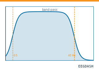 Preprocess EEG and create windowsFilter, resample, epoch — and persist the windowed dataset.
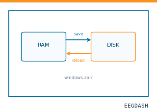 Save and reload prepared dataCache a windowed dataset to disk and reattach it without recompute.
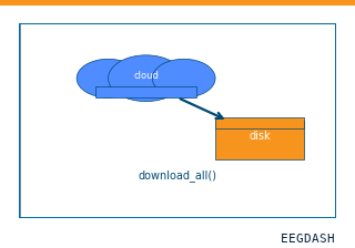 Download a dataset locallyPrefetch BIDS files to a local cache and validate the layout.
Swap any load_dataset(...) call for nm000369 to reproduce the tutorial on this dataset.
Citation
Arjen Stolk, Loek Brinkman, Mariska J. Vansteensel, Erik Aarnoutse, Frans S.S. Leijten, … (2019). Sensorimotor alpha and beta ECoG during movement imagery (Stolk et al. 2019, shared participants S4-S6). 10.82901/nemar.nm000369
Provenance
¹Contributed to nemar in BIDS format.
²Curated & ingested by the EEGDash catalog; see CITATION.cff for canonical reference.
³Persistent identifier: 10.82901/nemar.nm000369.
See Also#
eegdash.dataset.EEGDashDataseteegdash.dataset