
NM000173: eeg dataset, 15 subjects#
Motor Imagery ataset from Ofner et al 2017
Access recordings and metadata through EEGDash.
Citation: Patrick Ofner, Andreas Schwarz, Joana Pereira, Gernot R. Müller-Putz (2019). Motor Imagery ataset from Ofner et al 2017. 10.82901/nemar.nm000173
Modality: eeg Subjects: 15 Recordings: 300 License: CC-BY-4.0 Source: nemar
Metadata: Complete (100%)
15-participant EEG dataset — Motor Imagery ataset from Ofner et al 2017.
Quickstart#
Install
pip install eegdash
Access the data
from eegdash.dataset import NM000173

dataset = NM000173(cache_dir="./data")
# Get the raw object of the first recording
raw = dataset.datasets[0].raw
print(raw.info)
Filter by subject
dataset = NM000173(cache_dir="./data", subject="01")
Advanced query
dataset = NM000173(
    cache_dir="./data",
    query={"subject": {"$in": ["01", "02"]}},
)
Iterate recordings
for rec in dataset:
    print(rec.subject, rec.raw.info['sfreq'])
If you use this dataset in your research, please cite the original authors.
BibTeX
@dataset{nm000173,
  title = {Motor Imagery ataset from Ofner et al 2017},
  author = {Patrick Ofner and Andreas Schwarz and Joana Pereira and Gernot R. Müller-Putz},
  doi = {10.82901/nemar.nm000173},
  url = {https://doi.org/10.82901/nemar.nm000173},
}
About This Dataset#
Motor Imagery ataset from Ofner et al 2017.
Schema: HED 8.4.0 | Browse: https://www.hedtags.org/hed-schema-browser
Motor Imagery ataset from Ofner et al 2017
right_elbow_flexion
View full README
Motor Imagery ataset from Ofner et al 2017
right_elbow_flexion
     ├─ Sensory-event, Experimental-stimulus, Visual-presentation
     └─ Agent-action
        └─ Imagine
           ├─ Flex
           └─ Right, Elbow

right_elbow_extension
     ├─ Sensory-event, Experimental-stimulus, Visual-presentation
     └─ Agent-action
        └─ Imagine
           ├─ Stretch
           └─ Right, Elbow

right_supination
     ├─ Sensory-event, Experimental-stimulus, Visual-presentation
     └─ Agent-action
        └─ Imagine
           ├─ Turn
           ├─ Right, Forearm
           └─ Label/supination

right_pronation
     ├─ Sensory-event, Experimental-stimulus, Visual-presentation
     └─ Agent-action
        └─ Imagine
           ├─ Turn
           ├─ Right, Forearm
           └─ Label/pronation

right_hand_close
     ├─ Sensory-event, Experimental-stimulus, Visual-presentation
     └─ Agent-action
        └─ Imagine
           ├─ Close
           └─ Right, Hand

right_hand_open
     ├─ Sensory-event, Experimental-stimulus, Visual-presentation
     └─ Agent-action
        └─ Imagine
           ├─ Open
           └─ Right, Hand

rest
├─ Sensory-event
├─ Experimental-stimulus
├─ Visual-presentation
└─ Rest
Paradigm-Specific Parameters
Detected paradigm: motor_imagery
Imagery tasks: elbow_flexion, elbow_extension, forearm_supination, forearm_pronation, hand_open, hand_close
Data Structure
Trials: 420
Trials per class: elbow_flexion=60, elbow_extension=60, forearm_supination=60, forearm_pronation=60, hand_open=60, hand_close=60, rest=60
Trials context: per_session
Preprocessing
Preprocessing applied: False
Signal Processing
Classifiers: sLDA
Feature extraction: time-domain signals, discriminative spatial patterns (DSP)
Frequency bands: analyzed=[0.3, 3.0] Hz
Spatial filters: sLORETA source localization
Cross-Validation
Method: 10x10-fold cross-validation
Folds: 10
Evaluation type: within-session
Performance (Original Study)
Mov Vs Mov Me: 55.0
Mov Vs Rest Me: 87.0
Mov Vs Mov Mi: 27.0
Mov Vs Rest Mi: 73.0
BCI Application
Applications: neuroprosthesis, robotic_arm
Environment: laboratory
Online feedback: False
Tags
Pathology: Healthy
Modality: Motor
Type: Motor Imagery, Motor Execution
Documentation
DOI: 10.1371/journal.pone.0182578
Associated paper DOI: 10.1371/journal.pone.0182578
License: CC-BY-4.0
Investigators: Patrick Ofner, Andreas Schwarz, Joana Pereira, Gernot R. Müller-Putz
Senior author: Gernot R. Müller-Putz
Contact: gernot.mueller@tugraz.at
Institution: Graz University of Technology
Department: Institute of Neural Engineering, BCI-Lab
Country: AT
Repository: BNCI Horizon 2020
Publication year: 2017
Funding: H2020-643955 MoreGrasp; ERC Consolidator Grant ERC-681231 Feel Your Reach
Ethics approval: Medical University of Graz, approval number 28-108 ex 15/16
Acknowledgements: Data are available from the BNCI Horizon 2020 database at http://bnci-horizon-2020.eu/database/data-sets (accession number 001-2017) and from Zenodo at DOI 10.5281/zenodo.834976
Keywords: upper limb movements, EEG, motor imagery, movement execution, low-frequency, time-domain, BCI, neuroprosthesis
Abstract
How neural correlates of movements are represented in the human brain is of ongoing interest and has been researched with invasive and non-invasive methods. In this study, we analyzed the encoding of single upper limb movements in the time-domain of low-frequency electroencephalography (EEG) signals. Fifteen healthy subjects executed and imagined six different sustained upper limb movements. We classified these six movements and a rest class and obtained significant average classification accuracies of 55% (movement vs movement) and 87% (movement vs rest) for executed movements, and 27% and 73%, respectively, for imagined movements. Furthermore, we analyzed the classifier patterns in the source space and located the brain areas conveying discriminative movement information. The classifier patterns indicate that mainly premotor areas, primary motor cortex, somatosensory cortex and posterior parietal cortex convey discriminative movement information. The decoding of single upper limb movements is specially interesting in the context of a more natural non-invasive control of e.g., a motor neuroprosthesis or a robotic arm in highly motor disabled persons.
Methodology
Subjects performed 6 sustained upper limb movements (elbow flexion/extension, forearm supination/pronation, hand open/close) plus rest in two separate sessions (movement execution and motor imagery). EEG was recorded from 61 channels, filtered to 0.3-3 Hz, and classified using shrinkage LDA with discriminative spatial patterns. Source localization was performed using sLORETA. Classification employed both single time-point and time-window approaches with 10x10-fold cross-validation.
References
Ofner, P., Schwarz, A., Pereira, J. and Müller-Putz, G.R., 2017. Upper limb movements can be decoded from the time-domain of low-frequency EEG. PloS one, 12(8), p.e0182578. https://doi.org/10.1371/journal.pone.0182578 Appelhoff, S., Sanderson, M., Brooks, T., Vliet, M., Quentin, R., Holdgraf, C., Chaumon, M., Mikulan, E., Tavabi, K., Hochenberger, R., Welke, D., Brunner, C., Rockhill, A., Larson, E., Gramfort, A. and Jas, M. (2019). MNE-BIDS: Organizing electrophysiological data into the BIDS format and facilitating their analysis. Journal of Open Source Software 4: (1896). https://doi.org/10.21105/joss.01896 Pernet, C. R., Appelhoff, S., Gorgolewski, K. J., Flandin, G., Phillips, C., Delorme, A., Oostenveld, R. (2019). EEG-BIDS, an extension to the brain imaging data structure for electroencephalography. Scientific Data, 6, 103. https://doi.org/10.1038/s41597-019-0104-8 Generated by MOABB 1.5.0 (Mother of All BCI Benchmarks) NeuroTechX/moabb
NEMAR Metadata#
[](https://doi.org/10.82901/nemar.nm000173) # Motor Imagery ataset from Ofner et al 2017 Motor Imagery ataset from Ofner et al 2017. ## Dataset Overview - Code: Ofner2017 - Paradigm: imagery - DOI: 10.1371/journal.pone.0182578 - Subjects: 15 - Sessions per subject: 2 - Events: right_elbow_flexion=1536, right_elbow_extension=1537, right_supination=1538, right_pronation=1539, right_hand_close=1540, right_hand_open=1541, rest=1542 - Trial interval: [0, 3] s - Runs per session: 10 - Session IDs: movement_execution, motor_imagery - File format: gdf ## Acquisition - Sampling rate: 512.0 Hz - Number of channels: 61 - Channel types: eeg=61, eog=3, misc=32 - Channel names: C1, C2, C3, C4, C5, C6, CCP1h, CCP2h, CCP3h, CCP4h, CCP5h, CCP6h, CP1, CP2, CP3, CP4, CP5, CP6, CPP1h, CPP2h, CPP3h, CPP4h, CPP5h, CPP6h, CPz, Cz, F1, F2, F3, F4, FC1, FC2, FC3, FC4, FC5, FC6, FCC1h, FCC2h, FCC3h, FCC4h, FCC5h, FCC6h, FCz, FFC1h, FFC2h, FFC3h, FFC4h, FFC5h, FFC6h, FTT7h, FTT8h, Fz, P1, P2, P3, P4, PPO1h, PPO2h, Pz, TTP7h, TTP8h, armeodummy-0, armeodummy-1, armeodummy-10, armeodummy-11, armeodummy-12, armeodummy-2, armeodummy-3, armeodummy-4, armeodummy-5, armeodummy-6, armeodummy-7, armeodummy-8, armeodummy-9, eog-l, eog-m, eog-r, gesture, index_far, index_middle, index_near, litte_far, litte_near, middle_far, middle_near, middle_ring, pitch, ring_far, ring_little, ring_near, roll, thumb_far, thumb_index, thumb_near, thumb_palm, wrist_bend - Montage: standard_1005 - Hardware: g.tec medical engineering GmbH - Reference: right mastoid - Ground: AFz - Sensor type: active - Line frequency: 50.0 Hz - Online filters: 0.01-200 Hz bandpass (8th order Chebyshev), 50 Hz notch ## Participants - Number of subjects: 15 - Health status: healthy - Age: mean=27.0, std=5.0, min=22.0, max=40.0 - Gender distribution: female=9, male=6 - Handedness: {‘right’: 14, ‘left’: 1} - Species: human ## Experimental Protocol - Paradigm: imagery - Number of classes: 7 - Class labels: right_elbow_flexion, right_elbow_extension, right_supination, right_pronation, right_hand_close, right_hand_open, rest - Study design: Trial-based paradigm with sustained movements/motor imagery. Each trial: fixation cross at 0s, cue presentation at 2s, sustained movement/MI execution. Subjects performed both movement execution (ME) and motor imagery (MI) in separate sessions. - Feedback type: none - Stimulus type: visual cue - Synchronicity: synchronous - Mode: offline - Training/test split: False - Instructions: Subjects were instructed to execute sustained movements in ME session and perform kinesthetic motor imagery in MI session. For rest class, subjects were instructed to avoid any movement and to stay in the starting position. ## HED Event Annotations Schema: HED 8.4.0 | Browse: https://www.hedtags.org/hed-schema-browser ```
- right_elbow_flexion
├─ Sensory-event, Experimental-stimulus, Visual-presentation └─ Agent-action
- └─ Imagine
├─ Flex └─ Right, Elbow
- right_elbow_extension
├─ Sensory-event, Experimental-stimulus, Visual-presentation └─ Agent-action
- └─ Imagine
├─ Stretch └─ Right, Elbow
- right_supination
├─ Sensory-event, Experimental-stimulus, Visual-presentation └─ Agent-action
- └─ Imagine
├─ Turn ├─ Right, Forearm └─ Label/supination
- right_pronation
├─ Sensory-event, Experimental-stimulus, Visual-presentation └─ Agent-action
- └─ Imagine
├─ Turn ├─ Right, Forearm └─ Label/pronation
- right_hand_close
├─ Sensory-event, Experimental-stimulus, Visual-presentation └─ Agent-action
- └─ Imagine
├─ Close └─ Right, Hand
- right_hand_open
├─ Sensory-event, Experimental-stimulus, Visual-presentation └─ Agent-action
- └─ Imagine
├─ Open └─ Right, Hand
- rest
├─ Sensory-event ├─ Experimental-stimulus ├─ Visual-presentation └─ Rest
``` ## Paradigm-Specific Parameters - Detected paradigm: motor_imagery - Imagery tasks: elbow_flexion, elbow_extension, forearm_supination, forearm_pronation, hand_open, hand_close ## Data Structure - Trials: 420 - Trials per class: elbow_flexion=60, elbow_extension=60, forearm_supination=60, forearm_pronation=60, hand_open=60, hand_close=60, rest=60 - Trials context: per_session ## Preprocessing - Preprocessing applied: False ## Signal Processing - Classifiers: sLDA - Feature extraction: time-domain signals, discriminative spatial patterns (DSP) - Frequency bands: analyzed=[0.3, 3.0] Hz - Spatial filters: sLORETA source localization ## Cross-Validation - Method: 10x10-fold cross-validation - Folds: 10 - Evaluation type: within-session ## Performance (Original Study) - Mov Vs Mov Me: 55.0 - Mov Vs Rest Me: 87.0 - Mov Vs Mov Mi: 27.0 - Mov Vs Rest Mi: 73.0 ## BCI Application - Applications: neuroprosthesis, robotic_arm - Environment: laboratory - Online feedback: False ## Tags - Pathology: Healthy - Modality: Motor - Type: Motor Imagery, Motor Execution ## Documentation - DOI: 10.1371/journal.pone.0182578 - Associated paper DOI: 10.1371/journal.pone.0182578 - License: CC-BY-4.0 - Investigators: Patrick Ofner, Andreas Schwarz, Joana Pereira, Gernot R. Müller-Putz - Senior author: Gernot R. Müller-Putz - Contact: gernot.mueller@tugraz.at - Institution: Graz University of Technology - Department: Institute of Neural Engineering, BCI-Lab - Country: AT - Repository: BNCI Horizon 2020 - Data URL: https://bnci-horizon-2020.eu/database/data-sets - Publication year: 2017 - Funding: H2020-643955 MoreGrasp; ERC Consolidator Grant ERC-681231 Feel Your Reach - Ethics approval: Medical University of Graz, approval number 28-108 ex 15/16 - Acknowledgements: Data are available from the BNCI Horizon 2020 database at http://bnci-horizon-2020.eu/database/data-sets (accession number 001-2017) and from Zenodo at DOI 10.5281/zenodo.834976 - Keywords: upper limb movements, EEG, motor imagery, movement execution, low-frequency, time-domain, BCI, neuroprosthesis ## Abstract How neural correlates of movements are represented in the human brain is of ongoing interest and has been researched with invasive and non-invasive methods. In this study, we analyzed the encoding of single upper limb movements in the time-domain of low-frequency electroencephalography (EEG) signals. Fifteen healthy subjects executed and imagined six different sustained upper limb movements. We classified these six movements and a rest class and obtained significant average classification accuracies of 55% (movement vs movement) and 87% (movement vs rest) for executed movements, and 27% and 73%, respectively, for imagined movements. Furthermore, we analyzed the classifier patterns in the source space and located the brain areas conveying discriminative movement information. The classifier patterns indicate that mainly premotor areas, primary motor cortex, somatosensory cortex and posterior parietal cortex convey discriminative movement information. The decoding of single upper limb movements is specially interesting in the context of a more natural non-invasive control of e.g., a motor neuroprosthesis or a robotic arm in highly motor disabled persons. ## Methodology Subjects performed 6 sustained upper limb movements (elbow flexion/extension, forearm supination/pronation, hand open/close) plus rest in two separate sessions (movement execution and motor imagery). EEG was recorded from 61 channels, filtered to 0.3-3 Hz, and classified using shrinkage LDA with discriminative spatial patterns. Source localization was performed using sLORETA. Classification employed both single time-point and time-window approaches with 10x10-fold cross-validation. ## References Ofner, P., Schwarz, A., Pereira, J. and Müller-Putz, G.R., 2017. Upper limb movements can be decoded from the time-domain of low-frequency EEG. PloS one, 12(8), p.e0182578. https://doi.org/10.1371/journal.pone.0182578 Appelhoff, S., Sanderson, M., Brooks, T., Vliet, M., Quentin, R., Holdgraf, C., Chaumon, M., Mikulan, E., Tavabi, K., Hochenberger, R., Welke, D., Brunner, C., Rockhill, A., Larson, E., Gramfort, A. and Jas, M. (2019). MNE-BIDS: Organizing electrophysiological data into the BIDS format and facilitating their analysis. Journal of Open Source Software 4: (1896). https://doi.org/10.21105/joss.01896 Pernet, C. R., Appelhoff, S., Gorgolewski, K. J., Flandin, G., Phillips, C., Delorme, A., Oostenveld, R. (2019). EEG-BIDS, an extension to the brain imaging data structure for electroencephalography. Scientific Data, 6, 103. https://doi.org/10.1038/s41597-019-0104-8 — Generated by MOABB 1.5.0 (Mother of All BCI Benchmarks) NeuroTechX/moabb
License: CC-BY-4.0
Authors:
Patrick Ofner
Andreas Schwarz
Joana Pereira
Gernot R. Müller-Putz
Versions:
Version |
DOI |
Released |
|---|---|---|
|
Cohort#
Dataset Statistics#
Age distribution by gender (n=15, range 2014–2014 yr)
Channel counts: 61 ch (n=300 recordings)
Sampling frequencies: 512.0 Hz (n=300 recordings)
Total recording duration: 27 h
Signal · Electrodes & live trace#
Live trace viewer — sub-13 · ses-0execution · task-imagery · run-2
Showing one representative recording out of
15 subjects and 300 recordings in this dataset.
Browse the full set on OpenNeuro;
drop any other _eeg.{set,edf,bdf,vhdr} file onto the
viewer (or pass ?eeg=<url>) to inspect it.
Electrode layout — EEG · 61 sensors — 61 channels
NEMAR Processing Statistics#
The plots below are generated by NEMAR’s automated EEG pipeline. The histogram shows pipeline success for data cleaning and ICA decomposition, the percentage of data frames and EEG channels retained after artefact removal, line noise per channel (RMS, dB), and the age/gender distribution of participants.

HED event descriptors word cloud

Manifest#
File Explorer#
Browse the BIDS file structure of this dataset. Records are fetched on demand from the EEGDash catalog the first time you open the explorer.
Full dataset metadata table
Dataset ID |
|
Title |
Motor Imagery ataset from Ofner et al 2017 |
Author (year) |
|
Canonical |
— |
Importable as |
|
Year |
2019 |
Authors |
Patrick Ofner, Andreas Schwarz, Joana Pereira, Gernot R. Müller-Putz |
License |
CC-BY-4.0 |
Citation / DOI |
|
Source links |
Copy-paste BibTeX
@dataset{nm000173,
  title = {Motor Imagery ataset from Ofner et al 2017},
  author = {Patrick Ofner and Andreas Schwarz and Joana Pereira and Gernot R. Müller-Putz},
  doi = {10.82901/nemar.nm000173},
  url = {https://doi.org/10.82901/nemar.nm000173},
}
API Reference#
eegdash.datasetEEGDashDatasetNM000173 · Ofner2017eegdash/dataset/registry.py · [source ↗]- class eegdash.dataset.NM000173(cache_dir: str, query: dict | None = None, s3_bucket: str | None = None, **kwargs)[source]#
Motor Imagery ataset from Ofner et al 2017
- Study:
nm000173(NeMAR)- Author (year):
Ofner2017- Canonical:
—
Also importable as:
NM000173,Ofner2017.Modality:
eeg; Experiment type:Motor; Subject type:Healthy. Subjects: 15; recordings: 300; tasks: 1.- Parameters:
cache_dir (str | Path) – Directory where data are cached locally.
query (dict | None) – Additional MongoDB-style filters to AND with the dataset selection. Must not contain the key
dataset.s3_bucket (str | None) – Base S3 bucket used to locate the data.
**kwargs (dict) – Additional keyword arguments forwarded to
EEGDashDataset.
- data_dir#
Local dataset cache directory (
cache_dir / dataset_id).- Type:
Path
Notes
Each item is a recording; recording-level metadata are available via
dataset.description.querysupports MongoDB-style filters on fields inALLOWED_QUERY_FIELDSand is combined with the dataset filter. Dataset-specific caveats are not provided in the summary metadata.References
OpenNeuro dataset: https://openneuro.org/datasets/nm000173 NeMAR dataset: https://nemar.org/dataexplorer/detail?dataset_id=nm000173 DOI: https://doi.org/10.82901/nemar.nm000173
Examples
>>> from eegdash.dataset import NM000173
>>> dataset = NM000173(cache_dir="./data")
>>> recording = dataset[0]
>>> raw = recording.load()
- __init__(cache_dir: str, query: dict | None = None, s3_bucket: str | None = None, **kwargs)[source]#
- save(path: str, overwrite: bool = False, offset: int = 0)[source]#
Save datasets to files by creating one subdirectory for each dataset:
path/
    0/
        0-raw.fif | 0-epo.fif
        description.json
        raw_preproc_kwargs.json (if raws were preprocessed)
        window_kwargs.json (if this is a windowed dataset)
        window_preproc_kwargs.json  (if windows were preprocessed)
        target_name.json (if target_name is not None and dataset is raw)
    1/
        1-raw.fif | 1-epo.fif
        description.json
        raw_preproc_kwargs.json (if raws were preprocessed)
        window_kwargs.json (if this is a windowed dataset)
        window_preproc_kwargs.json  (if windows were preprocessed)
        target_name.json (if target_name is not None and dataset is raw)
- Parameters:
path (str) –
- Directory in which subdirectories are created to store
-raw.fif | -epo.fif and .json files to.
overwrite (bool) – Whether to delete old subdirectories that will be saved to in this call.
offset (int) – If provided, the integer is added to the id of the dataset in the concat. This is useful in the setting of very large datasets, where one dataset has to be processed and saved at a time to account for its original position.
BaseDataset from braindecode — windowed via create_windows_from_events.braindecodeDataLoader; supports parallel workers and on-the-fly augmentations.pytorchdatasets.load_dataset("EEGDash/nm000173").huggingface Find datasets with the EEGDash APIQuery the catalogue, filter by task or modality, list candidates.
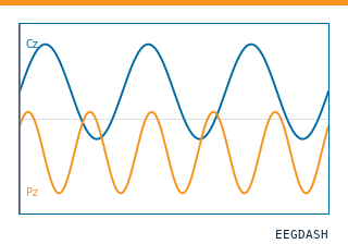 Load one EEG recordingResolve a single record to an MNE Raw with channels and events.
 EEG recording to PyTorch DataLoaderWrap braindecode windows in a DataLoader for model training.
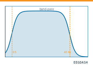 Preprocess EEG and create windowsFilter, resample, epoch — and persist the windowed dataset.
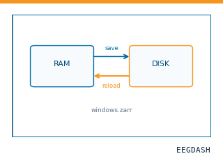 Save and reload prepared dataCache a windowed dataset to disk and reattach it without recompute.
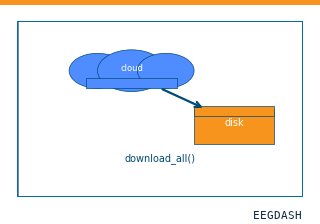 Download a dataset locallyPrefetch BIDS files to a local cache and validate the layout.
Swap any load_dataset(...) call for nm000173 to reproduce the tutorial on this dataset.
Citation
Patrick Ofner, Andreas Schwarz, Joana Pereira, Gernot R. Müller-Putz (2019). Motor Imagery ataset from Ofner et al 2017. 10.82901/nemar.nm000173
Provenance
¹Contributed to nemar in BIDS format.
²Curated & ingested by the EEGDash catalog; see CITATION.cff for canonical reference.
³Persistent identifier: 10.82901/nemar.nm000173.
See Also#
eegdash.dataset.EEGDashDataseteegdash.dataset