


NM000405: ieeg dataset, 16 subjects#
Intraoperative ECoG strip and subthalamic LFP during grip-force in Parkinson’s disease (Merk et al. 2022)
Access recordings and metadata through EEGDash.
Citation: Timon Merk, Victoria Peterson, Witold Lipski, Benjamin Blankertz, Robert S Turner, Ningfei Li, Andreas Horn, Robert Mark Richardson, Wolf-Julian Neumann (2022). Intraoperative ECoG strip and subthalamic LFP during grip-force in Parkinson’s disease (Merk et al. 2022). 10.82901/nemar.nm000405
Modality: ieeg Subjects: 16 Recordings: 73 License: CC0 Source: nemar
Metadata: Complete (100%)
16-participant iEEG dataset — Intraoperative ECoG strip and subthalamic LFP during grip-force in Parkinson's disease (Merk et al. 2022).
Quickstart#
Install
pip install eegdash
Access the data
from eegdash.dataset import NM000405

dataset = NM000405(cache_dir="./data")
# Get the raw object of the first recording
raw = dataset.datasets[0].raw
print(raw.info)
Filter by subject
dataset = NM000405(cache_dir="./data", subject="01")
Advanced query
dataset = NM000405(
    cache_dir="./data",
    query={"subject": {"$in": ["01", "02"]}},
)
Iterate recordings
for rec in dataset:
    print(rec.subject, rec.raw.info['sfreq'])
If you use this dataset in your research, please cite the original authors.
BibTeX
@dataset{nm000405,
  title = {Intraoperative ECoG strip and subthalamic LFP during grip-force in Parkinson's disease (Merk et al. 2022)},
  author = {Timon Merk and Victoria Peterson and Witold Lipski and Benjamin Blankertz and Robert S Turner and Ningfei Li and Andreas Horn and Robert Mark Richardson and Wolf-Julian Neumann},
  doi = {10.82901/nemar.nm000405},
  url = {https://doi.org/10.82901/nemar.nm000405},
}
About This Dataset#
Replication data for: Merk T. et al. (2022) “Electrocorticography is superior to subthalamic local field potentials
for movement decoding in Parkinson’s disease”, eLife 11:e75126, https://doi.org/10.7554/eLife.75126.
Source: Harvard Dataverse, https://doi.org/10.7910/DVN/IO2FLM (v1.0, 2022-04-19), licence CC0 1.0.
16 participants with Parkinson’s disease (University of Pittsburgh Medical Center), 73 runs of an intraoperative grip-force task recorded during bilateral STN-DBS lead implantation. Each run contains a subdural ECoG strip over sensorimotor cortex (ECOG_*), the four contacts of the DBS lead in the subthalamic nucleus (STN_*, where present), and grip-force channels (MOV_LEFT/MOV_RIGHT; MOV_*_CLEAN are the authors’ noise- and drift-cleaned force traces).
Intraoperative ECoG + STN LFP during grip force in Parkinson’s disease (Merk et al. 2022)
Sessions ses-left / ses-right are the hemispheres as labelled by the authors.
Why this is a derivative dataset
The paper states that ECoG and STN signals were filtered (0.3-7.5 kHz) and digitised at 30 kHz (Grapevine, Ripple Inc) and that “all signals were resampled to 1 kHz for offline analysis”. The released signals are these 1 kHz resampled
View full README
Intraoperative ECoG + STN LFP during grip force in Parkinson’s disease (Merk et al. 2022)
Sessions ses-left / ses-right are the hemispheres as labelled by the authors.
Why this is a derivative dataset
The paper states that ECoG and STN signals were filtered (0.3-7.5 kHz) and digitised at 30 kHz (Grapevine, Ripple Inc)
and that “all signals were resampled to 1 kHz for offline analysis”. The released signals are these 1 kHz resampled
signals, so this deposit is labelled DatasetType: derivative and every signal file carries desc-1kHz. Values are
exactly those of the Dataverse release (BrainVision float32); only file names and folders changed.
Changes relative to the Dataverse release
Files sorted into
sub-*/ses-*/ieeg/(Dataverse stores them flat). Data files renamed..._desc-1kHz_ieeg.*; the DataFile/MarkerFile lines in .vhdr/.vmrk follow the new names.Per-run
space-mni_electrodes.tsvtables merged into one per session (identical contacts; force channels dropped).*_ieeg.json: hardware/filter/reference facts added from the paper methods (the release said “BrainProducts”, which is the pybv writer default, not the amplifier).participants.tsv: the columncenter_ID(hospital DBS case numbers) is removed for privacy, also from the sourcedata copy.Not deposited:
ea_ui.mat,ea_ui-1.mat,ea_ui-2.mat(Lead-DBS GUI state files, no data).sourcedata/dataverse-IO2FLM/: all other original metadata files as released (incl. per-subjectsub-*_electrodes*.tsvin mm and the per-run electrode tables;-1suffixes are Dataverse duplicate-name renames).
Not stated by the source (left as n/a)
Age and handedness per participant (the paper’s Table 1 lists 11 patients and does not map onto these 16 IDs); the exact MNI template; anti-aliasing filter used for resampling.
Ethics
Informed consent was obtained under protocol #PRO13110420 approved by the Institutional Review Board of the University of Pittsburgh (paper methods).
Additional metadata and localisation (added 2026-10-08)
Compiled after the upload from the article, its supplement and the source deposit (each statement names its source). Text and sidecar metadata only; no data file was changed.
Recording system. Signals were acquired with a Grapevine neural interface processor (Ripple Inc): ECoG and STN signals filtered at 0.3–7.5 kHz, amplified and digitised at 30 kHz, then resampled to 1 kHz for offline analysis (paper, Methods “Electrophysiological recordings”). The deposit files are BrainVision (Manufacturer “BrainProducts” in the sidecar, i.e. the export format) at SamplingFrequency 1000 Hz with PowerLineFrequency 60 (all 73 *_ieeg.json). The deposit’s iEEGReference is n/a.
Reference scheme. The montage was referential, with the reference electrode in the scalp and the ground over the acromion process. STN LFPs from all four contacts were referenced offline in a bipolar montage (paper, Methods).
Electrode types. ECoG came from subdural strips placed temporarily through standard frontal burr holes near the coronal suture, aimed posteriorly toward the hand-knob motor cortex. Strips had six contacts (left n=5 patients, right n=3), eight contacts (left 3, right 3) or twenty-eight contacts (left 2, right 2). STN LFP came from the clinical DBS lead (Medtronic model 3389, 4 contacts). Details are in Supplementary file 1a of the paper. Source: paper, Methods.
Localisation method. For the subdural strips, preoperative MRI, intraoperative fluoroscopy and postoperative CT were aligned (Randazzo et al. 2016). CT and MRI were co-registered with SPM and rendered with Osirix v7.5 and FreeSurfer v5.3. Contacts were projected from the fluoroscopic image onto the cortical surface. The DBS contacts were reconstructed with Lead-DBS: preoperative MRI and postoperative CT were co-registered and normalised to MNI 2009b NLIN ASYM space. All contacts were “localized and transformed into MNI space” (paper, Methods “Electrode localization”; Figure 1B; coordinates in Figure 1—source data 1, elife-75126-fig1-data1.csv). The deposit has subject-level sub-XXX_electrodes*.tsv (mm, coordsystem “MNI”) for all 16 subjects. The session-level *_space-mni_electrodes.tsv of this dataset carry the same positions in metres; their column atlas_label_AAL3v1 adds an AAL3v1 atlas lookup of these coordinates (ieeg-atlas coord_regions.py, nearest labelled voxel; a derived label, not given by the authors). STN contacts are in the subthalamic nucleus per their channel names; AAL3 has no STN label, so their lookup names the nearest AAL3 structure.
NEMAR Metadata#
[](https://doi.org/10.82901/nemar.nm000405) # Intraoperative ECoG + STN LFP during grip force in Parkinson’s disease (Merk et al. 2022) Replication data for: Merk T. et al. (2022) “Electrocorticography is superior to subthalamic local field potentials for movement decoding in Parkinson’s disease”, eLife 11:e75126, https://doi.org/10.7554/eLife.75126. Source: Harvard Dataverse, https://doi.org/10.7910/DVN/IO2FLM (v1.0, 2022-04-19), licence CC0 1.0. 16 participants with Parkinson’s disease (University of Pittsburgh Medical Center), 73 runs of an intraoperative grip-force task recorded during bilateral STN-DBS lead implantation. Each run contains a subdural ECoG strip over sensorimotor cortex (ECOG_*), the four contacts of the DBS lead in the subthalamic nucleus (STN_*, where present), and grip-force channels (MOV_LEFT/MOV_RIGHT; MOV_*_CLEAN are the authors’ noise- and drift-cleaned force traces). Sessions ses-left / ses-right are the hemispheres as labelled by the authors. ## Why this is a derivative dataset The paper states that ECoG and STN signals were filtered (0.3-7.5 kHz) and digitised at 30 kHz (Grapevine, Ripple Inc) and that “all signals were resampled to 1 kHz for offline analysis”. The released signals are these 1 kHz resampled signals, so this deposit is labelled DatasetType: derivative and every signal file carries desc-1kHz. Values are exactly those of the Dataverse release (BrainVision float32); only file names and folders changed. ## Changes relative to the Dataverse release - Files sorted into sub-*/ses-*/ieeg/ (Dataverse stores them flat). Data files renamed …_desc-1kHz_ieeg.*; the
DataFile/MarkerFile lines in .vhdr/.vmrk follow the new names.
Per-run space-mni_electrodes.tsv tables merged into one per session (identical contacts; force channels dropped).
*_ieeg.json: hardware/filter/reference facts added from the paper methods (the release said “BrainProducts”, which is the pybv writer default, not the amplifier).
participants.tsv: the column center_ID (hospital DBS case numbers) is removed for privacy, also from the sourcedata copy.
Not deposited: ea_ui.mat, ea_ui-1.mat, ea_ui-2.mat (Lead-DBS GUI state files, no data).
sourcedata/dataverse-IO2FLM/: all other original metadata files as released (incl. per-subject sub-*_electrodes*.tsv in mm and the per-run electrode tables; -1 suffixes are Dataverse duplicate-name renames).
## Not stated by the source (left as n/a) Age and handedness per participant (the paper’s Table 1 lists 11 patients and does not map onto these 16 IDs); the exact MNI template; anti-aliasing filter used for resampling. ## Ethics Informed consent was obtained under protocol #PRO13110420 approved by the Institutional Review Board of the University of Pittsburgh (paper methods). ## Additional metadata and localisation (added 2026-10-08) Compiled after the upload from the article, its supplement and the source deposit (each statement names its source). Text and sidecar metadata only; no data file was changed. Recording system. Signals were acquired with a Grapevine neural interface processor (Ripple Inc): ECoG and STN signals filtered at 0.3–7.5 kHz, amplified and digitised at 30 kHz, then resampled to 1 kHz for offline analysis (paper, Methods “Electrophysiological recordings”). The deposit files are BrainVision (Manufacturer “BrainProducts” in the sidecar, i.e. the export format) at SamplingFrequency 1000 Hz with PowerLineFrequency 60 (all 73 *_ieeg.json). The deposit’s iEEGReference is n/a. Reference scheme. The montage was referential, with the reference electrode in the scalp and the ground over the acromion process. STN LFPs from all four contacts were referenced offline in a bipolar montage (paper, Methods). Electrode types. ECoG came from subdural strips placed temporarily through standard frontal burr holes near the coronal suture, aimed posteriorly toward the hand-knob motor cortex. Strips had six contacts (left n=5 patients, right n=3), eight contacts (left 3, right 3) or twenty-eight contacts (left 2, right 2). STN LFP came from the clinical DBS lead (Medtronic model 3389, 4 contacts). Details are in Supplementary file 1a of the paper. Source: paper, Methods. Localisation method. For the subdural strips, preoperative MRI, intraoperative fluoroscopy and postoperative CT were aligned (Randazzo et al. 2016). CT and MRI were co-registered with SPM and rendered with Osirix v7.5 and FreeSurfer v5.3. Contacts were projected from the fluoroscopic image onto the cortical surface. The DBS contacts were reconstructed with Lead-DBS: preoperative MRI and postoperative CT were co-registered and normalised to MNI 2009b NLIN ASYM space. All contacts were “localized and transformed into MNI space” (paper, Methods “Electrode localization”; Figure 1B; coordinates in Figure 1—source data 1, elife-75126-fig1-data1.csv). The deposit has subject-level sub-XXX_electrodes*.tsv (mm, coordsystem “MNI”) for all 16 subjects. The session-level *_space-mni_electrodes.tsv of this dataset carry the same positions in metres; their column atlas_label_AAL3v1 adds an AAL3v1 atlas lookup of these coordinates (ieeg-atlas coord_regions.py, nearest labelled voxel; a derived label, not given by the authors). STN contacts are in the subthalamic nucleus per their channel names; AAL3 has no STN label, so their lookup names the nearest AAL3 structure.
License: CC0
Authors:
Timon Merk
Victoria Peterson
Witold Lipski
Benjamin Blankertz
Robert S Turner
… and 4 more
Versions:
Version |
DOI |
Released |
|---|---|---|
|
Cohort#
Dataset Statistics#
Sex composition
Channel counts (ch)
Sampling frequencies: 1000.0 Hz (n=73 recordings)
Total recording duration: 5 h 41 min
Signal · Electrodes & live trace#
Live trace viewer — sub-000 · ses-right · task-force · run-1
Showing one representative recording out of
16 subjects and 73 recordings in this dataset.
Browse the full set on OpenNeuro;
drop any other _ieeg.{set,edf,bdf,vhdr} file onto the
viewer (or pass ?ieeg=<url>) to inspect it.
Electrode layout — iEEG · 9 sensors — 9 channels
NEMAR Processing Statistics#
The plots below are generated by NEMAR’s automated EEG pipeline. The histogram shows pipeline success for data cleaning and ICA decomposition, the percentage of data frames and EEG channels retained after artefact removal, line noise per channel (RMS, dB), and the age/gender distribution of participants.

HED event descriptors word cloud

Manifest#
File Explorer#
Browse the BIDS file structure of this dataset. Records are fetched on demand from the EEGDash catalog the first time you open the explorer.
Full dataset metadata table
Dataset ID |
|
Title |
Intraoperative ECoG strip and subthalamic LFP during grip-force in Parkinson’s disease (Merk et al. 2022) |
Author (year) |
— |
Canonical |
— |
Importable as |
|
Year |
2022 |
Authors |
Timon Merk, Victoria Peterson, Witold Lipski, Benjamin Blankertz, Robert S Turner, Ningfei Li, Andreas Horn, Robert Mark Richardson, Wolf-Julian Neumann |
License |
CC0 |
Citation / DOI |
|
Source links |
OpenNeuro | NeMAR | Source URL |
Copy-paste BibTeX
@dataset{nm000405,
  title = {Intraoperative ECoG strip and subthalamic LFP during grip-force in Parkinson's disease (Merk et al. 2022)},
  author = {Timon Merk and Victoria Peterson and Witold Lipski and Benjamin Blankertz and Robert S Turner and Ningfei Li and Andreas Horn and Robert Mark Richardson and Wolf-Julian Neumann},
  doi = {10.82901/nemar.nm000405},
  url = {https://doi.org/10.82901/nemar.nm000405},
}
API Reference#
eegdash.datasetEEGDashDataset- class eegdash.dataset.NM000405(cache_dir: str, query: dict | None = None, s3_bucket: str | None = None, **kwargs)[source]#
Intraoperative ECoG strip and subthalamic LFP during grip-force in Parkinson’s disease (Merk et al. 2022)
- Study:
nm000405(NeMAR)- Author (year):
—
- Canonical:
—
Also importable as:
NM000405.Modality:
ieeg; Subject type:Unknown. Subjects: 16; recordings: 73; tasks: 1.- Parameters:
cache_dir (str | Path) – Directory where data are cached locally.
query (dict | None) – Additional MongoDB-style filters to AND with the dataset selection. Must not contain the key
dataset.s3_bucket (str | None) – Base S3 bucket used to locate the data.
**kwargs (dict) – Additional keyword arguments forwarded to
EEGDashDataset.
- data_dir#
Local dataset cache directory (
cache_dir / dataset_id).- Type:
Path
Notes
Each item is a recording; recording-level metadata are available via
dataset.description.querysupports MongoDB-style filters on fields inALLOWED_QUERY_FIELDSand is combined with the dataset filter. Dataset-specific caveats are not provided in the summary metadata.References
OpenNeuro dataset: https://openneuro.org/datasets/nm000405 NeMAR dataset: https://nemar.org/dataexplorer/detail?dataset_id=nm000405 DOI: https://doi.org/10.82901/nemar.nm000405
Examples
>>> from eegdash.dataset import NM000405
>>> dataset = NM000405(cache_dir="./data")
>>> recording = dataset[0]
>>> raw = recording.load()
- __init__(cache_dir: str, query: dict | None = None, s3_bucket: str | None = None, **kwargs)[source]#
- save(path: str, overwrite: bool = False, offset: int = 0)[source]#
Save datasets to files by creating one subdirectory for each dataset:
path/
    0/
        0-raw.fif | 0-epo.fif
        description.json
        raw_preproc_kwargs.json (if raws were preprocessed)
        window_kwargs.json (if this is a windowed dataset)
        window_preproc_kwargs.json  (if windows were preprocessed)
        target_name.json (if target_name is not None and dataset is raw)
    1/
        1-raw.fif | 1-epo.fif
        description.json
        raw_preproc_kwargs.json (if raws were preprocessed)
        window_kwargs.json (if this is a windowed dataset)
        window_preproc_kwargs.json  (if windows were preprocessed)
        target_name.json (if target_name is not None and dataset is raw)
- Parameters:
path (str) –
- Directory in which subdirectories are created to store
-raw.fif | -epo.fif and .json files to.
overwrite (bool) – Whether to delete old subdirectories that will be saved to in this call.
offset (int) – If provided, the integer is added to the id of the dataset in the concat. This is useful in the setting of very large datasets, where one dataset has to be processed and saved at a time to account for its original position.
BaseDataset from braindecode — windowed via create_windows_from_events.braindecodeDataLoader; supports parallel workers and on-the-fly augmentations.pytorch Find datasets with the EEGDash APIQuery the catalogue, filter by task or modality, list candidates.
 Load one EEG recordingResolve a single record to an MNE Raw with channels and events.
 EEG recording to PyTorch DataLoaderWrap braindecode windows in a DataLoader for model training.
 Preprocess EEG and create windowsFilter, resample, epoch — and persist the windowed dataset.
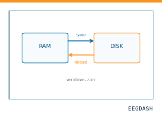 Save and reload prepared dataCache a windowed dataset to disk and reattach it without recompute.
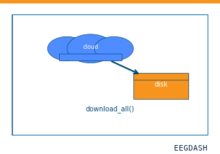 Download a dataset locallyPrefetch BIDS files to a local cache and validate the layout.
Swap any load_dataset(...) call for nm000405 to reproduce the tutorial on this dataset.
Citation
Timon Merk, Victoria Peterson, Witold Lipski, Benjamin Blankertz, Robert S Turner, … (2022). Intraoperative ECoG strip and subthalamic LFP during grip-force in Parkinson's disease (Merk et al. 2022). 10.82901/nemar.nm000405
Provenance
¹Contributed to nemar in BIDS format.
²Curated & ingested by the EEGDash catalog; see CITATION.cff for canonical reference.
³Persistent identifier: 10.82901/nemar.nm000405.
See Also#
eegdash.dataset.EEGDashDataseteegdash.dataset