


NM000374: ieeg dataset, 11 subjects#
Electrophysiological signatures underlying variability in human memory consolidation: raw intracranial EEG (learning and sleep TMR)
Access recordings and metadata through EEGDash.
Citation: Wei Duan, Zhansheng Xu, Dong Chen, Jing Wang, Jiali Liu, Zheng Tan, Xue Xiao, Pengcheng Lv, Mengyang Wang, Ken A. Paller, Nikolai Axmacher, Liang Wang (2025). Electrophysiological signatures underlying variability in human memory consolidation: raw intracranial EEG (learning and sleep TMR). 10.82901/nemar.nm000374
Modality: ieeg Subjects: 11 Recordings: 22 License: CC-BY-4.0 Source: nemar
Metadata: Complete (100%)
11-participant iEEG dataset — Electrophysiological signatures underlying variability in human memory consolidation: raw intracranial EEG (learning and sleep TMR).
Quickstart#
Install
pip install eegdash
Access the data
from eegdash.dataset import NM000374

dataset = NM000374(cache_dir="./data")
# Get the raw object of the first recording
raw = dataset.datasets[0].raw
print(raw.info)
Filter by subject
dataset = NM000374(cache_dir="./data", subject="01")
Advanced query
dataset = NM000374(
    cache_dir="./data",
    query={"subject": {"$in": ["01", "02"]}},
)
Iterate recordings
for rec in dataset:
    print(rec.subject, rec.raw.info['sfreq'])
If you use this dataset in your research, please cite the original authors.
BibTeX
@dataset{nm000374,
  title = {Electrophysiological signatures underlying variability in human memory consolidation: raw intracranial EEG (learning and sleep TMR)},
  author = {Wei Duan and Zhansheng Xu and Dong Chen and Jing Wang and Jiali Liu and Zheng Tan and Xue Xiao and Pengcheng Lv and Mengyang Wang and Ken A. Paller and Nikolai Axmacher and Liang Wang},
  doi = {10.82901/nemar.nm000374},
  url = {https://doi.org/10.82901/nemar.nm000374},
}
About This Dataset#
Intracranial EEG (stereo-EEG depth electrodes) from 11 patients with medically refractory temporal lobe epilepsy
(Sanbo Brain Hospital, Beijing) recorded during (1) learning of object–location associations and (2) targeted memory reactivation (TMR) with sounds during NREM sleep. Re-packaged in iEEG-BIDS from the authors’ public release (Zenodo record 14885382, concept DOI 10.5281/zenodo.14885381, “Raw data of article: Electrophysiological signatures underlying variability in human memory consolidation”, CC BY 4.0). The Zenodo record describes itself as “Raw intracranial electrophysiological signals and behavioral data from 11 participants. All data processed using MATLAB 2019b.” Reference article: Duan W, Xu Z, Chen D, Wang J, Liu J, Tan Z, Xiao X, Lv P, Wang M, Paller KA, Axmacher N, Wang L (2025). Electrophysiological signatures underlying variability in human memory consolidation. Nature Communications 16:2472. https://doi.org/10.1038/s41467-025-57766-x (PMC11903871). Processed data and analysis code: https://doi.org/10.5281/zenodo.14583770. Ripple-detection code used by the authors: https://doi.org/10.5281/zenodo.3259369.
Eleven patients (2 females and 9 males; mean age 23.9 ± 5.4 years, SD) with medically refractory temporal lobe epilepsy,
stereotactically implanted with depth contacts to identify epileptogenic zones, recruited at Beijing Sanbo Brain Hospital; they participated voluntarily without compensation, and all reported normal or corrected-to-normal visual acuity and normal colour vision (Duan et al. 2025, Methods). Recording years are not stated in the paper or the release.
Electrophysiological signatures underlying variability in human memory consolidation — raw intracranial EEG
Overview
The release and the paper give sex and age only at group level; participants.tsv therefore lists n/a for per-subject
age/sex and carries the per-subject recording-site information of Supplementary Table S1 (total recording sites,
hippocampal contacts, electrode laterality, seizure onset zone and laterality) and the per-subject TMR cue counts of
View full README
Electrophysiological signatures underlying variability in human memory consolidation — raw intracranial EEG
Overview
The release and the paper give sex and age only at group level; participants.tsv therefore lists n/a for per-subject
age/sex and carries the per-subject recording-site information of Supplementary Table S1 (total recording sites,
hippocampal contacts, electrode laterality, seizure onset zone and laterality) and the per-subject TMR cue counts of
Supplementary Table S3 (total cues and cues per sleep stage N3/N2/N1/wake; number of presentations per cued sound).
n_electrode_groups (number of electrode labels in the channel names) is derived from the release itself.
Subject mapping: release file subNN_raw_data.mat = sub-NN = paper Subject ID NN. Evidence: Table S1’s “total
recording sites” equals the channel count of each subject’s release file for all 11 subjects (checked by the converter);
the number of mark_tmr markers is lowest for sub-01 (150) and sub-02 (230) and 300 for all others, consistent with the
Methods statement that the first two participants of Table S3 heard each cued sound 3 and 4 times instead of 6; and
right-only implants in Table S1 (subjects 1, 5, 8, 9, 10) have only primed electrode labels while the left-only implant
(subject 7) has only unprimed labels.
Task
task-learning: object–location association learning. 50 objects (small squares with 2.3 cm sides) were associated with locations on a grid shown on a monitor (13.6 cm square); each object was paired with a characteristic sound (e.g. goblet with breaking sound). After a preview (items shown at their locations; participants instructed to remember the location of each item), participants placed each item (self-paced; button press to confirm), then saw the correct location for 3000 ms as feedback. Rounds (random order) continued until all objects were placed within 3.4 cm of the correct location on two consecutive rounds; correctly placed objects dropped out. Learning took 29.9 ± 9.4 min on average. The authors extracted the segment from 10 s before the first stimulus to 10 s after the last stimulus.task-sleeptmr: about 40 min after learning, a pre-sleep test (all 50 objects, no feedback) was given; participants then slept with lights off and low-intensity white noise (~55 dB SPL) from a Bluetooth speaker ~0.5 m from the head. When the participant entered NREM stage 2 or 3 (detected with scalp EEG), 25 cued sounds (paired during learning; chosen so that pre-sleep accuracy was matched for cued and uncued objects) and 25 new control sounds (guitar strum) were presented ~5.5 s apart; each sound was presented 6 times (total TMR stimulation about 30 min) (3 and 4 times for the first two participants of the paper’s Table S3). Segment from 10 s before the first to 10 s after the last stimulus. Offline AASM sleep staging (30-s windows) of the cue periods is summarised per subject in Table S3 (participants.tsv).
Pre-sleep and post-sleep tests (5.9 ± 0.6 and 5.8 ± 1.1 min) are not part of the release’s iEEG arrays.
Acquisition
Nicolet system (128 channels, 512 Hz; Thermo Nicolet Corporation). Depth electrodes with 8–16 contacts (2 mm long, 0.8 mm diameter, 1.5 mm spacing; Huake-Hengsheng Medical Technology, Beijing), robot-assisted implantation by clinical need. No seizure occurred during the task period and sleep (paper). For sleep staging the authors also placed scalp EEG electrodes (F3, F4, C3, C4, O1, O2, A1, A2; a nearby electrode was substituted for clinical reasons in 6 patients) and two EOG electrodes at the outer canthi; these scalp/EOG channels are not in the release. Contact localisation in the paper (post-implantation CT co-registered to pre-operative T1 MRI with FreeSurfer v6.0.0, mapped to MNI space) was not released.
Ground and acquisition filters are not documented.
Preprocessing already applied by the source
None documented beyond the authors’ segment extraction (learning and TMR periods). The paper’s analysis re-referencing (common average; nearby white-matter contact for hippocampal contacts) and notch filtering (50/100/150 Hz) were NOT applied to these data.
What was converted, and how
Each subNN_raw_data.mat (MATLAB 7.3) holds Raw_ieeg_data_learning, Raw_ieeg_data_tmr (channels × samples, float64),
label, fs (512), mark_learning, mark_tmr, Behavioral_data (2 × N). Each array became one BrainVision run
(IEEE float32, resolution 1: the file value is the stored value rounded to float32). The round-trip check found
the BrainVision values identical to the stored float64 values for all 22 runs (the stored values are float32-representable). The original
.mat files are copied byte-identically under sourcedata/zenodo-14885382/ (with the Zenodo record JSON), so the exact
float64 values and Behavioral_data remain available. No filtering, resampling, re-referencing or channel removal.
Files
participants.tsv/participants.json: per-subject Table S1 and Table S3 fields (see above).sub-NN/ieeg/sub-NN_task-{learning,sleeptmr}_ieeg.{vhdr,vmrk,eeg,json},_channels.tsv,_events.tsv.sub-NN/ieeg/sub-NN_space-Other_electrodes.tsv/_coordsystem.json: contact names and electrode labels only (x, y, z = n/a, because no coordinates are released).sourcedata/zenodo-14885382/: original .mat files and Zenodo record JSON;sourcedata/b2zen_provenance_IEEG040.json.
Known caveats
Units: the release does not state a physical unit. Values have the amplitude of microvolt-scale iEEG (median |x| ≈ 16–32, typical SD 27–68) and are labelled µV. Treat the unit as the authors’ export unit, inferred, not documented.
Reference: not documented in the release (“raw” export). The paper’s analysis re-referencing (common average; nearby white-matter contact for hippocampal contacts) and notch filtering (50/100/150 Hz) were NOT applied to these data.
Channel names are kept exactly as in the release (e.g.
A' 01). An earlier version of this README stated that a prime marks the left hemisphere “in the usual SEEG naming”; in this cohort the release labels and Supplementary Table S1 point the other way (right-only implants carry only primed labels, the left-only implant only unprimed labels), so do not infer hemisphere from the prime without checking. No electrode coordinates are included in the raw release;electrodes.tsvlists contact names with x/y/z = n/a.Events:
mark_learning/mark_tmrare stored sample indices (MATLAB 1-based). They are written toevents.tsv(sample= value − 1,onset= (value − 1)/512 s, plussource_value) and as BrainVision markers. The release does not label marker types (which item, cued vs. uncued, trial phase), so all markers havetrial_type = stimulus_marker. The number ofmark_learningentries differs from the number ofBehavioral_datacolumns (e.g. sub-01: 404 vs 402; per-subject counts insourcedata/b2zen_provenance_IEEG040.json); their correspondence is not documented and is not guessed here. Themark_tmrcounts (150, 230, then 300 for sub-03 to sub-11) are not explained in the release (sub-02: 230 markers, whereas 50 sounds × 4 presentations would give 200).Per-subject age, sex, handedness and recording dates are not available from the paper or the release.
How to load
from mne_bids import BIDSPath, read_raw_bids
bp = BIDSPath(root=".", subject="01", task="sleeptmr", datatype="ieeg")
raw = read_raw_bids(bp)          # BrainVision, 512 Hz, SEEG channels
events = raw.annotations         # stimulus_marker annotations from events.tsv
Citation
Duan W, Xu Z, et al. (2025) Electrophysiological signatures underlying variability in human memory consolidation.
Nat Commun 16:2472. doi:10.1038/s41467-025-57766-x; data: doi:10.5281/zenodo.14885382.
Provenance / sources
Zenodo record 14885382 (REST metadata JSON under sourcedata/); Duan et al. 2025 full text (Europe PMC PMC11903871:
Methods, Acknowledgements, Data and Code availability) and Supplementary Information (Tables S1 and S3). Metadata enrichment 2026-10-07 (see CHANGES).
Licence
CC BY 4.0, as stated on the Zenodo record (metadata license id cc-by-4.0).
Ethics approval
Verbatim from Duan W, Xu Z, Chen D, Wang J, et al. (2025). Electrophysiological signatures underlying variability in human memory consolidation. Nature Communications 16:2472. https://doi.org/10.1038/s41467-025-57766-x, Methods, “Participants”:
Informed consent was obtained from all participants and study procedures were approved by the ethical committee of Beijing Sanbo brain hospital.
NEMAR Metadata#
[](https://doi.org/10.82901/nemar.nm000374) # Electrophysiological signatures underlying variability in human memory consolidation — raw intracranial EEG ## Overview Intracranial EEG (stereo-EEG depth electrodes) from 11 patients with medically refractory temporal lobe epilepsy (Sanbo Brain Hospital, Beijing) recorded during (1) learning of object–location associations and (2) targeted memory reactivation (TMR) with sounds during NREM sleep. Re-packaged in iEEG-BIDS from the authors’ public release (Zenodo record [14885382](https://doi.org/10.5281/zenodo.14885382), concept DOI 10.5281/zenodo.14885381, “Raw data of article: Electrophysiological signatures underlying variability in human memory consolidation”, CC BY 4.0). The Zenodo record describes itself as “Raw intracranial electrophysiological signals and behavioral data from 11 participants. All data processed using MATLAB 2019b.” Reference article: Duan W, Xu Z, Chen D, Wang J, Liu J, Tan Z, Xiao X, Lv P, Wang M, Paller KA, Axmacher N, Wang L (2025). Electrophysiological signatures underlying variability in human memory consolidation. Nature Communications 16:2472. https://doi.org/10.1038/s41467-025-57766-x (PMC11903871). Processed data and analysis code: https://doi.org/10.5281/zenodo.14583770. Ripple-detection code used by the authors: https://doi.org/10.5281/zenodo.3259369. ## Participants / cohort Eleven patients (2 females and 9 males; mean age 23.9 ± 5.4 years, SD) with medically refractory temporal lobe epilepsy, stereotactically implanted with depth contacts to identify epileptogenic zones, recruited at Beijing Sanbo Brain Hospital; they participated voluntarily without compensation, and all reported normal or corrected-to-normal visual acuity and normal colour vision (Duan et al. 2025, Methods). Recording years are not stated in the paper or the release. The release and the paper give sex and age only at group level; participants.tsv therefore lists n/a for per-subject age/sex and carries the per-subject recording-site information of Supplementary Table S1 (total recording sites, hippocampal contacts, electrode laterality, seizure onset zone and laterality) and the per-subject TMR cue counts of Supplementary Table S3 (total cues and cues per sleep stage N3/N2/N1/wake; number of presentations per cued sound). n_electrode_groups (number of electrode labels in the channel names) is derived from the release itself. Subject mapping: release file subNN_raw_data.mat = sub-NN = paper Subject ID NN. Evidence: Table S1’s “total recording sites” equals the channel count of each subject’s release file for all 11 subjects (checked by the converter); the number of mark_tmr markers is lowest for sub-01 (150) and sub-02 (230) and 300 for all others, consistent with the Methods statement that the first two participants of Table S3 heard each cued sound 3 and 4 times instead of 6; and right-only implants in Table S1 (subjects 1, 5, 8, 9, 10) have only primed electrode labels while the left-only implant (subject 7) has only unprimed labels. ## Task - task-learning: object–location association learning. 50 objects (small squares with 2.3 cm sides) were associated with
locations on a grid shown on a monitor (13.6 cm square); each object was paired with a characteristic sound (e.g. goblet with breaking sound). After a preview (items shown at their locations; participants instructed to remember the location of each item), participants placed each item (self-paced; button press to confirm), then saw the correct location for 3000 ms as feedback. Rounds (random order) continued until all objects were placed within 3.4 cm of the correct location on two consecutive rounds; correctly placed objects dropped out. Learning took 29.9 ± 9.4 min on average. The authors extracted the segment from 10 s before the first stimulus to 10 s after the last stimulus.
task-sleeptmr: about 40 min after learning, a pre-sleep test (all 50 objects, no feedback) was given; participants then slept with lights off and low-intensity white noise (~55 dB SPL) from a Bluetooth speaker ~0.5 m from the head. When the participant entered NREM stage 2 or 3 (detected with scalp EEG), 25 cued sounds (paired during learning; chosen so that pre-sleep accuracy was matched for cued and uncued objects) and 25 new control sounds (guitar strum) were presented ~5.5 s apart; each sound was presented 6 times (total TMR stimulation about 30 min) (3 and 4 times for the first two participants of the paper’s Table S3). Segment from 10 s before the first to 10 s after the last stimulus. Offline AASM sleep staging (30-s windows) of the cue periods is summarised per subject in Table S3 (participants.tsv).
Pre-sleep and post-sleep tests (5.9 ± 0.6 and 5.8 ± 1.1 min) are not part of the release’s iEEG arrays. ## Acquisition Nicolet system (128 channels, 512 Hz; Thermo Nicolet Corporation). Depth electrodes with 8–16 contacts (2 mm long, 0.8 mm diameter, 1.5 mm spacing; Huake-Hengsheng Medical Technology, Beijing), robot-assisted implantation by clinical need. No seizure occurred during the task period and sleep (paper). For sleep staging the authors also placed scalp EEG electrodes (F3, F4, C3, C4, O1, O2, A1, A2; a nearby electrode was substituted for clinical reasons in 6 patients) and two EOG electrodes at the outer canthi; these scalp/EOG channels are not in the release. Contact localisation in the paper (post-implantation CT co-registered to pre-operative T1 MRI with FreeSurfer v6.0.0, mapped to MNI space) was not released. Ground and acquisition filters are not documented. ## Preprocessing already applied by the source None documented beyond the authors’ segment extraction (learning and TMR periods). The paper’s analysis re-referencing (common average; nearby white-matter contact for hippocampal contacts) and notch filtering (50/100/150 Hz) were NOT applied to these data. ## What was converted, and how Each subNN_raw_data.mat (MATLAB 7.3) holds Raw_ieeg_data_learning, Raw_ieeg_data_tmr (channels × samples, float64), label, fs (512), mark_learning, mark_tmr, Behavioral_data (2 × N). Each array became one BrainVision run (IEEE float32, resolution 1: the file value is the stored value rounded to float32). The round-trip check found the BrainVision values identical to the stored float64 values for all 22 runs (the stored values are float32-representable). The original .mat files are copied byte-identically under sourcedata/zenodo-14885382/ (with the Zenodo record JSON), so the exact float64 values and Behavioral_data remain available. No filtering, resampling, re-referencing or channel removal. ## Files - participants.tsv / participants.json: per-subject Table S1 and Table S3 fields (see above). - sub-NN/ieeg/sub-NN_task-{learning,sleeptmr}_ieeg.{vhdr,vmrk,eeg,json}, _channels.tsv, _events.tsv. - sub-NN/ieeg/sub-NN_space-Other_electrodes.tsv / _coordsystem.json: contact names and electrode labels only
(x, y, z = n/a, because no coordinates are released).
sourcedata/zenodo-14885382/: original .mat files and Zenodo record JSON; sourcedata/b2zen_provenance_IEEG040.json.
## Known caveats - Units: the release does not state a physical unit. Values have the amplitude of microvolt-scale iEEG (median |x|
≈ 16–32, typical SD 27–68) and are labelled µV. Treat the unit as the authors’ export unit, inferred, not documented.
Reference: not documented in the release (“raw” export). The paper’s analysis re-referencing (common average; nearby white-matter contact for hippocampal contacts) and notch filtering (50/100/150 Hz) were NOT applied to these data.
Channel names are kept exactly as in the release (e.g. A’ 01). An earlier version of this README stated that a prime marks the left hemisphere “in the usual SEEG naming”; in this cohort the release labels and Supplementary Table S1 point the other way (right-only implants carry only primed labels, the left-only implant only unprimed labels), so do not infer hemisphere from the prime without checking. No electrode coordinates are included in the raw release; electrodes.tsv lists contact names with x/y/z = n/a.
Events: mark_learning / mark_tmr are stored sample indices (MATLAB 1-based). They are written to events.tsv (sample = value − 1, onset = (value − 1)/512 s, plus source_value) and as BrainVision markers. The release does not label marker types (which item, cued vs. uncued, trial phase), so all markers have trial_type = stimulus_marker. The number of mark_learning entries differs from the number of Behavioral_data columns (e.g. sub-01: 404 vs 402; per-subject counts in sourcedata/b2zen_provenance_IEEG040.json); their correspondence is not documented and is not guessed here. The mark_tmr counts (150, 230, then 300 for sub-03 to sub-11) are not explained in the release (sub-02: 230 markers, whereas 50 sounds × 4 presentations would give 200).
Per-subject age, sex, handedness and recording dates are not available from the paper or the release.
## How to load
`python
from mne_bids import BIDSPath, read_raw_bids
bp = BIDSPath(root=".", subject="01", task="sleeptmr", datatype="ieeg")
raw = read_raw_bids(bp)          # BrainVision, 512 Hz, SEEG channels
events = raw.annotations         # stimulus_marker annotations from events.tsv
`
## Citation
Duan W, Xu Z, et al. (2025) Electrophysiological signatures underlying variability in human memory consolidation.
Nat Commun 16:2472. doi:10.1038/s41467-025-57766-x; data: doi:10.5281/zenodo.14885382.
## Provenance / sources
Zenodo record 14885382 (REST metadata JSON under sourcedata/); Duan et al. 2025 full text (Europe PMC PMC11903871:
Methods, Acknowledgements, Data and Code availability) and Supplementary Information (Tables S1 and S3). Metadata
enrichment 2026-10-07 (see CHANGES).
## Licence
CC BY 4.0, as stated on the Zenodo record (metadata license id cc-by-4.0).
## Ethics approval
Verbatim from Duan W, Xu Z, Chen D, Wang J, et al. (2025). Electrophysiological signatures underlying variability in human memory consolidation. Nature Communications 16:2472. https://doi.org/10.1038/s41467-025-57766-x, Methods, “Participants”:
> Informed consent was obtained from all participants and study procedures were approved by the ethical committee of Beijing Sanbo brain hospital.
License: CC-BY-4.0
Authors:
Wei Duan
Zhansheng Xu
Dong Chen
Jing Wang
Jiali Liu
… and 7 more
Versions:
Version |
DOI |
Released |
|---|---|---|
|
Cohort#
Dataset Statistics#
Channel counts (ch)
Sampling frequencies: 512.0 Hz (n=22 recordings)
Total recording duration: 10 h 49 min
Signal · Electrodes & live trace#
Live trace viewer — sub-04 · task-learning
Showing one representative recording out of
11 subjects and 22 recordings in this dataset.
Browse the full set on OpenNeuro;
drop any other _ieeg.{set,edf,bdf,vhdr} file onto the
viewer (or pass ?ieeg=<url>) to inspect it.
No scalp electrode layout is currently indexed for this dataset. Once the eegdash montage registry ingests it, the interactive viewer will appear here automatically.
NEMAR Processing Statistics#
The plots below are generated by NEMAR’s automated EEG pipeline. The histogram shows pipeline success for data cleaning and ICA decomposition, the percentage of data frames and EEG channels retained after artefact removal, line noise per channel (RMS, dB), and the age/gender distribution of participants.

HED event descriptors word cloud

Manifest#
File Explorer#
Browse the BIDS file structure of this dataset. Records are fetched on demand from the EEGDash catalog the first time you open the explorer.
Full dataset metadata table
Dataset ID |
|
Title |
Electrophysiological signatures underlying variability in human memory consolidation: raw intracranial EEG (learning and sleep TMR) |
Author (year) |
— |
Canonical |
— |
Importable as |
|
Year |
2025 |
Authors |
Wei Duan, Zhansheng Xu, Dong Chen, Jing Wang, Jiali Liu, Zheng Tan, Xue Xiao, Pengcheng Lv, Mengyang Wang, Ken A. Paller, Nikolai Axmacher, Liang Wang |
License |
CC-BY-4.0 |
Citation / DOI |
|
Source links |
OpenNeuro | NeMAR | Source URL |
Copy-paste BibTeX
@dataset{nm000374,
  title = {Electrophysiological signatures underlying variability in human memory consolidation: raw intracranial EEG (learning and sleep TMR)},
  author = {Wei Duan and Zhansheng Xu and Dong Chen and Jing Wang and Jiali Liu and Zheng Tan and Xue Xiao and Pengcheng Lv and Mengyang Wang and Ken A. Paller and Nikolai Axmacher and Liang Wang},
  doi = {10.82901/nemar.nm000374},
  url = {https://doi.org/10.82901/nemar.nm000374},
}
API Reference#
eegdash.datasetEEGDashDataset- class eegdash.dataset.NM000374(cache_dir: str, query: dict | None = None, s3_bucket: str | None = None, **kwargs)[source]#
Electrophysiological signatures underlying variability in human memory consolidation: raw intracranial EEG (learning and sleep TMR)
- Study:
nm000374(NeMAR)- Author (year):
—
- Canonical:
—
Also importable as:
NM000374.Modality:
ieeg; Subject type:Unknown. Subjects: 11; recordings: 22; tasks: 2.- Parameters:
cache_dir (str | Path) – Directory where data are cached locally.
query (dict | None) – Additional MongoDB-style filters to AND with the dataset selection. Must not contain the key
dataset.s3_bucket (str | None) – Base S3 bucket used to locate the data.
**kwargs (dict) – Additional keyword arguments forwarded to
EEGDashDataset.
- data_dir#
Local dataset cache directory (
cache_dir / dataset_id).- Type:
Path
Notes
Each item is a recording; recording-level metadata are available via
dataset.description.querysupports MongoDB-style filters on fields inALLOWED_QUERY_FIELDSand is combined with the dataset filter. Dataset-specific caveats are not provided in the summary metadata.References
OpenNeuro dataset: https://openneuro.org/datasets/nm000374 NeMAR dataset: https://nemar.org/dataexplorer/detail?dataset_id=nm000374 DOI: https://doi.org/10.82901/nemar.nm000374
Examples
>>> from eegdash.dataset import NM000374
>>> dataset = NM000374(cache_dir="./data")
>>> recording = dataset[0]
>>> raw = recording.load()
- __init__(cache_dir: str, query: dict | None = None, s3_bucket: str | None = None, **kwargs)[source]#
- save(path: str, overwrite: bool = False, offset: int = 0)[source]#
Save datasets to files by creating one subdirectory for each dataset:
path/
    0/
        0-raw.fif | 0-epo.fif
        description.json
        raw_preproc_kwargs.json (if raws were preprocessed)
        window_kwargs.json (if this is a windowed dataset)
        window_preproc_kwargs.json  (if windows were preprocessed)
        target_name.json (if target_name is not None and dataset is raw)
    1/
        1-raw.fif | 1-epo.fif
        description.json
        raw_preproc_kwargs.json (if raws were preprocessed)
        window_kwargs.json (if this is a windowed dataset)
        window_preproc_kwargs.json  (if windows were preprocessed)
        target_name.json (if target_name is not None and dataset is raw)
- Parameters:
path (str) –
- Directory in which subdirectories are created to store
-raw.fif | -epo.fif and .json files to.
overwrite (bool) – Whether to delete old subdirectories that will be saved to in this call.
offset (int) – If provided, the integer is added to the id of the dataset in the concat. This is useful in the setting of very large datasets, where one dataset has to be processed and saved at a time to account for its original position.
BaseDataset from braindecode — windowed via create_windows_from_events.braindecodeDataLoader; supports parallel workers and on-the-fly augmentations.pytorch Find datasets with the EEGDash APIQuery the catalogue, filter by task or modality, list candidates.
 Load one EEG recordingResolve a single record to an MNE Raw with channels and events.
 EEG recording to PyTorch DataLoaderWrap braindecode windows in a DataLoader for model training.
 Preprocess EEG and create windowsFilter, resample, epoch — and persist the windowed dataset.
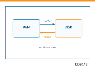 Save and reload prepared dataCache a windowed dataset to disk and reattach it without recompute.
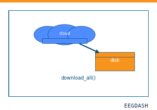 Download a dataset locallyPrefetch BIDS files to a local cache and validate the layout.
Swap any load_dataset(...) call for nm000374 to reproduce the tutorial on this dataset.
Citation
Wei Duan, Zhansheng Xu, Dong Chen, Jing Wang, Jiali Liu, … (2025). Electrophysiological signatures underlying variability in human memory consolidation: raw intracranial EEG (learning and sleep TMR). 10.82901/nemar.nm000374
Provenance
¹Contributed to nemar in BIDS format.
²Curated & ingested by the EEGDash catalog; see CITATION.cff for canonical reference.
³Persistent identifier: 10.82901/nemar.nm000374.
See Also#
eegdash.dataset.EEGDashDataseteegdash.dataset