


NM000403: ieeg dataset, 28 subjects#
Face-selective ventral occipito-temporal responses to fast periodic visual stimulation, SEEG (Jonas et al., 2016) (derivative)
Access recordings and metadata through EEGDash.
Citation: Jacques Jonas, Corentin Jacques, Joan Liu-Shuang, Hélène Brissart, Sophie Colnat-Coulbois, Louis Maillard, Bruno Rossion (2016). Face-selective ventral occipito-temporal responses to fast periodic visual stimulation, SEEG (Jonas et al., 2016) (derivative). 10.82901/nemar.nm000403
Modality: ieeg Subjects: 28 Recordings: 56 License: CC0-1.0 Source: nemar
Metadata: Complete (100%)
28-participant iEEG dataset — Face-selective ventral occipito-temporal responses to fast periodic visual stimulation, SEEG (Jonas et al., 2016) (derivative).
Quickstart#
Install
pip install eegdash
Access the data
from eegdash.dataset import NM000403

dataset = NM000403(cache_dir="./data")
# Get the raw object of the first recording
raw = dataset.datasets[0].raw
print(raw.info)
Filter by subject
dataset = NM000403(cache_dir="./data", subject="01")
Advanced query
dataset = NM000403(
    cache_dir="./data",
    query={"subject": {"$in": ["01", "02"]}},
)
Iterate recordings
for rec in dataset:
    print(rec.subject, rec.raw.info['sfreq'])
If you use this dataset in your research, please cite the original authors.
BibTeX
@dataset{nm000403,
  title = {Face-selective ventral occipito-temporal responses to fast periodic visual stimulation, SEEG (Jonas et al., 2016) (derivative)},
  author = {Jacques Jonas and Corentin Jacques and Joan Liu-Shuang and Hélène Brissart and Sophie Colnat-Coulbois and Louis Maillard and Bruno Rossion},
  doi = {10.82901/nemar.nm000403},
  url = {https://doi.org/10.82901/nemar.nm000403},
}
About This Dataset#
SEEG from 28 right-handed patients with refractory epilepsy (University Hospital of Nancy, Dec 2012 - Mar 2015) during
fast periodic visual stimulation: natural object images at 6 Hz with faces as every fifth image (1.2 Hz) in the periodic condition, and the same images with faces at random positions in the non-periodic control condition. THIS IS A DERIVATIVE DATASET: the release contains the authors’ Letswave files, low-pass filtered at 30 Hz (Butterworth order 4) and cut into sequences (README), not the raw Micromed recordings.
intracerebral potentials. doi:10.5061/dryad.5f9v7 (version 1, 2017-06-08).
License: CC0 1.0 (Dryad).
Face-selective ventral occipito-temporal map with intracerebral recordings (Jonas et al., 2016) - DERIVATIVE
Article: PNAS 113(28):E4088-E4097 (2016), doi:10.1073/pnas.1522033113 (PMC4948344).
All 6 Dryad files (3 zips + 3 identical READMEs) were downloaded through the Dryad API and matched the md5 digests.
Contents
sub-P<nn>/ieeg/sub-P<nn>_task-<faceperiodic|facenonperiodic>_run-1_ieeg.*: 56 files, 28
View full README
Face-selective ventral occipito-temporal map with intracerebral recordings (Jonas et al., 2016) - DERIVATIVE
Article: PNAS 113(28):E4088-E4097 (2016), doi:10.1073/pnas.1522033113 (PMC4948344).
All 6 Dryad files (3 zips + 3 identical READMEs) were downloaded through the Dryad API and matched the md5 digests.
Contents
sub-P<nn>/ieeg/sub-P<nn>_task-<faceperiodic|facenonperiodic>_run-1_ieeg.*: 56 files, 28 participants, 2.05 h. Each file holds the 1-4 sequences of one condition back-to-back (one BrainVision segment per sequence;RecordingTypeepoched,EpochLength= sequence length). Values are the release’s float32 values, unchanged (MNE read-back matches). Sampling rate 512 Hz (Letswave header).Channels: labels from the Letswave header. Contacts are typed SEEG; scalp electrodes recorded with the SEEG (labels
s<10-20 name>, e.g. sC3) EEG; ECG*, PULS+/BEAT+/SpO2+ (physiological monitors, MISC) and MKR* (marker inputs, TRIG) as labelled. Unit µV is assumed (Letswave import of Micromed TRC; not stated in the release).Electrode positions: the README says per-participant Talairach and MNI coordinate files are in each participant’s folder, but THEY ARE NOT IN THE DEPOSIT. Only contact labels exist;
electrodes.tsvlists the SEEG contact names with x, y, z = n/a.events.tsv: the Letswave events (code, sequence, latency) of each file; codes are not documented in the release.Letswave history per file is in each
ieeg.json(SoftwareFilters). 4 files also went through LW_downsample (to 512 Hz) according to their history.sourcedata/dryad-5f9v7-deidentified/: the per-participant .lw5/.mat files and READ_ME.txt of the three zips, de-identified (below).DEIDENTIFICATION_MANIFEST.tsvlists original and new sha-256 per file.NOT included: the
Stimuli/folder (face and object JPEG images, 750 files over the three zips). The article states that the actual face images could not be shown for copyright reasons; they remain available from the Dryad record.
Privacy
Some Letswave event codes began with a patient code formed from letters of the patient’s name (e.g. a pattern like ‘ABCDE1_greyscale_sinstim_Localiser_6Hz_ObjectsFaces_Per_Grey_1’). That prefix was replaced by ‘seq’ in events.tsv and in the de-identified .lw5 headers (8 distinct codes; a byte search of the whole output for these codes found 0 remaining occurrences). The original Dryad files still contain them.
Letswave history dates and MAT text-header dates in sourcedata are reduced to month and year (day -> 01). The .lw5 headers were rewritten with scipy (savemat, MATLAB v5, compressed); their content is otherwise unchanged.
Age and sex are given only for the cohort (mean age 30.5 +- 4.4 years; 15 female).
Additional metadata and localisation (added 2026-10-08)
Compiled after the upload from the article, its supplement and the source deposit (each statement names its source). Text and sidecar metadata only; no data file was changed.
Sources: P = Jonas et al. 2016, PNAS 113(28):E4088-E4097, doi:10.1073/pnas.1522033113 (read on pnas.org; SI is integrated in the article page). R = deposit README (README_for_Jonas_et_al_PNAS_2016_Part_*.txt, identical in all three parts). F = deposit files (Letswave .lw5 headers read in a Voyager Job). Recording. Each SEEG electrode is a 0.8 mm diameter cylinder with 8-15 contacts, 2 mm long, 1.5 mm apart edge to edge (3.5 mm centre to centre). Recording used a 256-channel amplifier at 512 Hz. Original files were Micromed TRC (R). Reference: a midline prefrontal scalp electrode (FPz) in 21 participants, or an intracerebral white-matter contact in 7 participants (P, Methods). The per-participant reference is not given. Preprocessing in the deposit. Data were imported into Letswave 5 and low-pass filtered at 30 Hz (Butterworth, order 4). Sequences were segmented from 2 s after onset to about 65 s (an integer number of 1.2 Hz cycles), and the sequences of each condition were merged into one file (2 or 4 periodic, 1 or 2 nonperiodic). No FFT or other analysis was applied. Data dimensions are [sequences, channels, 1, 1, 1, time] (R). The header history is LW_importTRC → LW_merge_epochs → LW_butter_lowpass, with xstep 1/512 s (F). Channels. header.chanlocs holds labels only: topo_enabled = 0 and there are no coordinates (F). Besides intracerebral contacts named by electrode letter and number (e.g. L’1, TB3), the files contain scalp EEG channels with an s prefix (e.g. sFz), ECG/PULS+/BEAT+/SpO2+/MKR channels, and placeholder channels (e.g. el237, zz193, xx152). Localisation.**Contacts were labelled in each participant’s own anatomy from gyri and sulci landmarks (CoS, OTS, MFS, posterior tip of the hippocampus, anterior tip of the parieto-occipital sulcus; Fig. S2). In a separate analysis, MRIs were normalised to obtain Talairach and MNI coordinates (P, Methods “Contact Localization in the Individual Anatomy”). Talairach and MNI coordinate .txt files for each participant are described in R (“Recording contacts coordinates … reported in a txt file in each participant’s folder”) but are not present** in the three zips (F: the zips contain only the .lw5/.mat pairs, READ_ME.txt and the stimuli). The paper reports only region-mean coordinates of face-selective contacts (Table S1) and the number of face-selective contacts per region (Table 1).
Regions per participant (as published; no coordinates exist)
| participant | region (as stated) | hemisphere | contacts | source |
|---|---|---|---|---|
| all (cohort; 11 participants) | VMO: ventromedial occipital (occipital CoS, lingual gyrus, calcarine sulcus, cuneus, occipital pole) | L | 89 | doi:10.1073/pnas.1522033113, Table 1 (face-selective contacts only) |
| all (cohort; 11 participants) | VMO: ventromedial occipital (occipital CoS, lingual gyrus, calcarine sulcus, cuneus, occipital pole) | R | 50 | doi:10.1073/pnas.1522033113, Table 1 (face-selective contacts only) |
| all (cohort; 11 participants) | IOG: inferior occipital gyrus | L | 26 | doi:10.1073/pnas.1522033113, Table 1 (face-selective contacts only) |
| all (cohort; 11 participants) | IOG: inferior occipital gyrus | R | 36 | doi:10.1073/pnas.1522033113, Table 1 (face-selective contacts only) |
| all (cohort; 17 participants) | medFG: medial fusiform gyrus and adjacent CoS | L | 40 | doi:10.1073/pnas.1522033113, Table 1 (face-selective contacts only) |
| all (cohort; 17 participants) | medFG: medial fusiform gyrus and adjacent CoS | R | 30 | doi:10.1073/pnas.1522033113, Table 1 (face-selective contacts only) |
| all (cohort; 17 participants) | latFG: lateral fusiform gyrus and adjacent OTS | L | 30 | doi:10.1073/pnas.1522033113, Table 1 (face-selective contacts only) |
| all (cohort; 17 participants) | latFG: lateral fusiform gyrus and adjacent OTS | R | 33 | doi:10.1073/pnas.1522033113, Table 1 (face-selective contacts only) |
| all (cohort; 13 participants) | MTG/ITG: posterior middle/inferior temporal gyri | L | 30 | doi:10.1073/pnas.1522033113, Table 1 (face-selective contacts only) |
| all (cohort; 13 participants) | MTG/ITG: posterior middle/inferior temporal gyri | R | 25 | doi:10.1073/pnas.1522033113, Table 1 (face-selective contacts only) |
| all (cohort; 18 participants) | antCoS: anterior collateral sulcus | L | 33 | doi:10.1073/pnas.1522033113, Table 1 (face-selective contacts only) |
| all (cohort; 18 participants) | antCoS: anterior collateral sulcus | R | 23 | doi:10.1073/pnas.1522033113, Table 1 (face-selective contacts only) |
| all (cohort; 7 participants) | antFG: anterior fusiform gyrus | L | 10 | doi:10.1073/pnas.1522033113, Table 1 (face-selective contacts only) |
| all (cohort; 7 participants) | antFG: anterior fusiform gyrus | R | 11 | doi:10.1073/pnas.1522033113, Table 1 (face-selective contacts only) |
| all (cohort; 15 participants) | antOTS: anterior occipito-temporal sulcus | L | 35 | doi:10.1073/pnas.1522033113, Table 1 (face-selective contacts only) |
| all (cohort; 15 participants) | antOTS: anterior occipito-temporal sulcus | R | 17 | doi:10.1073/pnas.1522033113, Table 1 (face-selective contacts only) |
| all (cohort; 12 participants) | antMTG/ITG: anterior middle/inferior temporal gyri | L | 20 | doi:10.1073/pnas.1522033113, Table 1 (face-selective contacts only) |
| all (cohort; 12 participants) | antMTG/ITG: anterior middle/inferior temporal gyri | R | 17 | doi:10.1073/pnas.1522033113, Table 1 (face-selective contacts only) |
NEMAR Metadata#
[](https://doi.org/10.82901/nemar.nm000403) # Face-selective ventral occipito-temporal map with intracerebral recordings (Jonas et al., 2016) - DERIVATIVE SEEG from 28 right-handed patients with refractory epilepsy (University Hospital of Nancy, Dec 2012 - Mar 2015) during fast periodic visual stimulation: natural object images at 6 Hz with faces as every fifth image (1.2 Hz) in the periodic condition, and the same images with faces at random positions in the non-periodic control condition. THIS IS A DERIVATIVE DATASET: the release contains the authors’ Letswave files, low-pass filtered at 30 Hz (Butterworth order 4) and cut into sequences (README), not the raw Micromed recordings. ## Source - Dryad: Jacques Jonas, Corentin Jacques, Joan Liu-Shuang, Hélène Brissart, Sophie Colnat-Coulbois, Louis Maillard, Bruno Rossion. Data from: A face-selective ventral occipito-temporal map of the human brain with
intracerebral potentials. doi:10.5061/dryad.5f9v7 (version 1, 2017-06-08). License: CC0 1.0 (Dryad).
Article: PNAS 113(28):E4088-E4097 (2016), doi:10.1073/pnas.1522033113 (PMC4948344).
All 6 Dryad files (3 zips + 3 identical READMEs) were downloaded through the Dryad API and matched the md5 digests.
## Contents - sub-P<nn>/ieeg/sub-P<nn>_task-<faceperiodic|facenonperiodic>_run-1_ieeg.*: 56 files, 28
participants, 2.05 h. Each file holds the 1-4 sequences of one condition back-to-back (one BrainVision segment per sequence; RecordingType epoched, EpochLength = sequence length). Values are the release’s float32 values, unchanged (MNE read-back matches). Sampling rate 512 Hz (Letswave header).
Channels: labels from the Letswave header. Contacts are typed SEEG; scalp electrodes recorded with the SEEG (labels s<10-20 name>, e.g. sC3) EEG; ECG*, PULS+/BEAT+/SpO2+ (physiological monitors, MISC) and MKR* (marker inputs, TRIG) as labelled. Unit µV is assumed (Letswave import of Micromed TRC; not stated in the release).
Electrode positions: the README says per-participant Talairach and MNI coordinate files are in each participant’s folder, but THEY ARE NOT IN THE DEPOSIT. Only contact labels exist; electrodes.tsv lists the SEEG contact names with x, y, z = n/a.
events.tsv: the Letswave events (code, sequence, latency) of each file; codes are not documented in the release.
Letswave history per file is in each ieeg.json (SoftwareFilters). 4 files also went through LW_downsample (to 512 Hz) according to their history.
sourcedata/dryad-5f9v7-deidentified/: the per-participant .lw5/.mat files and READ_ME.txt of the three zips, de-identified (below). DEIDENTIFICATION_MANIFEST.tsv lists original and new sha-256 per file.
NOT included: the Stimuli/ folder (face and object JPEG images, 750 files over the three zips). The article states that the actual face images could not be shown for copyright reasons; they remain available from the Dryad record.
## Privacy - Some Letswave event codes began with a patient code formed from letters of the patient’s name (e.g. a pattern
like ‘ABCDE1_greyscale_sinstim_Localiser_6Hz_ObjectsFaces_Per_Grey_1’). That prefix was replaced by ‘seq’ in events.tsv and in the de-identified .lw5 headers (8 distinct codes; a byte search of the whole output for these codes found 0 remaining occurrences). The original Dryad files still contain them.
Letswave history dates and MAT text-header dates in sourcedata are reduced to month and year (day -> 01). The .lw5 headers were rewritten with scipy (savemat, MATLAB v5, compressed); their content is otherwise unchanged.
Age and sex are given only for the cohort (mean age 30.5 +- 4.4 years; 15 female).
## Additional metadata and localisation (added 2026-10-08) Compiled after the upload from the article, its supplement and the source deposit (each statement names its source). Text and sidecar metadata only; no data file was changed. Sources: P = Jonas et al. 2016, PNAS 113(28):E4088-E4097, doi:10.1073/pnas.1522033113 (read on pnas.org; SI is integrated in the article page). R = deposit README (README_for_Jonas_et_al_PNAS_2016_Part_*.txt, identical in all three parts). F = deposit files (Letswave .lw5 headers read in a Voyager Job). Recording. Each SEEG electrode is a 0.8 mm diameter cylinder with 8-15 contacts, 2 mm long, 1.5 mm apart edge to edge (3.5 mm centre to centre). Recording used a 256-channel amplifier at 512 Hz. Original files were Micromed TRC (R). Reference: a midline prefrontal scalp electrode (FPz) in 21 participants, or an intracerebral white-matter contact in 7 participants (P, Methods). The per-participant reference is not given. Preprocessing in the deposit. Data were imported into Letswave 5 and low-pass filtered at 30 Hz (Butterworth, order 4). Sequences were segmented from 2 s after onset to about 65 s (an integer number of 1.2 Hz cycles), and the sequences of each condition were merged into one file (2 or 4 periodic, 1 or 2 nonperiodic). No FFT or other analysis was applied. Data dimensions are [sequences, channels, 1, 1, 1, time] (R). The header history is LW_importTRC → LW_merge_epochs → LW_butter_lowpass, with xstep 1/512 s (F). Channels. header.chanlocs holds labels only: topo_enabled = 0 and there are no coordinates (F). Besides intracerebral contacts named by electrode letter and number (e.g. L’1, TB3), the files contain scalp EEG channels with an s prefix (e.g. sFz), ECG/PULS+/BEAT+/SpO2+/MKR channels, and placeholder channels (e.g. el237, zz193, xx152). Localisation. Contacts were labelled in each participant’s own anatomy from gyri and sulci landmarks (CoS, OTS, MFS, posterior tip of the hippocampus, anterior tip of the parieto-occipital sulcus; Fig. S2). In a separate analysis, MRIs were normalised to obtain Talairach and MNI coordinates (P, Methods “Contact Localization in the Individual Anatomy”). Talairach and MNI coordinate .txt files for each participant are described in R (“Recording contacts coordinates … reported in a txt file in each participant’s folder”) but are not present in the three zips (F: the zips contain only the .lw5/.mat pairs, READ_ME.txt and the stimuli). The paper reports only region-mean coordinates of face-selective contacts (Table S1) and the number of face-selective contacts per region (Table 1). ### Regions per participant (as published; no coordinates exist) | participant | region (as stated) | hemisphere | contacts | source | |---|—|---|—|---| | all (cohort; 11 participants) | VMO: ventromedial occipital (occipital CoS, lingual gyrus, calcarine sulcus, cuneus, occipital pole) | L | 89 | doi:10.1073/pnas.1522033113, Table 1 (face-selective contacts only) | | all (cohort; 11 participants) | VMO: ventromedial occipital (occipital CoS, lingual gyrus, calcarine sulcus, cuneus, occipital pole) | R | 50 | doi:10.1073/pnas.1522033113, Table 1 (face-selective contacts only) | | all (cohort; 11 participants) | IOG: inferior occipital gyrus | L | 26 | doi:10.1073/pnas.1522033113, Table 1 (face-selective contacts only) | | all (cohort; 11 participants) | IOG: inferior occipital gyrus | R | 36 | doi:10.1073/pnas.1522033113, Table 1 (face-selective contacts only) | | all (cohort; 17 participants) | medFG: medial fusiform gyrus and adjacent CoS | L | 40 | doi:10.1073/pnas.1522033113, Table 1 (face-selective contacts only) | | all (cohort; 17 participants) | medFG: medial fusiform gyrus and adjacent CoS | R | 30 | doi:10.1073/pnas.1522033113, Table 1 (face-selective contacts only) | | all (cohort; 17 participants) | latFG: lateral fusiform gyrus and adjacent OTS | L | 30 | doi:10.1073/pnas.1522033113, Table 1 (face-selective contacts only) | | all (cohort; 17 participants) | latFG: lateral fusiform gyrus and adjacent OTS | R | 33 | doi:10.1073/pnas.1522033113, Table 1 (face-selective contacts only) | | all (cohort; 13 participants) | MTG/ITG: posterior middle/inferior temporal gyri | L | 30 | doi:10.1073/pnas.1522033113, Table 1 (face-selective contacts only) | | all (cohort; 13 participants) | MTG/ITG: posterior middle/inferior temporal gyri | R | 25 | doi:10.1073/pnas.1522033113, Table 1 (face-selective contacts only) | | all (cohort; 18 participants) | antCoS: anterior collateral sulcus | L | 33 | doi:10.1073/pnas.1522033113, Table 1 (face-selective contacts only) | | all (cohort; 18 participants) | antCoS: anterior collateral sulcus | R | 23 | doi:10.1073/pnas.1522033113, Table 1 (face-selective contacts only) | | all (cohort; 7 participants) | antFG: anterior fusiform gyrus | L | 10 | doi:10.1073/pnas.1522033113, Table 1 (face-selective contacts only) | | all (cohort; 7 participants) | antFG: anterior fusiform gyrus | R | 11 | doi:10.1073/pnas.1522033113, Table 1 (face-selective contacts only) | | all (cohort; 15 participants) | antOTS: anterior occipito-temporal sulcus | L | 35 | doi:10.1073/pnas.1522033113, Table 1 (face-selective contacts only) | | all (cohort; 15 participants) | antOTS: anterior occipito-temporal sulcus | R | 17 | doi:10.1073/pnas.1522033113, Table 1 (face-selective contacts only) | | all (cohort; 12 participants) | antMTG/ITG: anterior middle/inferior temporal gyri | L | 20 | doi:10.1073/pnas.1522033113, Table 1 (face-selective contacts only) | | all (cohort; 12 participants) | antMTG/ITG: anterior middle/inferior temporal gyri | R | 17 | doi:10.1073/pnas.1522033113, Table 1 (face-selective contacts only) |
License: CC0-1.0
Authors:
Jacques Jonas
Corentin Jacques
Joan Liu-Shuang
Hélène Brissart
Sophie Colnat-Coulbois
… and 2 more
Versions:
Version |
DOI |
Released |
|---|---|---|
|
Cohort#
Dataset Statistics#
Channel counts (ch)
Sampling frequencies: 512.0 Hz (n=56 recordings)
Total recording duration: 2 h 3 min
Signal · Electrodes & live trace#
Live trace viewer — sub-P03 · task-faceperiodic · run-1
Showing one representative recording out of
28 subjects and 56 recordings in this dataset.
Browse the full set on OpenNeuro;
drop any other _ieeg.{set,edf,bdf,vhdr} file onto the
viewer (or pass ?ieeg=<url>) to inspect it.
No scalp electrode layout is currently indexed for this dataset. Once the eegdash montage registry ingests it, the interactive viewer will appear here automatically.
NEMAR Processing Statistics#
The plots below are generated by NEMAR’s automated EEG pipeline. The histogram shows pipeline success for data cleaning and ICA decomposition, the percentage of data frames and EEG channels retained after artefact removal, line noise per channel (RMS, dB), and the age/gender distribution of participants.

HED event descriptors word cloud

Manifest#
File Explorer#
Browse the BIDS file structure of this dataset. Records are fetched on demand from the EEGDash catalog the first time you open the explorer.
Full dataset metadata table
Dataset ID |
|
Title |
Face-selective ventral occipito-temporal responses to fast periodic visual stimulation, SEEG (Jonas et al., 2016) (derivative) |
Author (year) |
— |
Canonical |
— |
Importable as |
|
Year |
2016 |
Authors |
Jacques Jonas, Corentin Jacques, Joan Liu-Shuang, Hélène Brissart, Sophie Colnat-Coulbois, Louis Maillard, Bruno Rossion |
License |
CC0-1.0 |
Citation / DOI |
|
Source links |
OpenNeuro | NeMAR | Source URL |
Copy-paste BibTeX
@dataset{nm000403,
  title = {Face-selective ventral occipito-temporal responses to fast periodic visual stimulation, SEEG (Jonas et al., 2016) (derivative)},
  author = {Jacques Jonas and Corentin Jacques and Joan Liu-Shuang and Hélène Brissart and Sophie Colnat-Coulbois and Louis Maillard and Bruno Rossion},
  doi = {10.82901/nemar.nm000403},
  url = {https://doi.org/10.82901/nemar.nm000403},
}
API Reference#
eegdash.datasetEEGDashDataset- class eegdash.dataset.NM000403(cache_dir: str, query: dict | None = None, s3_bucket: str | None = None, **kwargs)[source]#
Face-selective ventral occipito-temporal responses to fast periodic visual stimulation, SEEG (Jonas et al., 2016) (derivative)
- Study:
nm000403(NeMAR)- Author (year):
—
- Canonical:
—
Also importable as:
NM000403.Modality:
ieeg; Subject type:Unknown. Subjects: 28; recordings: 56; tasks: 2.- Parameters:
cache_dir (str | Path) – Directory where data are cached locally.
query (dict | None) – Additional MongoDB-style filters to AND with the dataset selection. Must not contain the key
dataset.s3_bucket (str | None) – Base S3 bucket used to locate the data.
**kwargs (dict) – Additional keyword arguments forwarded to
EEGDashDataset.
- data_dir#
Local dataset cache directory (
cache_dir / dataset_id).- Type:
Path
Notes
Each item is a recording; recording-level metadata are available via
dataset.description.querysupports MongoDB-style filters on fields inALLOWED_QUERY_FIELDSand is combined with the dataset filter. Dataset-specific caveats are not provided in the summary metadata.References
OpenNeuro dataset: https://openneuro.org/datasets/nm000403 NeMAR dataset: https://nemar.org/dataexplorer/detail?dataset_id=nm000403 DOI: https://doi.org/10.82901/nemar.nm000403
Examples
>>> from eegdash.dataset import NM000403
>>> dataset = NM000403(cache_dir="./data")
>>> recording = dataset[0]
>>> raw = recording.load()
- __init__(cache_dir: str, query: dict | None = None, s3_bucket: str | None = None, **kwargs)[source]#
- save(path: str, overwrite: bool = False, offset: int = 0)[source]#
Save datasets to files by creating one subdirectory for each dataset:
path/
    0/
        0-raw.fif | 0-epo.fif
        description.json
        raw_preproc_kwargs.json (if raws were preprocessed)
        window_kwargs.json (if this is a windowed dataset)
        window_preproc_kwargs.json  (if windows were preprocessed)
        target_name.json (if target_name is not None and dataset is raw)
    1/
        1-raw.fif | 1-epo.fif
        description.json
        raw_preproc_kwargs.json (if raws were preprocessed)
        window_kwargs.json (if this is a windowed dataset)
        window_preproc_kwargs.json  (if windows were preprocessed)
        target_name.json (if target_name is not None and dataset is raw)
- Parameters:
path (str) –
- Directory in which subdirectories are created to store
-raw.fif | -epo.fif and .json files to.
overwrite (bool) – Whether to delete old subdirectories that will be saved to in this call.
offset (int) – If provided, the integer is added to the id of the dataset in the concat. This is useful in the setting of very large datasets, where one dataset has to be processed and saved at a time to account for its original position.
BaseDataset from braindecode — windowed via create_windows_from_events.braindecodeDataLoader; supports parallel workers and on-the-fly augmentations.pytorch Find datasets with the EEGDash APIQuery the catalogue, filter by task or modality, list candidates.
 Load one EEG recordingResolve a single record to an MNE Raw with channels and events.
 EEG recording to PyTorch DataLoaderWrap braindecode windows in a DataLoader for model training.
 Preprocess EEG and create windowsFilter, resample, epoch — and persist the windowed dataset.
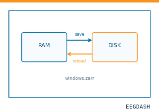 Save and reload prepared dataCache a windowed dataset to disk and reattach it without recompute.
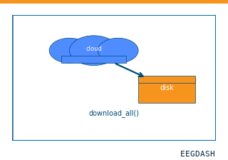 Download a dataset locallyPrefetch BIDS files to a local cache and validate the layout.
Swap any load_dataset(...) call for nm000403 to reproduce the tutorial on this dataset.
Citation
Jacques Jonas, Corentin Jacques, Joan Liu-Shuang, Hélène Brissart, Sophie Colnat-Coulbois, … (2016). Face-selective ventral occipito-temporal responses to fast periodic visual stimulation, SEEG (Jonas et al., 2016) (derivative). 10.82901/nemar.nm000403
Provenance
¹Contributed to nemar in BIDS format.
²Curated & ingested by the EEGDash catalog; see CITATION.cff for canonical reference.
³Persistent identifier: 10.82901/nemar.nm000403.
See Also#
eegdash.dataset.EEGDashDataseteegdash.dataset