


NM000386: ieeg dataset, 6 subjects#
The involvement of the human prefrontal cortex in the emergence of visual awareness (Fang et al., 2024): sEEG, 6 patients
Access recordings and metadata through EEGDash.
Citation: Zepeng Fang, Yuanyuan Dang, Zhipei Ling, Yongzheng Han, Hulin Zhao, Xin Xu, Mingsha Zhang (2024). The involvement of the human prefrontal cortex in the emergence of visual awareness (Fang et al., 2024): sEEG, 6 patients. 10.82901/nemar.nm000386
Modality: ieeg Subjects: 6 Recordings: 41 License: CC0-1.0 Source: nemar
Metadata: Complete (100%)
6-participant iEEG dataset — The involvement of the human prefrontal cortex in the emergence of visual awareness (Fang et al., 2024): sEEG, 6 patients.
Quickstart#
Install
pip install eegdash
Access the data
from eegdash.dataset import NM000386

dataset = NM000386(cache_dir="./data")
# Get the raw object of the first recording
raw = dataset.datasets[0].raw
print(raw.info)
Filter by subject
dataset = NM000386(cache_dir="./data", subject="01")
Advanced query
dataset = NM000386(
    cache_dir="./data",
    query={"subject": {"$in": ["01", "02"]}},
)
Iterate recordings
for rec in dataset:
    print(rec.subject, rec.raw.info['sfreq'])
If you use this dataset in your research, please cite the original authors.
BibTeX
@dataset{nm000386,
  title = {The involvement of the human prefrontal cortex in the emergence of visual awareness (Fang et al., 2024): sEEG, 6 patients},
  author = {Zepeng Fang and Yuanyuan Dang and Zhipei Ling and Yongzheng Han and Hulin Zhao and Xin Xu and Mingsha Zhang},
  doi = {10.82901/nemar.nm000386},
  url = {https://doi.org/10.82901/nemar.nm000386},
}
About This Dataset#
Raw stereo-EEG of 6 adult male patients with drug-resistant epilepsy (Department of Neurosurgery, PLA General Hospital,
Beijing) performing a visual awareness task with saccadic report, with per-trial behaviour, defaced T1 MRI, and (in sourcedata) post-implantation CT and the eye-position/behaviour files of the patients and of 10 healthy controls.
for patients 1, 5 and 6, one hemisphere for patients 2-4 (participants.tsv). Patients were on their usual
anti-epileptic medication. Electrode placement was based on clinical requirements only.
Human prefrontal cortex and the emergence of visual awareness: sEEG (Fang et al., 2024)
Healthy controls: 10 (4 males, 6 females, 27.80 ± 3.92 years), behaviour/eye position only (no neural data); their files are in
sourcedata/dryad-p8cz8w9xp/HealthyControls/(C01-C10, release order).
Task (article, ‘Experimental task’)
Fixation (white cross) for 600 ms, then a 2° Gabor grating for 50 ms at 7° eccentricity, contralateral to the implanted
View full README
Human prefrontal cortex and the emergence of visual awareness: sEEG (Fang et al., 2024)
Healthy controls: 10 (4 males, 6 females, 27.80 ± 3.92 years), behaviour/eye position only (no neural data); their files are in
sourcedata/dryad-p8cz8w9xp/HealthyControls/(C01-C10, release order).
Task (article, ‘Experimental task’)
Fixation (white cross) for 600 ms, then a 2° Gabor grating for 50 ms at 7° eccentricity, contralateral to the implanted hemisphere (right side for the bilateral patients 1, 5, 6). In 70% of trials the contrast tracks the perceptual threshold (1 up/1 down staircase), in 10% it is well above threshold, in 20% no grating is shown. After another 600 ms the fixation point turns green or red and two saccade targets appear at 10° left and right. Seen + green -> right target, seen + red -> left target; not seen inverts the rule. 180 trials per session, ITI 800 ms. Patients’ stimuli: 24-inch 144 Hz screen; eye position by an infrared eye tracker (Jsmz EM2000C) at 1 kHz; controls: 27-inch 120 Hz screen, EyeLink 1000 at 1 kHz. MATLAB + Psychtoolbox-3.
Recording
Depth electrodes (SINOVATION, 8-20 contacts, 0.8 mm diameter, 2 mm length, 1.5 mm spacing), NEURACLE amplifier, 1 kHz, hardware filter 1-250 Hz and 50 Hz notch, reference: a contact in white matter. 108-254 channels per recording.
Source
Dryad doi:10.5061/dryad.p8cz8w9xp (version 6, 2024-01-18), CC0 1.0. Authors: Zepeng Fang, Yuanyuan Dang, Zhipei Ling, Yongzheng Han, Hulin Zhao, Xin Xu, Mingsha Zhang (Beijing Normal University / PLA General Hospital).
Article: Fang et al. (2024) eLife 13:RP89076, doi:10.7554/eLife.89076.
Ethics (verbatim from the article)
“All subjects provided informed consent to participate in this study. The Ethics Committee of Chinese PLA General Hospital approved the experimental procedures (approval numbers S2022-457-01).”
Conversion
Each release session (
SEEG Data/Pn/SessionK/data.bdf) is one run. BDF is not an iEEG-BIDS format, so the data are written as BrainVision with 32-bit integer samples holding the exact BDF digital values, and per-channel resolution = the BDF gain. The BDF digital range is asymmetric (-8388608..8388607 for a symmetric physical range), so the BDF physical value = digital x gain + half a digital step (about 0.045 µV); that constant is not representable in BrainVision and is dropped. The digital values are exact (checked by round-trip).sub-04: the BDF has the label LDELT1 twice (channels 1 and 251, different signals); channel 251 is named LDELT1_ch251 (original label in channels.tsv column source_label). No filtering or resampling.
Events come from
evt.bdf(Neuracle ‘Trigger-In:<code>’ annotations). The onsets are used as data latencies, as Neuracle’s own reader does; the two files’ header start times (1-s resolution) differ by 0-1 s. Codes are kept verbatim (value). The authors do not document them;event_namegives our reading, checked against the release behaviour files (seetask-visualawareness_events.json).Per-trial behaviour from the release MAT files (stimulus presence, contrast, colour cue, saccade direction, awareness report, completion, fixation break) is added to every event of the trial. A recording is matched to its MAT file only when the number of trial starts (code 101) equals the number of MAT trials and the trigger intervals agree with the MAT event times (99th percentile < 25 ms; observed < 9 ms): 39 of 41 runs. Not matched (behaviour columns n/a; raw MAT files in sourcedata): sub-03 run-11 (117 trial starts in the recording vs 180 MAT trials) and sub-04 run-06 (178 vs 180).
P5 ‘Session6-7’ is a single file in the release (two sessions in one recording) and is kept as one run.
Channels named DELT (present for some patients) are typed MISC: the name suggests deltoid EMG but the release does not say. All other channels are SEEG. No electrode coordinates are released (only defaced MRI/CT).
Dates: year and month kept, day set to 01 (scans.tsv; BDF headers and MAT-file header creation dates in sourcedata; times of day kept). Patient initials and recording dates in release file names were removed from sourcedata names (see
sourcedata/dryad-p8cz8w9xp/MANIFEST.tsv, which lists the sha256 of every original file).Images: the authors’ defaced T1 MRI is in
anat/(T1w). Defacing was checked by surface rendering for all 12 images. CT has no raw BIDS suffix and is kept in sourcedata; the CT of P5 is excluded because its defacing could not be verified (it remains available from Dryad).Behaviour/eye position (.mat, per session) for patients and the 10 healthy controls (behaviour only, no neural data) are in sourcedata with neutral names; their struct fields are described in
sourcedata/dryad-p8cz8w9xp/README.md(the release README, byte for byte).The release zip itself is not redistributed: its file names carry patient initials and recording dates, and the BDF and MAT headers carry full recording dates. Every original file is in sourcedata under a neutral name, byte-identical except for those header dates;
MANIFEST.tsvlists the sha256 of each original release file and what was changed.Age per patient is not given (article: 32.33 ± 4.75 years, mean ± SEM); all six are male (article).
How to load
import mne, pandas as pd
raw = mne.io.read_raw_brainvision("sub-01/ieeg/sub-01_task-visualawareness_run-01_ieeg.vhdr", preload=True)  # volts
ev = pd.read_csv("sub-01/ieeg/sub-01_task-visualawareness_run-01_events.tsv", sep="\t")
cues = ev[ev.event_name == "grating_on"]  # one row per trial with stim_presence/contrast/awareness
MNE-BIDS: mne_bids.read_raw_bids(BIDSPath(root=".", subject="01", task="visualawareness", run="01", datatype="ieeg")).
Behaviour MAT files: scipy.io.loadmat(path, squeeze_me=True, struct_as_record=False)["Trial"].
References
Fang Z, Dang Y, Ling Z, Han Y, Zhao H, Xu X, Zhang M (2024). The involvement of the human prefrontal cortex in the emergence of visual awareness. eLife 13:RP89076. doi:10.7554/eLife.89076
Data: Dryad doi:10.5061/dryad.p8cz8w9xp (CC0 1.0).
NEMAR Metadata#
[](https://doi.org/10.82901/nemar.nm000386) # Human prefrontal cortex and the emergence of visual awareness: sEEG (Fang et al., 2024) Raw stereo-EEG of 6 adult male patients with drug-resistant epilepsy (Department of Neurosurgery, PLA General Hospital, Beijing) performing a visual awareness task with saccadic report, with per-trial behaviour, defaced T1 MRI, and (in sourcedata) post-implantation CT and the eye-position/behaviour files of the patients and of 10 healthy controls. ## Cohort (article, Methods ‘Data acquisition’) - Patients: 6 (6 males, 32.33 ± 4.75 years, mean ± SEM; per-patient ages not given). Electrodes in both hemispheres
for patients 1, 5 and 6, one hemisphere for patients 2-4 (participants.tsv). Patients were on their usual anti-epileptic medication. Electrode placement was based on clinical requirements only.
Healthy controls: 10 (4 males, 6 females, 27.80 ± 3.92 years), behaviour/eye position only (no neural data); their files are in sourcedata/dryad-p8cz8w9xp/HealthyControls/ (C01-C10, release order).
## Task (article, ‘Experimental task’) Fixation (white cross) for 600 ms, then a 2° Gabor grating for 50 ms at 7° eccentricity, contralateral to the implanted hemisphere (right side for the bilateral patients 1, 5, 6). In 70% of trials the contrast tracks the perceptual threshold (1 up/1 down staircase), in 10% it is well above threshold, in 20% no grating is shown. After another 600 ms the fixation point turns green or red and two saccade targets appear at 10° left and right. Seen + green -> right target, seen + red -> left target; not seen inverts the rule. 180 trials per session, ITI 800 ms. Patients’ stimuli: 24-inch 144 Hz screen; eye position by an infrared eye tracker (Jsmz EM2000C) at 1 kHz; controls: 27-inch 120 Hz screen, EyeLink 1000 at 1 kHz. MATLAB + Psychtoolbox-3. ## Recording Depth electrodes (SINOVATION, 8-20 contacts, 0.8 mm diameter, 2 mm length, 1.5 mm spacing), NEURACLE amplifier, 1 kHz, hardware filter 1-250 Hz and 50 Hz notch, reference: a contact in white matter. 108-254 channels per recording. ## Source - Dryad doi:10.5061/dryad.p8cz8w9xp (version 6, 2024-01-18), CC0 1.0. Authors: Zepeng Fang, Yuanyuan Dang, Zhipei Ling,
Yongzheng Han, Hulin Zhao, Xin Xu, Mingsha Zhang (Beijing Normal University / PLA General Hospital).
Article: Fang et al. (2024) eLife 13:RP89076, doi:10.7554/eLife.89076.
## Ethics (verbatim from the article) “All subjects provided informed consent to participate in this study. The Ethics Committee of Chinese PLA General Hospital approved the experimental procedures (approval numbers S2022-457-01).” ## Conversion - Each release session (SEEG Data/Pn/SessionK/data.bdf) is one run. BDF is not an iEEG-BIDS format, so the data are
written as BrainVision with 32-bit integer samples holding the exact BDF digital values, and per-channel resolution = the BDF gain. The BDF digital range is asymmetric (-8388608..8388607 for a symmetric physical range), so the BDF physical value = digital x gain + half a digital step (about 0.045 µV); that constant is not representable in BrainVision and is dropped. The digital values are exact (checked by round-trip).
sub-04: the BDF has the label LDELT1 twice (channels 1 and 251, different signals); channel 251 is named LDELT1_ch251 (original label in channels.tsv column source_label). No filtering or resampling.
Events come from evt.bdf (Neuracle ‘Trigger-In:<code>’ annotations). The onsets are used as data latencies, as Neuracle’s own reader does; the two files’ header start times (1-s resolution) differ by 0-1 s. Codes are kept verbatim (value). The authors do not document them; event_name gives our reading, checked against the release behaviour files (see task-visualawareness_events.json).
Per-trial behaviour from the release MAT files (stimulus presence, contrast, colour cue, saccade direction, awareness report, completion, fixation break) is added to every event of the trial. A recording is matched to its MAT file only when the number of trial starts (code 101) equals the number of MAT trials and the trigger intervals agree with the MAT event times (99th percentile < 25 ms; observed < 9 ms): 39 of 41 runs. Not matched (behaviour columns n/a; raw MAT files in sourcedata): sub-03 run-11 (117 trial starts in the recording vs 180 MAT trials) and sub-04 run-06 (178 vs 180).
P5 ‘Session6-7’ is a single file in the release (two sessions in one recording) and is kept as one run.
Channels named DELT (present for some patients) are typed MISC: the name suggests deltoid EMG but the release does not say. All other channels are SEEG. No electrode coordinates are released (only defaced MRI/CT).
Dates: year and month kept, day set to 01 (scans.tsv; BDF headers and MAT-file header creation dates in sourcedata; times of day kept). Patient initials and recording dates in release file names were removed from sourcedata names (see sourcedata/dryad-p8cz8w9xp/MANIFEST.tsv, which lists the sha256 of every original file).
Images: the authors’ defaced T1 MRI is in anat/ (T1w). Defacing was checked by surface rendering for all 12 images. CT has no raw BIDS suffix and is kept in sourcedata; the CT of P5 is excluded because its defacing could not be verified (it remains available from Dryad).
Behaviour/eye position (.mat, per session) for patients and the 10 healthy controls (behaviour only, no neural data) are in sourcedata with neutral names; their struct fields are described in sourcedata/dryad-p8cz8w9xp/README.md (the release README, byte for byte).
The release zip itself is not redistributed: its file names carry patient initials and recording dates, and the BDF and MAT headers carry full recording dates. Every original file is in sourcedata under a neutral name, byte-identical except for those header dates; MANIFEST.tsv lists the sha256 of each original release file and what was changed.
Age per patient is not given (article: 32.33 ± 4.75 years, mean ± SEM); all six are male (article).
## How to load
`python
import mne, pandas as pd
raw = mne.io.read_raw_brainvision("sub-01/ieeg/sub-01_task-visualawareness_run-01_ieeg.vhdr", preload=True)  # volts
ev = pd.read_csv("sub-01/ieeg/sub-01_task-visualawareness_run-01_events.tsv", sep="\t")
cues = ev[ev.event_name == "grating_on"]  # one row per trial with stim_presence/contrast/awareness
`
MNE-BIDS: mne_bids.read_raw_bids(BIDSPath(root=”.”, subject=”01”, task=”visualawareness”, run=”01”, datatype=”ieeg”)).
Behaviour MAT files: scipy.io.loadmat(path, squeeze_me=True, struct_as_record=False)[“Trial”].
## References
- Fang Z, Dang Y, Ling Z, Han Y, Zhao H, Xu X, Zhang M (2024). The involvement of the human prefrontal cortex in the
emergence of visual awareness. eLife 13:RP89076. doi:10.7554/eLife.89076
Data: Dryad doi:10.5061/dryad.p8cz8w9xp (CC0 1.0).
License: CC0-1.0
Authors:
Zepeng Fang
Yuanyuan Dang
Zhipei Ling
Yongzheng Han
Hulin Zhao
… and 2 more
Versions:
Version |
DOI |
Released |
|---|---|---|
|
Cohort#
Dataset Statistics#
Sex composition
Channel counts (ch)
Sampling frequencies: 1000.0 Hz (n=41 recordings)
Total recording duration: 9 h 2 min
Signal · Electrodes & live trace#
Live trace viewer — sub-04 · task-visualawareness · run-04
Showing one representative recording out of
6 subjects and 41 recordings in this dataset.
Browse the full set on OpenNeuro;
drop any other _ieeg.{set,edf,bdf,vhdr} file onto the
viewer (or pass ?ieeg=<url>) to inspect it.
No scalp electrode layout is currently indexed for this dataset. Once the eegdash montage registry ingests it, the interactive viewer will appear here automatically.
NEMAR Processing Statistics#
The plots below are generated by NEMAR’s automated EEG pipeline. The histogram shows pipeline success for data cleaning and ICA decomposition, the percentage of data frames and EEG channels retained after artefact removal, line noise per channel (RMS, dB), and the age/gender distribution of participants.

HED event descriptors word cloud

Manifest#
File Explorer#
Browse the BIDS file structure of this dataset. Records are fetched on demand from the EEGDash catalog the first time you open the explorer.
Full dataset metadata table
Dataset ID |
|
Title |
The involvement of the human prefrontal cortex in the emergence of visual awareness (Fang et al., 2024): sEEG, 6 patients |
Author (year) |
— |
Canonical |
— |
Importable as |
|
Year |
2024 |
Authors |
Zepeng Fang, Yuanyuan Dang, Zhipei Ling, Yongzheng Han, Hulin Zhao, Xin Xu, Mingsha Zhang |
License |
CC0-1.0 |
Citation / DOI |
|
Source links |
OpenNeuro | NeMAR | Source URL |
Copy-paste BibTeX
@dataset{nm000386,
  title = {The involvement of the human prefrontal cortex in the emergence of visual awareness (Fang et al., 2024): sEEG, 6 patients},
  author = {Zepeng Fang and Yuanyuan Dang and Zhipei Ling and Yongzheng Han and Hulin Zhao and Xin Xu and Mingsha Zhang},
  doi = {10.82901/nemar.nm000386},
  url = {https://doi.org/10.82901/nemar.nm000386},
}
API Reference#
eegdash.datasetEEGDashDataset- class eegdash.dataset.NM000386(cache_dir: str, query: dict | None = None, s3_bucket: str | None = None, **kwargs)[source]#
The involvement of the human prefrontal cortex in the emergence of visual awareness (Fang et al., 2024): sEEG, 6 patients
- Study:
nm000386(NeMAR)- Author (year):
—
- Canonical:
—
Also importable as:
NM000386.Modality:
ieeg; Subject type:Unknown. Subjects: 6; recordings: 41; tasks: 1.- Parameters:
cache_dir (str | Path) – Directory where data are cached locally.
query (dict | None) – Additional MongoDB-style filters to AND with the dataset selection. Must not contain the key
dataset.s3_bucket (str | None) – Base S3 bucket used to locate the data.
**kwargs (dict) – Additional keyword arguments forwarded to
EEGDashDataset.
- data_dir#
Local dataset cache directory (
cache_dir / dataset_id).- Type:
Path
Notes
Each item is a recording; recording-level metadata are available via
dataset.description.querysupports MongoDB-style filters on fields inALLOWED_QUERY_FIELDSand is combined with the dataset filter. Dataset-specific caveats are not provided in the summary metadata.References
OpenNeuro dataset: https://openneuro.org/datasets/nm000386 NeMAR dataset: https://nemar.org/dataexplorer/detail?dataset_id=nm000386 DOI: https://doi.org/10.82901/nemar.nm000386
Examples
>>> from eegdash.dataset import NM000386
>>> dataset = NM000386(cache_dir="./data")
>>> recording = dataset[0]
>>> raw = recording.load()
- __init__(cache_dir: str, query: dict | None = None, s3_bucket: str | None = None, **kwargs)[source]#
- save(path: str, overwrite: bool = False, offset: int = 0)[source]#
Save datasets to files by creating one subdirectory for each dataset:
path/
    0/
        0-raw.fif | 0-epo.fif
        description.json
        raw_preproc_kwargs.json (if raws were preprocessed)
        window_kwargs.json (if this is a windowed dataset)
        window_preproc_kwargs.json  (if windows were preprocessed)
        target_name.json (if target_name is not None and dataset is raw)
    1/
        1-raw.fif | 1-epo.fif
        description.json
        raw_preproc_kwargs.json (if raws were preprocessed)
        window_kwargs.json (if this is a windowed dataset)
        window_preproc_kwargs.json  (if windows were preprocessed)
        target_name.json (if target_name is not None and dataset is raw)
- Parameters:
path (str) –
- Directory in which subdirectories are created to store
-raw.fif | -epo.fif and .json files to.
overwrite (bool) – Whether to delete old subdirectories that will be saved to in this call.
offset (int) – If provided, the integer is added to the id of the dataset in the concat. This is useful in the setting of very large datasets, where one dataset has to be processed and saved at a time to account for its original position.
BaseDataset from braindecode — windowed via create_windows_from_events.braindecodeDataLoader; supports parallel workers and on-the-fly augmentations.pytorch Find datasets with the EEGDash APIQuery the catalogue, filter by task or modality, list candidates.
 Load one EEG recordingResolve a single record to an MNE Raw with channels and events.
 EEG recording to PyTorch DataLoaderWrap braindecode windows in a DataLoader for model training.
 Preprocess EEG and create windowsFilter, resample, epoch — and persist the windowed dataset.
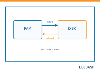 Save and reload prepared dataCache a windowed dataset to disk and reattach it without recompute.
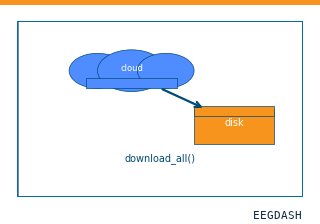 Download a dataset locallyPrefetch BIDS files to a local cache and validate the layout.
Swap any load_dataset(...) call for nm000386 to reproduce the tutorial on this dataset.
Citation
Zepeng Fang, Yuanyuan Dang, Zhipei Ling, Yongzheng Han, Hulin Zhao, … (2024). The involvement of the human prefrontal cortex in the emergence of visual awareness (Fang et al., 2024): sEEG, 6 patients. 10.82901/nemar.nm000386
Provenance
¹Contributed to nemar in BIDS format.
²Curated & ingested by the EEGDash catalog; see CITATION.cff for canonical reference.
³Persistent identifier: 10.82901/nemar.nm000386.
See Also#
eegdash.dataset.EEGDashDataseteegdash.dataset