


NM000460: ieeg dataset, 1 subjects#
Spiking activity (MUA) and LFP from two microwires in human V2/V3, one patient, stimulus-locked epochs of 7 visual experiments (Self et al. 2016; derivative)
Access recordings and metadata through EEGDash.
Citation: Matthew W. Self, Judith C. Peters, Jessy K. Possel, Joel Reithler, Rainer Goebel, Peterjan Ris, Danique Jeurissen, Leila Reddy, Steven Claus, Johannes C. Baayen, Pieter R. Roelfsema (2016). Spiking activity (MUA) and LFP from two microwires in human V2/V3, one patient, stimulus-locked epochs of 7 visual experiments (Self et al. 2016; derivative). 10.82901/nemar.nm000460
Modality: ieeg Subjects: 1 Recordings: 7 License: CC-BY-4.0 Source: nemar
Metadata: Complete (100%)
1-participant iEEG dataset — Spiking activity (MUA) and LFP from two microwires in human V2/V3, one patient, stimulus-locked epochs of 7 visual experiments (Self et al. 2016; derivative).
Quickstart#
Install
pip install eegdash
Access the data
from eegdash.dataset import NM000460

dataset = NM000460(cache_dir="./data")
# Get the raw object of the first recording
raw = dataset.datasets[0].raw
print(raw.info)
Filter by subject
dataset = NM000460(cache_dir="./data", subject="01")
Advanced query
dataset = NM000460(
    cache_dir="./data",
    query={"subject": {"$in": ["01", "02"]}},
)
Iterate recordings
for rec in dataset:
    print(rec.subject, rec.raw.info['sfreq'])
If you use this dataset in your research, please cite the original authors.
BibTeX
@dataset{nm000460,
  title = {Spiking activity (MUA) and LFP from two microwires in human V2/V3, one patient, stimulus-locked epochs of 7 visual experiments (Self et al. 2016; derivative)},
  author = {Matthew W. Self and Judith C. Peters and Jessy K. Possel and Joel Reithler and Rainer Goebel and Peterjan Ris and Danique Jeurissen and Leila Reddy and Steven Claus and Johannes C. Baayen and Pieter R. Roelfsema},
  doi = {10.82901/nemar.nm000460},
  url = {https://doi.org/10.82901/nemar.nm000460},
}
About This Dataset#
It repackages the public Open Science Framework registration “The effects of context and attention on human early visual
cortex.” (Matthew Self, registered February 2016, doi:10.17605/OSF.IO/BRCZY, https://osf.io/brczy/, licence CC-By Attribution 4.0 International). The deposit holds the data of Self et al. 2016, PLoS Biology, doi:10.1371/journal.pbio.1002420. The same files are also listed on EBRAINS; this copy was made from the OSF release.
One patient, a 35-year-old woman with late-onset epilepsy, normal IQ and no reported visual deficits. She had 15 depth electrodes
(AdTech) implanted under general anaesthesia by frameless stereotaxy. One electrode in visual cortex carried a microwire bundle
(40 um platinum-iridium side-wires between the clinical macro-contacts). Recordings from the microwires started 2 days after
surgery and continued for 7 more days. Two microwires, E6 and E7, recorded visually driven spiking activity. Their location is
left occipital, V2/V3 per fMRI retinotopy: the paper estimated it on post-explantation fMRI retinotopic maps of the left
hemisphere. No electrode coordinates were released (MRI/CT only on request from the Data Access Committee of the Netherlands
Institute for Neuroscience), so electrodes.tsv has x/y/z = n/a plus the stated region, hemisphere, material and the
post-explantation impedances (E6 158 kOhm, E7 115 kOhm).
Spiking activity and LFP in human early visual cortex (V2/V3), one patient, stimulus-locked epochs: DERIVATIVE dataset
This is a DERIVATIVE dataset: the signals are epoched around stimulus onset. No continuous recording was released.
Acquisition (paper): NeuraLynx HS-9 unity-gain head-stage, skull-screw ground, 32.5 kHz. E6/E7 re-referenced to the average of microwires E0-E4 (E5 excluded because of noise). From this the authors made three signals, all released at FsD = 930.1714 Hz: - LFP: down-sampled to 930 Hz, band-pass 1-200 Hz (2nd-order zero-phase Butterworth), 50 Hz removed by fitting and
subtracting a sine per trial. Channel type SEEG (microwire LFP).
View full README
Spiking activity and LFP in human early visual cortex (V2/V3), one patient, stimulus-locked epochs: DERIVATIVE dataset
This is a DERIVATIVE dataset: the signals are epoched around stimulus onset. No continuous recording was released.
Acquisition (paper): NeuraLynx HS-9 unity-gain head-stage, skull-screw ground, 32.5 kHz. E6/E7 re-referenced to the average of microwires E0-E4 (E5 excluded because of noise). From this the authors made three signals, all released at FsD = 930.1714 Hz: - LFP: down-sampled to 930 Hz, band-pass 1-200 Hz (2nd-order zero-phase Butterworth), 50 Hz removed by fitting and
subtracting a sine per trial. Channel type SEEG (microwire LFP).
MUA: multi-unit envelope. 500 Hz-5 kHz band-pass, rectified, down-sampled to 930 Hz, low-pass < 200 Hz. Channel type MISC.
MUAT: thresholded MUA (MUAt). Channel type MISC.
The OSF document says MUA and MUAT are not normalised or baseline-corrected. Units are not stated anywhere, so channels.tsv says
n/a. The BrainVision header carries no unit, so MNE reads the numbers as microvolts; for MUA and MUAT that is wrong.
Experiments (one task per OSF file)
Visual stimuli on a laptop LCD (60 Hz, 66 cm). Fixation within 2 deg near the top-left of the screen, else the trial was aborted. Eye position was recorded with an EyeLink (1000 Hz).
| task | OSF file | epochs | epoch length (time re stimulus onset) | layout |
|---|---|---|---|---|
| `rfmapping` | RFmapping.mat | 1210 | 0.601 s (-0.200 to +0.400 s) | both electrodes in one run (trial-wise LFP residual correlation E6-E7 median 0.88 vs -0.10 shuffled within condition) |
| `orientation` | ORItuning.mat | 398 | 1.500 s (-0.399 to +1.100 s) | both electrodes in one run (trial-wise LFP residual correlation E6-E7 median 0.89 vs -0.14 shuffled within condition) |
| `spatialfreq` | SFtuning.mat | 100 | 1.100 s (-0.399 to +0.700 s) | both electrodes in one run (trial-wise LFP residual correlation E6-E7 median 0.90 vs -0.04 shuffled within condition) |
| `contrast` | CONTRASTtuning.mat | 320 | 1.100 s (-0.399 to +0.700 s) | both electrodes in one run (trial-wise LFP residual correlation E6-E7 median 0.88 vs -0.05 shuffled within condition) |
| `size` | SIZEtuning.mat | 200 | 1.300 s (-0.499 to +0.800 s) | both electrodes in one run (trial-wise LFP residual correlation E6-E7 median 0.90 vs -0.04 shuffled within condition) |
| `context` | CONTEXT.mat | 840 | 1.000 s (-0.399 to +0.600 s) | both electrodes in one run (identical eye traces; trial-wise LFP residual correlation E6-E7 median 0.88 vs -0.03 shuffled within condition) |
| `curvetracing` | CURVET.mat | 314 | 2.500 s (-0.499 to +2.000 s) | both electrodes in one run (identical eye traces; trial-wise LFP residual correlation E6-E7 median 0.89 vs -0.01 shuffled within condition) |
Channels per run: E6_LFP, E7_LFP, E6_MUA, E7_MUA, E6_MUAT, E7_MUAT (a per-electrode run has only its own three).
In the OSF files every trial appears once per electrode (rows = electrode x trial, DETS column 1 = electrode). We merged the
two electrodes’ rows into one run only after checking that they come from the same trials: identical DETS sequence, plus
identical eye traces (CONTEXT, CURVET) or a trial-wise LFP correlation far above a within-condition shuffle.
Files and how to read them
sub-01/ieeg/sub-01_task-<task>_ieeg.vhdr: BrainVision, IEEE float32, the epochs of one experiment concatenated in the OSF row order **(``RecordingType`` epoched, ``EpochLength`` in the sidecar). Epoch boundaries are discontinuities: do not filter across them.** Reshape to (epochs, channels, samples) or cut atepoch_startin events.tsv.events.tsv: one row per epoch at stimulus onset (t = 0 of the authors’ time baseTD), with everyDETScolumn under a readable name (events.json) and atrial_typetext. The context condition names are those of the OSF document (ORI 1 = 45 deg, ORI 2 = 135 deg, Sz 1 = 4 deg, Sz 2 = 6 deg). The paper’s Methods give 60 and 150 deg for the central grating; we kept the deposit’s wording.Totals: 1 participant, 7 experiments, 3382 epochs, 1.020 h of epoch time.
float64 -> float32: max absolute deviation 1.52e-05. The files read back bit-exactly and through
mne.io.read_raw_brainvision.Not converted: the eye traces (EYEX, EYEY, deg from fixation, 1000 Hz, own time base
eyetb) of CONTEXT and CURVET. Their epochs are 1.000 s / 2.500 s at 1000 Hz against 0.9998 s / 2.4995 s of neural data, so concatenating them would not line up. They are insourcedata/osf-brczy/CONTEXT.matandCURVET.mat.
Analysis notes from the paper
Trials with eye position more than 1 deg from fixation (10-15%) were discarded in the context and attention analyses. That
selection is not applied here: all deposited epochs are included. The curve-tracing events keep the deposit’s correct column as is (values 0, 1, 2; the OSF document gives no code list).
Ethics
Quoted verbatim from Self et al. 2016 (Materials and Methods, “Surgical Procedures and Patient Details”):
The study was approved by the medical ethics board of the Free University Medical Center (protocol 2009/194). The patient gave written informed consent to participate in the study.
sourcedata/osf-brczy/
The 8 OSF files (“Archive of OSF Storage”): the 7 .mat files and “Details of the dataset.docx” (the authors’ description of the
variables and DETS columns). osf_manifest.json lists the original OSF sizes and md5 hashes, which matched on download. Two
privacy-only changes:
1. The MAT text header (“Created on: <weekday> <month> <day> <time> <year>”) keeps only month and year; the day is set to 01.
The data bytes are unchanged, and every variable reloads identically.
The docx creation/modification dates in docProps/core.xml are set to day 01. The document text is unchanged.
So md5 of these copies differs from OSF; get the byte-identical originals from https://osf.io/brczy/.
Licence and citation
CC BY 4.0 (OSF licence “CC-By Attribution 4.0 International”, 2016), kept. Cite the paper (Self, M. W., Peters, J. C., Possel, J. K., Reithler, J., Goebel, R., Ris, P., Jeurissen, D., Reddy, L., Claus, S., Baayen, J. C. & Roelfsema, P. R. The Effects of Context and Attention on Spiking Activity in Human Early Visual Cortex. PLoS Biology 14(3): e1002420 (2016). doi:10.1371/journal.pbio.1002420) and the data (Self, M. W. The effects of context and attention on human early visual cortex. Open Science Framework registration, 2016. doi:10.17605/OSF.IO/BRCZY).
The OSF document also asks users to contact the corresponding author (Matthew Self, Netherlands Institute for Neuroscience) so that analyses do not overlap.
Changes made here (CC BY “indicate if changes were made”): format conversion to BIDS/BrainVision float32, events and sidecars from the DETS tables and the paper, header dates in sourcedata reduced to month/year. No signal processing.
Funding (paper): NWO (OnderzoeksTalent 406-12-072; Brain and Cognition 433-09-208; ALW 823-02-010), EU FP7 (Marie-Curie PITN-GA-2011-290011; ERC advanced grants 339490 and 269853; HBP 604102), Neuroscience Campus Amsterdam proof-of-concept grant.
NEMAR Metadata#
[](https://doi.org/10.82901/nemar.nm000460) # Spiking activity and LFP in human early visual cortex (V2/V3), one patient, stimulus-locked epochs: DERIVATIVE dataset This is a DERIVATIVE dataset: the signals are epoched around stimulus onset. No continuous recording was released. It repackages the public Open Science Framework registration “The effects of context and attention on human early visual cortex.” (Matthew Self, registered February 2016, doi:10.17605/OSF.IO/BRCZY, https://osf.io/brczy/, licence CC-By Attribution 4.0 International). The deposit holds the data of Self et al. 2016, PLoS Biology, doi:10.1371/journal.pbio.1002420. The same files are also listed on EBRAINS; this copy was made from the OSF release. ## Participant and recording (from the paper) One patient, a 35-year-old woman with late-onset epilepsy, normal IQ and no reported visual deficits. She had 15 depth electrodes (AdTech) implanted under general anaesthesia by frameless stereotaxy. One electrode in visual cortex carried a microwire bundle (40 um platinum-iridium side-wires between the clinical macro-contacts). Recordings from the microwires started 2 days after surgery and continued for 7 more days. Two microwires, E6 and E7, recorded visually driven spiking activity. Their location is left occipital, V2/V3 per fMRI retinotopy: the paper estimated it on post-explantation fMRI retinotopic maps of the left hemisphere. No electrode coordinates were released (MRI/CT only on request from the Data Access Committee of the Netherlands Institute for Neuroscience), so electrodes.tsv has x/y/z = n/a plus the stated region, hemisphere, material and the post-explantation impedances (E6 158 kOhm, E7 115 kOhm). Acquisition (paper): NeuraLynx HS-9 unity-gain head-stage, skull-screw ground, 32.5 kHz. E6/E7 re-referenced to the average of microwires E0-E4 (E5 excluded because of noise). From this the authors made three signals, all released at FsD = 930.1714 Hz: - LFP: down-sampled to 930 Hz, band-pass 1-200 Hz (2nd-order zero-phase Butterworth), 50 Hz removed by fitting and
subtracting a sine per trial. Channel type SEEG (microwire LFP).
MUA: multi-unit envelope. 500 Hz-5 kHz band-pass, rectified, down-sampled to 930 Hz, low-pass < 200 Hz. Channel type MISC.
MUAT: thresholded MUA (MUAt). Channel type MISC.
The OSF document says MUA and MUAT are not normalised or baseline-corrected. Units are not stated anywhere, so channels.tsv says n/a. The BrainVision header carries no unit, so MNE reads the numbers as microvolts; for MUA and MUAT that is wrong. ## Experiments (one task per OSF file) Visual stimuli on a laptop LCD (60 Hz, 66 cm). Fixation within 2 deg near the top-left of the screen, else the trial was aborted. Eye position was recorded with an EyeLink (1000 Hz). | task | OSF file | epochs | epoch length (time re stimulus onset) | layout | |---|—|---|—|---| | rfmapping | RFmapping.mat | 1210 | 0.601 s (-0.200 to +0.400 s) | both electrodes in one run (trial-wise LFP residual correlation E6-E7 median 0.88 vs -0.10 shuffled within condition) | | orientation | ORItuning.mat | 398 | 1.500 s (-0.399 to +1.100 s) | both electrodes in one run (trial-wise LFP residual correlation E6-E7 median 0.89 vs -0.14 shuffled within condition) | | spatialfreq | SFtuning.mat | 100 | 1.100 s (-0.399 to +0.700 s) | both electrodes in one run (trial-wise LFP residual correlation E6-E7 median 0.90 vs -0.04 shuffled within condition) | | contrast | CONTRASTtuning.mat | 320 | 1.100 s (-0.399 to +0.700 s) | both electrodes in one run (trial-wise LFP residual correlation E6-E7 median 0.88 vs -0.05 shuffled within condition) | | size | SIZEtuning.mat | 200 | 1.300 s (-0.499 to +0.800 s) | both electrodes in one run (trial-wise LFP residual correlation E6-E7 median 0.90 vs -0.04 shuffled within condition) | | context | CONTEXT.mat | 840 | 1.000 s (-0.399 to +0.600 s) | both electrodes in one run (identical eye traces; trial-wise LFP residual correlation E6-E7 median 0.88 vs -0.03 shuffled within condition) | | curvetracing | CURVET.mat | 314 | 2.500 s (-0.499 to +2.000 s) | both electrodes in one run (identical eye traces; trial-wise LFP residual correlation E6-E7 median 0.89 vs -0.01 shuffled within condition) | Channels per run: E6_LFP, E7_LFP, E6_MUA, E7_MUA, E6_MUAT, E7_MUAT (a per-electrode run has only its own three). In the OSF files every trial appears once per electrode (rows = electrode x trial, DETS column 1 = electrode). We merged the two electrodes’ rows into one run only after checking that they come from the same trials: identical DETS sequence, plus identical eye traces (CONTEXT, CURVET) or a trial-wise LFP correlation far above a within-condition shuffle. ## Files and how to read them - sub-01/ieeg/sub-01_task-<task>_ieeg.vhdr: BrainVision, IEEE float32, the epochs of one experiment **concatenated in the
OSF row order** (RecordingType epoched, EpochLength in the sidecar). Epoch boundaries are discontinuities: do not filter across them. Reshape to (epochs, channels, samples) or cut at epoch_start in events.tsv.
events.tsv: one row per epoch at stimulus onset (t = 0 of the authors’ time base TD), with every DETS column under a readable name (events.json) and a trial_type text. The context condition names are those of the OSF document (ORI 1 = 45 deg, ORI 2 = 135 deg, Sz 1 = 4 deg, Sz 2 = 6 deg). The paper’s Methods give 60 and 150 deg for the central grating; we kept the deposit’s wording.
Totals: 1 participant, 7 experiments, 3382 epochs, 1.020 h of epoch time.
float64 -> float32: max absolute deviation 1.52e-05. The files read back bit-exactly and through mne.io.read_raw_brainvision.
Not converted: the eye traces (EYEX, EYEY, deg from fixation, 1000 Hz, own time base eyetb) of CONTEXT and CURVET. Their epochs are 1.000 s / 2.500 s at 1000 Hz against 0.9998 s / 2.4995 s of neural data, so concatenating them would not line up. They are in sourcedata/osf-brczy/CONTEXT.mat and CURVET.mat.
## Analysis notes from the paper Trials with eye position more than 1 deg from fixation (10-15%) were discarded in the context and attention analyses. That selection is not applied here: all deposited epochs are included. The curve-tracing events keep the deposit’s correct column as is (values 0, 1, 2; the OSF document gives no code list). ## Ethics Quoted verbatim from Self et al. 2016 (Materials and Methods, “Surgical Procedures and Patient Details”): > The study was approved by the medical ethics board of the Free University Medical Center (protocol 2009/194). The patient gave written informed consent to participate in the study. ## sourcedata/osf-brczy/ The 8 OSF files (“Archive of OSF Storage”): the 7 .mat files and “Details of the dataset.docx” (the authors’ description of the variables and DETS columns). osf_manifest.json lists the original OSF sizes and md5 hashes, which matched on download. Two privacy-only changes: 1. The MAT text header (“Created on: <weekday> <month> <day> <time> <year>”) keeps only month and year; the day is set to 01.
The data bytes are unchanged, and every variable reloads identically.
2. The docx creation/modification dates in docProps/core.xml are set to day 01. The document text is unchanged. So md5 of these copies differs from OSF; get the byte-identical originals from https://osf.io/brczy/. ## Licence and citation CC BY 4.0 (OSF licence “CC-By Attribution 4.0 International”, 2016), kept. Cite the paper (Self, M. W., Peters, J. C., Possel, J. K., Reithler, J., Goebel, R., Ris, P., Jeurissen, D., Reddy, L., Claus, S., Baayen, J. C. & Roelfsema, P. R. The Effects of Context and Attention on Spiking Activity in Human Early Visual Cortex. PLoS Biology 14(3): e1002420 (2016). doi:10.1371/journal.pbio.1002420) and the data (Self, M. W. The effects of context and attention on human early visual cortex. Open Science Framework registration, 2016. doi:10.17605/OSF.IO/BRCZY). The OSF document also asks users to contact the corresponding author (Matthew Self, Netherlands Institute for Neuroscience) so that analyses do not overlap. Changes made here (CC BY “indicate if changes were made”): format conversion to BIDS/BrainVision float32, events and sidecars from the DETS tables and the paper, header dates in sourcedata reduced to month/year. No signal processing. Funding (paper): NWO (OnderzoeksTalent 406-12-072; Brain and Cognition 433-09-208; ALW 823-02-010), EU FP7 (Marie-Curie PITN-GA-2011-290011; ERC advanced grants 339490 and 269853; HBP 604102), Neuroscience Campus Amsterdam proof-of-concept grant.
License: CC-BY-4.0
Authors:
Matthew W. Self
Judith C. Peters
Jessy K. Possel
Joel Reithler
Rainer Goebel
… and 6 more
Versions:
Version |
DOI |
Released |
|---|---|---|
|
Cohort#
Dataset Statistics#
Age distribution by gender (n=1, range 35–35 yr, mean 35.0 yr)
Sex composition
Channel counts: 6 ch (n=7 recordings)
Sampling frequencies: 930.1714285714286 Hz (n=7 recordings)
Total recording duration: 1 h 1 min
Signal · Electrodes & live trace#
Live trace viewer — sub-01 · task-curvetracing
Showing one representative recording out of
1 subjects and 7 recordings in this dataset.
Browse the full set on OpenNeuro;
drop any other _ieeg.{set,edf,bdf,vhdr} file onto the
viewer (or pass ?ieeg=<url>) to inspect it.
No scalp electrode layout is currently indexed for this dataset. Once the eegdash montage registry ingests it, the interactive viewer will appear here automatically.
NEMAR Processing Statistics#
The plots below are generated by NEMAR’s automated EEG pipeline. The histogram shows pipeline success for data cleaning and ICA decomposition, the percentage of data frames and EEG channels retained after artefact removal, line noise per channel (RMS, dB), and the age/gender distribution of participants.

HED event descriptors word cloud

Manifest#
File Explorer#
Browse the BIDS file structure of this dataset. Records are fetched on demand from the EEGDash catalog the first time you open the explorer.
Full dataset metadata table
Dataset ID |
|
Title |
Spiking activity (MUA) and LFP from two microwires in human V2/V3, one patient, stimulus-locked epochs of 7 visual experiments (Self et al. 2016; derivative) |
Author (year) |
— |
Canonical |
— |
Importable as |
|
Year |
2016 |
Authors |
Matthew W. Self, Judith C. Peters, Jessy K. Possel, Joel Reithler, Rainer Goebel, Peterjan Ris, Danique Jeurissen, Leila Reddy, Steven Claus, Johannes C. Baayen, Pieter R. Roelfsema |
License |
CC-BY-4.0 |
Citation / DOI |
|
Source links |
OpenNeuro | NeMAR | Source URL |
Copy-paste BibTeX
@dataset{nm000460,
  title = {Spiking activity (MUA) and LFP from two microwires in human V2/V3, one patient, stimulus-locked epochs of 7 visual experiments (Self et al. 2016; derivative)},
  author = {Matthew W. Self and Judith C. Peters and Jessy K. Possel and Joel Reithler and Rainer Goebel and Peterjan Ris and Danique Jeurissen and Leila Reddy and Steven Claus and Johannes C. Baayen and Pieter R. Roelfsema},
  doi = {10.82901/nemar.nm000460},
  url = {https://doi.org/10.82901/nemar.nm000460},
}
API Reference#
eegdash.datasetEEGDashDataset- class eegdash.dataset.NM000460(cache_dir: str, query: dict | None = None, s3_bucket: str | None = None, **kwargs)[source]#
Spiking activity (MUA) and LFP from two microwires in human V2/V3, one patient, stimulus-locked epochs of 7 visual experiments (Self et al. 2016; derivative)
- Study:
nm000460(NeMAR)- Author (year):
—
- Canonical:
—
Also importable as:
NM000460.Modality:
ieeg; Subject type:Unknown. Subjects: 1; recordings: 7; tasks: 7.- Parameters:
cache_dir (str | Path) – Directory where data are cached locally.
query (dict | None) – Additional MongoDB-style filters to AND with the dataset selection. Must not contain the key
dataset.s3_bucket (str | None) – Base S3 bucket used to locate the data.
**kwargs (dict) – Additional keyword arguments forwarded to
EEGDashDataset.
- data_dir#
Local dataset cache directory (
cache_dir / dataset_id).- Type:
Path
Notes
Each item is a recording; recording-level metadata are available via
dataset.description.querysupports MongoDB-style filters on fields inALLOWED_QUERY_FIELDSand is combined with the dataset filter. Dataset-specific caveats are not provided in the summary metadata.References
OpenNeuro dataset: https://openneuro.org/datasets/nm000460 NeMAR dataset: https://nemar.org/dataexplorer/detail?dataset_id=nm000460 DOI: https://doi.org/10.82901/nemar.nm000460
Examples
>>> from eegdash.dataset import NM000460
>>> dataset = NM000460(cache_dir="./data")
>>> recording = dataset[0]
>>> raw = recording.load()
- __init__(cache_dir: str, query: dict | None = None, s3_bucket: str | None = None, **kwargs)[source]#
- save(path: str, overwrite: bool = False, offset: int = 0)[source]#
Save datasets to files by creating one subdirectory for each dataset:
path/
    0/
        0-raw.fif | 0-epo.fif
        description.json
        raw_preproc_kwargs.json (if raws were preprocessed)
        window_kwargs.json (if this is a windowed dataset)
        window_preproc_kwargs.json  (if windows were preprocessed)
        target_name.json (if target_name is not None and dataset is raw)
    1/
        1-raw.fif | 1-epo.fif
        description.json
        raw_preproc_kwargs.json (if raws were preprocessed)
        window_kwargs.json (if this is a windowed dataset)
        window_preproc_kwargs.json  (if windows were preprocessed)
        target_name.json (if target_name is not None and dataset is raw)
- Parameters:
path (str) –
- Directory in which subdirectories are created to store
-raw.fif | -epo.fif and .json files to.
overwrite (bool) – Whether to delete old subdirectories that will be saved to in this call.
offset (int) – If provided, the integer is added to the id of the dataset in the concat. This is useful in the setting of very large datasets, where one dataset has to be processed and saved at a time to account for its original position.
BaseDataset from braindecode — windowed via create_windows_from_events.braindecodeDataLoader; supports parallel workers and on-the-fly augmentations.pytorch Find datasets with the EEGDash APIQuery the catalogue, filter by task or modality, list candidates.
 Load one EEG recordingResolve a single record to an MNE Raw with channels and events.
 EEG recording to PyTorch DataLoaderWrap braindecode windows in a DataLoader for model training.
 Preprocess EEG and create windowsFilter, resample, epoch — and persist the windowed dataset.
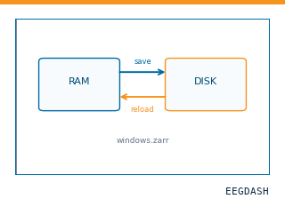 Save and reload prepared dataCache a windowed dataset to disk and reattach it without recompute.
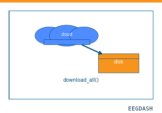 Download a dataset locallyPrefetch BIDS files to a local cache and validate the layout.
Swap any load_dataset(...) call for nm000460 to reproduce the tutorial on this dataset.
Citation
Matthew W. Self, Judith C. Peters, Jessy K. Possel, Joel Reithler, Rainer Goebel, … (2016). Spiking activity (MUA) and LFP from two microwires in human V2/V3, one patient, stimulus-locked epochs of 7 visual experiments (Self et al. 2016; derivative). 10.82901/nemar.nm000460
Provenance
¹Contributed to nemar in BIDS format.
²Curated & ingested by the EEGDash catalog; see CITATION.cff for canonical reference.
³Persistent identifier: 10.82901/nemar.nm000460.
See Also#
eegdash.dataset.EEGDashDataseteegdash.dataset