


NM000372: ieeg dataset, 1 subjects#
Network of autoscopic hallucinations elicited by intracerebral stimulation of periventricular nodular heterotopia: SEEG during 43 Hz and single-pulse stimulation (1 patient)
Access recordings and metadata through EEGDash.
Citation: Flavius Ionut Bratu, Irina Oane, Andrei Barborica, Cristian Donos, Constantin Pistol, Andrei Daneasa, Camelia Lentoiu, Ioana Mindruta (2021). Network of autoscopic hallucinations elicited by intracerebral stimulation of periventricular nodular heterotopia: SEEG during 43 Hz and single-pulse stimulation (1 patient). 10.82901/nemar.nm000372
Modality: ieeg Subjects: 1 Recordings: 2 License: CC0-1.0 Source: nemar
Metadata: Complete (100%)
1-participant iEEG dataset — Network of autoscopic hallucinations elicited by intracerebral stimulation of periventricular nodular heterotopia: SEEG during 43 Hz and single-pulse stimulation (1 patient).
Quickstart#
Install
pip install eegdash
Access the data
from eegdash.dataset import NM000372

dataset = NM000372(cache_dir="./data")
# Get the raw object of the first recording
raw = dataset.datasets[0].raw
print(raw.info)
Filter by subject
dataset = NM000372(cache_dir="./data", subject="01")
Advanced query
dataset = NM000372(
    cache_dir="./data",
    query={"subject": {"$in": ["01", "02"]}},
)
Iterate recordings
for rec in dataset:
    print(rec.subject, rec.raw.info['sfreq'])
If you use this dataset in your research, please cite the original authors.
BibTeX
@dataset{nm000372,
  title = {Network of autoscopic hallucinations elicited by intracerebral stimulation of periventricular nodular heterotopia: SEEG during 43 Hz and single-pulse stimulation (1 patient)},
  author = {Flavius Ionut Bratu and Irina Oane and Andrei Barborica and Cristian Donos and Constantin Pistol and Andrei Daneasa and Camelia Lentoiu and Ioana Mindruta},
  doi = {10.82901/nemar.nm000372},
  url = {https://doi.org/10.82901/nemar.nm000372},
}
About This Dataset#
Stereo-EEG from one drug-resistant epilepsy patient (release code SEEG84) with electrodes covering a right posterior
temporal periventricular nodular heterotopia (PVNH) and adjacent cortex. 43 Hz electrical stimulation of the PVNH (contacts D08-D09) elicited autoscopic hallucinations; single-pulse electrical stimulation (SPES) was used to map effective connectivity.
The study (Bratu et al. 2021, Cortex 145:285-294; published as a case report) combined four connectivity modalities around
the PVNH: SPES-evoked potentials (resting-state effective connectivity), high-frequency stimulation with alternating polarity pulses time-locked to the clinical effect (symptom-related effective connectivity), non-linear regression (h2) functional connectivity after stimulation, and deterministic fiber tracking (structural connectivity). The multi-modal analysis identified a network involving the PVNH, occipital and temporal neocortex, fusiform gyrus and parietal cortex (article abstract).
Autoscopic hallucinations elicited by stimulation of periventricular nodular heterotopia (SEEG, 1 patient)
Overview
Participant
One patient with drug-resistant epilepsy explored with SEEG (stereo-electroencephalography) because of a right
View full README
Autoscopic hallucinations elicited by stimulation of periventricular nodular heterotopia (SEEG, 1 patient)
Overview
Participant
One patient with drug-resistant epilepsy explored with SEEG (stereo-electroencephalography) because of a right posterior temporal PVNH (article abstract).
Implantation in this release: 16 depth electrodes, 246 contacts with coordinates; 13 right-hemisphere electrodes (198 contacts) and 3 left-hemisphere electrodes F’, X’, Y’ (48 contacts), from the authors’ Contact_coordinates.xlsx.
Clinical centre: the clinical authors are at the Epilepsy Monitoring Unit, Emergency University Hospital Bucharest, Romania (author affiliations); the release does not state the recording site or the recording year.
From the article full text (section 2, Patient and methods): a 32-year-old right-handed male with no relevant medical history and epilepsy since the age of 13; drug-resistant, considered a surgical candidate. Video-EEG: asynchronous interictal discharges in both temporal posterior regions, ictal electrical onset in the right posterior quadrant. MRI: right temporal posterior PVNH; FDG-PET: right temporal hypometabolism. SEEG epileptogenic zone: the PVNH, the right posterior hippocampus and temporo-basal regions.
Implantation per the article: 15 Dixi Medical depth electrodes (12-18 contacts, 2 mm contact length, 3.5 mm spacing, 0.8 mm diameter), 214 intracranial contacts, of which 126 cortical contacts were selected for continuous recording; right temporo-parieto-occipital coverage with contralateral “sentinel” electrodes. The release coordinates list 16 electrodes / 246 contacts (see Known caveats).
The article does not state the recording year.
Task / stimulation protocols
task-hfs: 43 Hz high-frequency electrical stimulation (alternating polarity pulses) of contacts D08-D09 in the PVNH at 0.25-0.6 mA with clinical testing; autoscopic hallucinations (“body perception”, mainly involving the face and upper trunk) were elicited at >= 0.5 mA (marker labels; abstract). The authors’ analysis windows (prestim / stim / poststim) are in the events.task-spes: single-pulse electrical stimulation of D08-D09 (marker ‘SPES D08 D09’); stimulation-evoked potentials.Stimulation protocol (article, sections 2, 2.1.2, 2.1.3): bipolar stimulation with a programmable clinical stimulator (Guideline 4000LP+, FHC, Bowdoin, ME). HFS: 43.3 Hz square, biphasic, alternating polarity 1 msec pulses on adjacent contacts for 5 sec, .25-3 mA in .25 or .1 mA steps, while the patient was reading a text aloud; 8 stimulation trials were performed in D08-09 (section 3.1). SPES: 20 pulses per trial, 15 sec inter-stimulus interval, 3 msec pulse duration, .25-5 mA in .25 mA steps.
Acquisition
4096 Hz, 148 channels per recording: 128 SEEG channels (127 contacts listed in the coordinate table plus one channel labelled C90 that has no coordinates), 16 DC inputs, TRIG, and the OSAT/PR/Pleth monitor channels (typed SEEG in the source header, MISC here). Recording system per the article: 128-channel XLTek Quantum (Natus, Middleton, WI). Hardware filters, reference and ground are not stated in the release or the article (n/a). Power line 50 Hz.
Source
Dryad: Flavius Ionut Bratu, Irina Oane, Andrei Barborica, Cristian Donos, Constantin Pistol, Andrei Daneasa, Camelia Lentoiu, Ioana Mindruta. Data for: Network of autoscopic hallucinations elicited by intracerebral stimulations of periventricular nodular heterotopia: an SEEG study. doi:10.5061/dryad.7h44j0ztk (version 7, 2021-09-23). License: CC0 1.0 (Dryad record
https://spdx.org/licenses/CC0-1.0.html; Zenodo replica 5524947 also cc-zero). Article: Cortex (2021), doi:10.1016/j.cortex.2021.08.018. Analysis code: doi:10.5281/zenodo.5524902.Acquired from the Zenodo replica of the Dryad record; Autoscopic.zip and ReadMe.txt matched the Dryad sha-256 digests.
Contents
sub-01/ieeg/sub-01_task-hfs_run-1_ieeg.*: recording with the 43 Hz stimulation trains (4096 Hz, 148 channels: 128 SEEG channels, 16 DC inputs, TRIG, and OSAT/PR/Pleth monitor channels).sub-01/ieeg/sub-01_task-spes_run-1_ieeg.*: recording with single-pulse stimulation (4096 Hz, 148 channels).*_events.tsv: AnyWave markers (stimulation site, intensity and clinical response, analysis windows).*_channels.tsv: status=bad for the channels in the authors’ AnyWave.ades.badlist.sub-01_space-Other_electrodes.tsv: contact coordinates (mm) in the FreeSurfer surface RAS of the T1 image, with hemisphere and FreeSurfer label, from the authors’ Contact_coordinates.xlsx.sub-01/anat/sub-01_T1w.nii.gz: the authors’ defaced T1 (MRI_defaced.mgz), voxel data and affine unchanged. Deface check (surface renderings, six views): facial features are removed.sourcedata/dryad-7h44j0ztk-deidentified/: the release folder (scripts, ROI/tract files, coordinate and SPES spreadsheets, AnyWave sidecars, h2 connectivity results), without the.datsignal files (identical bytes are in the BIDS.eegfiles) and without the members whose file names or contents carry patient-identifying text (the DSI Studio diffusion source/fib/mapping files, the stereotactic plan.ppr, and one statistics spreadsheet).
Conversion / preprocessing
AnyWave ADES stores little-endian float32 samples, channel-multiplexed (AnyWave keeps SEEG amplitudes in µV; the magnitudes are consistent with µV, but the release does not state the unit); these bytes are copied unchanged as BrainVision
.eeg(IEEE_FLOAT_32, MULTIPLEXED, resolution 1, unit µV). No filtering, resampling, re-referencing or channel removal. The.ades.fltdisplay filter settings are reported inSoftwareFilters, not applied.Age, sex and handedness are not in the Dryad release; they come from the article full text. Diffusion MRI is not included (identifying file names; only derived tractography inputs were released).
Known caveats
Signal unit: µV by AnyWave convention; the release does not state the unit.
Recording year, reference and ground: not given in the release or the article (n/a).
Electrode counts differ between sources. All values are kept, with their sources: - Article (Bratu et al. 2021, section 2 “Patient and methods”): “15 depth electrodes with 12-18 contacts”; “From 214
contacts placed intracranially we selected 126 contacts placed in the cortex, to continuously record cortical activity”. Section 2.1.2: SEPs “were recorded in the 122 other contacts”.
Article Supplementary Material 1, Supplementary Table 1 (“Recording contact coordinates”): 126 contacts on 15 electrodes (A, B, C, D, F, I, K, L, P, Q, R, S, U, F’, Y’; none on X’).
Released coordinate table (Contact_coordinates.xlsx ->
sub-01_space-Other_electrodes.tsv): 246 contacts on 16 electrodes (the 15 above plus X’, 18 contacts). participants.tsv n_electrodes/n_contacts (16/246) count this table; n_electrodes_paper/n_contacts_paper/n_contacts_recorded_paper (15/214/126) are the article values.Released recordings (
*_channels.tsv, both runs): 128 SEEG channels = the 126 contacts of Supplementary Table 1 plus R11 (in the coordinate table, not in Supplementary Table 1) and C90 (no coordinates; label kept as in the source). No X’ contact was recorded.So the recordings support the article’s 15 electrodes and its recording montage (126 + 2 channels). The release does not reproduce the article’s 214 intracranial contacts; the coordinate table lists 246 contacts.
Competing interest stated in the article: Andrei Barborica is Vice-President and CTO of FHC Inc, the manufacturer of the electrical stimulator and stereotactic fixture used.
Diffusion MRI and five release members carrying patient-identifying text are not redistributed (see Contents).
How to load
from mne_bids import BIDSPath, read_raw_bids
bp = BIDSPath(root=".", subject="01", task="hfs", run="1", datatype="ieeg")  # or task="spes"
raw = read_raw_bids(bp)
Citation
Bratu FI, Oane I, Barborica A, Donos C, Pistol C, Daneasa A, Lentoiu C, Mindruta I (2021). Network of autoscopic hallucinations elicited by intracerebral stimulations of periventricular nodular heterotopia: an SEEG study. Cortex 145:285-294. doi:10.1016/j.cortex.2021.08.018. Data: doi:10.5061/dryad.7h44j0ztk.
Provenance of the metadata
Dryad record (API v2, version 7) and ReadMe.txt; release files (ADES headers, AnyWave markers/bad lists, Contact_coordinates.xlsx); article abstract (PubMed 34775265). Enriched 2026-10-07; second pass 2026-10-07 from the article full text (Bratu et al. 2021 Cortex, sections 2, 2.1.2, 2.1.3, 3.1, Funding, Acknowledgements and disclosures).
Ethics approval
Verbatim from Barborica A, Oane I, Donos C, Daneasa A, Mihai F, Pistol C, Dabu A, Roceanu A, Mindruta I (2022). Imaging the effective networks associated with cortical function through intracranial high-frequency stimulation. Human Brain Mapping 43(5):1657-1675. https://doi.org/10.1002/hbm.25749 (PMC8886668), “Ethics statement” and “Patient consent” sections. Note: the source article of this dataset (Bratu et al. 2021, Cortex 145:285-294) contains no ethics statement; this statement is from the companion study by the same Bucharest SEEG group (Emergency University Hospital Bucharest), same SEEG/high-frequency stimulation protocol, patients explored 2017-2021, and was designated as the applicable approval by the dataset curator (B. Aristimunha, 2026-10-07):
The study has been performed under Emergency University Hospital Bucharest ethical committee approval 32483/27.06.2018 and Bucharest University ethical committee approval CEC 45/11.06.2020. All patients signed a written informed consent, in accordance with the Declaration of Helsinki, for the recordings, stimulations and data sharing procedures.
NEMAR Metadata#
[](https://doi.org/10.82901/nemar.nm000372) # Autoscopic hallucinations elicited by stimulation of periventricular nodular heterotopia (SEEG, 1 patient) ## Overview Stereo-EEG from one drug-resistant epilepsy patient (release code SEEG84) with electrodes covering a right posterior temporal periventricular nodular heterotopia (PVNH) and adjacent cortex. 43 Hz electrical stimulation of the PVNH (contacts D08-D09) elicited autoscopic hallucinations; single-pulse electrical stimulation (SPES) was used to map effective connectivity. The study (Bratu et al. 2021, Cortex 145:285-294; published as a case report) combined four connectivity modalities around the PVNH: SPES-evoked potentials (resting-state effective connectivity), high-frequency stimulation with alternating polarity pulses time-locked to the clinical effect (symptom-related effective connectivity), non-linear regression (h2) functional connectivity after stimulation, and deterministic fiber tracking (structural connectivity). The multi-modal analysis identified a network involving the PVNH, occipital and temporal neocortex, fusiform gyrus and parietal cortex (article abstract). ## Participant - One patient with drug-resistant epilepsy explored with SEEG (stereo-electroencephalography) because of a right
posterior temporal PVNH (article abstract).
Implantation in this release: 16 depth electrodes, 246 contacts with coordinates; 13 right-hemisphere electrodes (198 contacts) and 3 left-hemisphere electrodes F’, X’, Y’ (48 contacts), from the authors’ Contact_coordinates.xlsx.
Clinical centre: the clinical authors are at the Epilepsy Monitoring Unit, Emergency University Hospital Bucharest, Romania (author affiliations); the release does not state the recording site or the recording year.
From the article full text (section 2, Patient and methods): a 32-year-old right-handed male with no relevant medical history and epilepsy since the age of 13; drug-resistant, considered a surgical candidate. Video-EEG: asynchronous interictal discharges in both temporal posterior regions, ictal electrical onset in the right posterior quadrant. MRI: right temporal posterior PVNH; FDG-PET: right temporal hypometabolism. SEEG epileptogenic zone: the PVNH, the right posterior hippocampus and temporo-basal regions.
Implantation per the article: 15 Dixi Medical depth electrodes (12-18 contacts, 2 mm contact length, 3.5 mm spacing, 0.8 mm diameter), 214 intracranial contacts, of which 126 cortical contacts were selected for continuous recording; right temporo-parieto-occipital coverage with contralateral “sentinel” electrodes. The release coordinates list 16 electrodes / 246 contacts (see Known caveats).
The article does not state the recording year.
## Task / stimulation protocols - task-hfs: 43 Hz high-frequency electrical stimulation (alternating polarity pulses) of contacts D08-D09 in the PVNH at
0.25-0.6 mA with clinical testing; autoscopic hallucinations (“body perception”, mainly involving the face and upper trunk) were elicited at >= 0.5 mA (marker labels; abstract). The authors’ analysis windows (prestim / stim / poststim) are in the events.
task-spes: single-pulse electrical stimulation of D08-D09 (marker ‘SPES D08 D09’); stimulation-evoked potentials.
Stimulation protocol (article, sections 2, 2.1.2, 2.1.3): bipolar stimulation with a programmable clinical stimulator (Guideline 4000LP+, FHC, Bowdoin, ME). HFS: 43.3 Hz square, biphasic, alternating polarity 1 msec pulses on adjacent contacts for 5 sec, .25-3 mA in .25 or .1 mA steps, while the patient was reading a text aloud; 8 stimulation trials were performed in D08-09 (section 3.1). SPES: 20 pulses per trial, 15 sec inter-stimulus interval, 3 msec pulse duration, .25-5 mA in .25 mA steps.
## Acquisition - 4096 Hz, 148 channels per recording: 128 SEEG channels (127 contacts listed in the coordinate table plus one channel
labelled C90 that has no coordinates), 16 DC inputs, TRIG, and the OSAT/PR/Pleth monitor channels (typed SEEG in the source header, MISC here). Recording system per the article: 128-channel XLTek Quantum (Natus, Middleton, WI). Hardware filters, reference and ground are not stated in the release or the article (n/a). Power line 50 Hz.
## Source - Dryad: Flavius Ionut Bratu, Irina Oane, Andrei Barborica, Cristian Donos, Constantin Pistol, Andrei Daneasa, Camelia Lentoiu, Ioana Mindruta. Data for: Network of autoscopic hallucinations elicited by intracerebral stimulations of
periventricular nodular heterotopia: an SEEG study. doi:10.5061/dryad.7h44j0ztk (version 7, 2021-09-23). License: CC0 1.0 (Dryad record https://spdx.org/licenses/CC0-1.0.html; Zenodo replica 5524947 also cc-zero). Article: Cortex (2021), doi:10.1016/j.cortex.2021.08.018. Analysis code: doi:10.5281/zenodo.5524902.
Acquired from the Zenodo replica of the Dryad record; Autoscopic.zip and ReadMe.txt matched the Dryad sha-256 digests.
## Contents - sub-01/ieeg/sub-01_task-hfs_run-1_ieeg.*: recording with the 43 Hz stimulation trains (4096 Hz, 148 channels:
128 SEEG channels, 16 DC inputs, TRIG, and OSAT/PR/Pleth monitor channels).
sub-01/ieeg/sub-01_task-spes_run-1_ieeg.*: recording with single-pulse stimulation (4096 Hz, 148 channels).
*_events.tsv: AnyWave markers (stimulation site, intensity and clinical response, analysis windows).
*_channels.tsv: status=bad for the channels in the authors’ AnyWave .ades.bad list.
sub-01_space-Other_electrodes.tsv: contact coordinates (mm) in the FreeSurfer surface RAS of the T1 image, with hemisphere and FreeSurfer label, from the authors’ Contact_coordinates.xlsx.
sub-01/anat/sub-01_T1w.nii.gz: the authors’ defaced T1 (MRI_defaced.mgz), voxel data and affine unchanged. Deface check (surface renderings, six views): facial features are removed.
sourcedata/dryad-7h44j0ztk-deidentified/: the release folder (scripts, ROI/tract files, coordinate and SPES spreadsheets, AnyWave sidecars, h2 connectivity results), without the .dat signal files (identical bytes are in the BIDS .eeg files) and without the members whose file names or contents carry patient-identifying text (the DSI Studio diffusion source/fib/mapping files, the stereotactic plan .ppr, and one statistics spreadsheet).
## Conversion / preprocessing - AnyWave ADES stores little-endian float32 samples, channel-multiplexed (AnyWave keeps SEEG amplitudes in µV; the
magnitudes are consistent with µV, but the release does not state the unit); these bytes are copied unchanged as BrainVision .eeg (IEEE_FLOAT_32, MULTIPLEXED, resolution 1, unit µV). No filtering, resampling, re-referencing or channel removal. The .ades.flt display filter settings are reported in SoftwareFilters, not applied.
Age, sex and handedness are not in the Dryad release; they come from the article full text. Diffusion MRI is not included (identifying file names; only derived tractography inputs were released).
## Known caveats - Signal unit: µV by AnyWave convention; the release does not state the unit. - Recording year, reference and ground: not given in the release or the article (n/a). - Electrode counts differ between sources. All values are kept, with their sources:
Article (Bratu et al. 2021, section 2 “Patient and methods”): “15 depth electrodes with 12-18 contacts”; “From 214 contacts placed intracranially we selected 126 contacts placed in the cortex, to continuously record cortical activity”. Section 2.1.2: SEPs “were recorded in the 122 other contacts”.
Article Supplementary Material 1, Supplementary Table 1 (“Recording contact coordinates”): 126 contacts on 15 electrodes (A, B, C, D, F, I, K, L, P, Q, R, S, U, F’, Y’; none on X’).
Released coordinate table (Contact_coordinates.xlsx -> sub-01_space-Other_electrodes.tsv): 246 contacts on 16 electrodes (the 15 above plus X’, 18 contacts). participants.tsv n_electrodes/n_contacts (16/246) count this table; n_electrodes_paper/n_contacts_paper/n_contacts_recorded_paper (15/214/126) are the article values.
Released recordings (*_channels.tsv, both runs): 128 SEEG channels = the 126 contacts of Supplementary Table 1 plus R11 (in the coordinate table, not in Supplementary Table 1) and C90 (no coordinates; label kept as in the source). No X’ contact was recorded.
So the recordings support the article’s 15 electrodes and its recording montage (126 + 2 channels). The release does not reproduce the article’s 214 intracranial contacts; the coordinate table lists 246 contacts.
Competing interest stated in the article: Andrei Barborica is Vice-President and CTO of FHC Inc, the manufacturer of the electrical stimulator and stereotactic fixture used.
Diffusion MRI and five release members carrying patient-identifying text are not redistributed (see Contents).
## How to load
`python
from mne_bids import BIDSPath, read_raw_bids
bp = BIDSPath(root=".", subject="01", task="hfs", run="1", datatype="ieeg")  # or task="spes"
raw = read_raw_bids(bp)
`
## Citation
Bratu FI, Oane I, Barborica A, Donos C, Pistol C, Daneasa A, Lentoiu C, Mindruta I (2021). Network of autoscopic
hallucinations elicited by intracerebral stimulations of periventricular nodular heterotopia: an SEEG study. Cortex
145:285-294. doi:10.1016/j.cortex.2021.08.018. Data: doi:10.5061/dryad.7h44j0ztk.
## Provenance of the metadata
Dryad record (API v2, version 7) and ReadMe.txt; release files (ADES headers, AnyWave markers/bad lists,
Contact_coordinates.xlsx); article abstract (PubMed 34775265). Enriched 2026-10-07; second pass 2026-10-07 from the article full text
(Bratu et al. 2021 Cortex, sections 2, 2.1.2, 2.1.3, 3.1, Funding, Acknowledgements and disclosures).
## Ethics approval
Verbatim from Barborica A, Oane I, Donos C, Daneasa A, Mihai F, Pistol C, Dabu A, Roceanu A, Mindruta I (2022). Imaging the effective networks associated with cortical function through intracranial high-frequency stimulation. Human Brain Mapping 43(5):1657-1675. https://doi.org/10.1002/hbm.25749 (PMC8886668), “Ethics statement” and “Patient consent” sections. Note: the source article of this dataset (Bratu et al. 2021, Cortex 145:285-294) contains no ethics statement; this statement is from the companion study by the same Bucharest SEEG group (Emergency University Hospital Bucharest), same SEEG/high-frequency stimulation protocol, patients explored 2017-2021, and was designated as the applicable approval by the dataset curator (B. Aristimunha, 2026-10-07):
> The study has been performed under Emergency University Hospital Bucharest ethical committee approval 32483/27.06.2018 and Bucharest University ethical committee approval CEC 45/11.06.2020. All patients signed a written informed consent, in accordance with the Declaration of Helsinki, for the recordings, stimulations and data sharing procedures.
License: CC0-1.0
Authors:
Flavius Ionut Bratu
Irina Oane
Andrei Barborica
Cristian Donos
Constantin Pistol
… and 3 more
Versions:
Version |
DOI |
Released |
|---|---|---|
|
Cohort#
Dataset Statistics#
Age distribution by gender (n=1, range 32–32 yr, mean 32.0 yr)
Sex composition
Channel counts: 148 ch (n=2 recordings)
Sampling frequencies: 4096.0 Hz (n=2 recordings)
Total recording duration: 16 min
Signal · Electrodes & live trace#
Live trace viewer — sub-01 · task-hfs · run-1
Showing one representative recording out of
1 subjects and 2 recordings in this dataset.
Browse the full set on OpenNeuro;
drop any other _ieeg.{set,edf,bdf,vhdr} file onto the
viewer (or pass ?ieeg=<url>) to inspect it.
Electrode layout — iEEG · 246 sensors — 246 channels
NEMAR Processing Statistics#
The plots below are generated by NEMAR’s automated EEG pipeline. The histogram shows pipeline success for data cleaning and ICA decomposition, the percentage of data frames and EEG channels retained after artefact removal, line noise per channel (RMS, dB), and the age/gender distribution of participants.

HED event descriptors word cloud

Manifest#
File Explorer#
Browse the BIDS file structure of this dataset. Records are fetched on demand from the EEGDash catalog the first time you open the explorer.
Full dataset metadata table
Dataset ID |
|
Title |
Network of autoscopic hallucinations elicited by intracerebral stimulation of periventricular nodular heterotopia: SEEG during 43 Hz and single-pulse stimulation (1 patient) |
Author (year) |
— |
Canonical |
— |
Importable as |
|
Year |
2021 |
Authors |
Flavius Ionut Bratu, Irina Oane, Andrei Barborica, Cristian Donos, Constantin Pistol, Andrei Daneasa, Camelia Lentoiu, Ioana Mindruta |
License |
CC0-1.0 |
Citation / DOI |
|
Source links |
OpenNeuro | NeMAR | Source URL |
Copy-paste BibTeX
@dataset{nm000372,
  title = {Network of autoscopic hallucinations elicited by intracerebral stimulation of periventricular nodular heterotopia: SEEG during 43 Hz and single-pulse stimulation (1 patient)},
  author = {Flavius Ionut Bratu and Irina Oane and Andrei Barborica and Cristian Donos and Constantin Pistol and Andrei Daneasa and Camelia Lentoiu and Ioana Mindruta},
  doi = {10.82901/nemar.nm000372},
  url = {https://doi.org/10.82901/nemar.nm000372},
}
API Reference#
eegdash.datasetEEGDashDataset- class eegdash.dataset.NM000372(cache_dir: str, query: dict | None = None, s3_bucket: str | None = None, **kwargs)[source]#
Network of autoscopic hallucinations elicited by intracerebral stimulation of periventricular nodular heterotopia: SEEG during 43 Hz and single-pulse stimulation (1 patient)
- Study:
nm000372(NeMAR)- Author (year):
—
- Canonical:
—
Also importable as:
NM000372.Modality:
ieeg; Subject type:Unknown. Subjects: 1; recordings: 2; tasks: 2.- Parameters:
cache_dir (str | Path) – Directory where data are cached locally.
query (dict | None) – Additional MongoDB-style filters to AND with the dataset selection. Must not contain the key
dataset.s3_bucket (str | None) – Base S3 bucket used to locate the data.
**kwargs (dict) – Additional keyword arguments forwarded to
EEGDashDataset.
- data_dir#
Local dataset cache directory (
cache_dir / dataset_id).- Type:
Path
Notes
Each item is a recording; recording-level metadata are available via
dataset.description.querysupports MongoDB-style filters on fields inALLOWED_QUERY_FIELDSand is combined with the dataset filter. Dataset-specific caveats are not provided in the summary metadata.References
OpenNeuro dataset: https://openneuro.org/datasets/nm000372 NeMAR dataset: https://nemar.org/dataexplorer/detail?dataset_id=nm000372 DOI: https://doi.org/10.82901/nemar.nm000372
Examples
>>> from eegdash.dataset import NM000372
>>> dataset = NM000372(cache_dir="./data")
>>> recording = dataset[0]
>>> raw = recording.load()
- __init__(cache_dir: str, query: dict | None = None, s3_bucket: str | None = None, **kwargs)[source]#
- save(path: str, overwrite: bool = False, offset: int = 0)[source]#
Save datasets to files by creating one subdirectory for each dataset:
path/
    0/
        0-raw.fif | 0-epo.fif
        description.json
        raw_preproc_kwargs.json (if raws were preprocessed)
        window_kwargs.json (if this is a windowed dataset)
        window_preproc_kwargs.json  (if windows were preprocessed)
        target_name.json (if target_name is not None and dataset is raw)
    1/
        1-raw.fif | 1-epo.fif
        description.json
        raw_preproc_kwargs.json (if raws were preprocessed)
        window_kwargs.json (if this is a windowed dataset)
        window_preproc_kwargs.json  (if windows were preprocessed)
        target_name.json (if target_name is not None and dataset is raw)
- Parameters:
path (str) –
- Directory in which subdirectories are created to store
-raw.fif | -epo.fif and .json files to.
overwrite (bool) – Whether to delete old subdirectories that will be saved to in this call.
offset (int) – If provided, the integer is added to the id of the dataset in the concat. This is useful in the setting of very large datasets, where one dataset has to be processed and saved at a time to account for its original position.
BaseDataset from braindecode — windowed via create_windows_from_events.braindecodeDataLoader; supports parallel workers and on-the-fly augmentations.pytorch Find datasets with the EEGDash APIQuery the catalogue, filter by task or modality, list candidates.
 Load one EEG recordingResolve a single record to an MNE Raw with channels and events.
 EEG recording to PyTorch DataLoaderWrap braindecode windows in a DataLoader for model training.
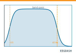 Preprocess EEG and create windowsFilter, resample, epoch — and persist the windowed dataset.
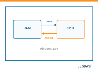 Save and reload prepared dataCache a windowed dataset to disk and reattach it without recompute.
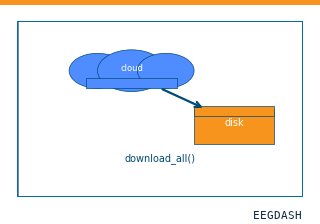 Download a dataset locallyPrefetch BIDS files to a local cache and validate the layout.
Swap any load_dataset(...) call for nm000372 to reproduce the tutorial on this dataset.
Citation
Flavius Ionut Bratu, Irina Oane, Andrei Barborica, Cristian Donos, Constantin Pistol, … (2021). Network of autoscopic hallucinations elicited by intracerebral stimulation of periventricular nodular heterotopia: SEEG during 43 Hz and single-pulse stimulation (1 patient). 10.82901/nemar.nm000372
Provenance
¹Contributed to nemar in BIDS format.
²Curated & ingested by the EEGDash catalog; see CITATION.cff for canonical reference.
³Persistent identifier: 10.82901/nemar.nm000372.
See Also#
eegdash.dataset.EEGDashDataseteegdash.dataset