


NM000394: ieeg dataset, 1 subjects#
Intracranial current source density (CSD) during epileptic seizures and interictal periods, one patient (Budapest)
Access recordings and metadata through EEGDash.
Citation: Dániel Fabó, Loránd Erőss, Zsigmond Benkő (20). Intracranial current source density (CSD) during epileptic seizures and interictal periods, one patient (Budapest). 10.82901/nemar.nm000394
Modality: ieeg Subjects: 1 Recordings: 34 License: CC-BY-NC-SA-4.0 Source: nemar
Metadata: Complete (100%)
1-participant iEEG dataset — Intracranial current source density (CSD) during epileptic seizures and interictal periods, one patient (Budapest).
Quickstart#
Install
pip install eegdash
Access the data
from eegdash.dataset import NM000394

dataset = NM000394(cache_dir="./data")
# Get the raw object of the first recording
raw = dataset.datasets[0].raw
print(raw.info)
Filter by subject
dataset = NM000394(cache_dir="./data", subject="01")
Advanced query
dataset = NM000394(
    cache_dir="./data",
    query={"subject": {"$in": ["01", "02"]}},
)
Iterate recordings
for rec in dataset:
    print(rec.subject, rec.raw.info['sfreq'])
If you use this dataset in your research, please cite the original authors.
BibTeX
@dataset{nm000394,
  title = {Intracranial current source density (CSD) during epileptic seizures and interictal periods, one patient (Budapest)},
  author = {Dániel Fabó and Loránd Erőss and Zsigmond Benkő},
  doi = {10.82901/nemar.nm000394},
  url = {https://doi.org/10.82901/nemar.nm000394},
}
About This Dataset#
This dataset contains intracranial Current Source Density (CSD) recordings of 18 epileptic seizures and 16 interictal
segments from an epileptic patient (text of the source README). This is a derivative dataset. The signals are current source density traces computed by the data authors from subdural ECoG; they are not raw electrode voltages. No raw ECoG is part of the source release.
A 20-year-old patient with drug-resistant epilepsy underwent subdural grid and strip implantation (ADTECH, 10 mm
inter-contact spacing) for pre-surgical evaluation. Video-EEG was recorded with a Micromed Brain-Quick System Evolution, referenced to the skull or mastoid, at 1024 Hz. CSD was computed at fronto-lateral (Fl1, Fl2), inferior-parietal (iP) and fronto-basal (Fb) sites. Patients consented to clinical investigation and surgery along institutional review board guidelines, in accordance with the Declaration of Helsinki (paper methods).
Intracranial current source density during epileptic seizures (Budapest)
Layout
sub-01/ieeg/*_task-seizure_run-XX_desc-csd_ieeg.vhdr: 18 seizure segments (source folderdata/seizure)sub-01/ieeg/*_task-interictal_run-XX_desc-csd_ieeg.vhdr: 16 interictal segments (source folderdata/control)each segment: 20480 samples x 4 channels (GrB6, GrE2, GrF4, FbB3); values copied unchanged from the CSV files as float32
sub-01/sub-01_scans.tsv: maps each run to its original CSV file
View full README
Intracranial current source density during epileptic seizures (Budapest)
Layout
sub-01/ieeg/*_task-seizure_run-XX_desc-csd_ieeg.vhdr: 18 seizure segments (source folderdata/seizure)sub-01/ieeg/*_task-interictal_run-XX_desc-csd_ieeg.vhdr: 16 interictal segments (source folderdata/control)each segment: 20480 samples x 4 channels (GrB6, GrE2, GrF4, FbB3); values copied unchanged from the CSV files as float32
sub-01/sub-01_scans.tsv: maps each run to its original CSV filesourcedata/gin-intracranial_csd/: the complete original repository content (README, LICENSE, CSV files), unchanged
Not stated by the source (left as n/a)
Units of the CSD values; onset times of the segments; meaning of the numbers in the CSV file names; mapping of the contact names (GrB6, GrE2, GrF4, FbB3) to the paper’s region labels; sex of the patient; filters.
Additional metadata and localisation (added 2026-10-08)
Compiled after the upload from the article, its supplement and the source deposit (each statement names its source). Text and sidecar metadata only; no data file was changed. Processing / reference. The deposited signals are CSD, not raw potentials: “CSD was computed (at Fl1, Fl2, iP and Fb), 1–30 Hz Fourier filtering (4th order Butterworth filter) and subsequent rank normalization was carried out” (App. B.2, p.28). The deposited CSV values are integers, so whether the stored files are before or after the filtering and rank normalisation is not stated. Electrode types. AD-TECH subdural strips and a grid with 10 mm inter-contact spacing were implanted through a craniotomy, guided by neuronavigation and fluoroscopy (App. B.2). The text describes them as “a subdural grid and two strip electrodes” (p.11). Localisation. Electrodes were identified on the post-implant CT with BioImage Suite, mapped to the pre-implant MRI with FSL FLIRT/BET2, and projected onto the FreeSurfer pial surface (Dykstra et al. 2012). Intraoperative photographs and electrical stimulation mapping (ESM) were used to corroborate the result (App. B.2). The four analysed sites are fronto-basal (Fb), frontal (Fl1), fronto-lateral (Fl2) and infero-parietal (iP), shown on the patient’s brain surface in Fig. 5F. Neither coordinates nor the patient MRI are published. Most of the high-frequency seizure activity was in Fb and Fl1. The frontal and fronto-basal regions were resected, and the patient was seizure-free for 1 year before a relapse (p.11).
Regions per participant (as published; no coordinates exist)
| participant | region (as stated) | hemisphere | contacts | source |
|---|---|---|---|---|
| sub-01 | fronto-basal (Fb) | n/a | 1 (CSD derivation) | arXiv:1808.10806v4, p.11 Results; Fig. 5A/F; App. B.2 |
| sub-01 | frontal (Fl1) | n/a | 1 (CSD derivation) | arXiv:1808.10806v4, p.11 Results; Fig. 5A/F; App. B.2 |
| sub-01 | fronto-lateral (Fl2) | n/a | 1 (CSD derivation) | arXiv:1808.10806v4, p.11 Results; Fig. 5A/F; App. B.2 |
| sub-01 | infero-parietal (iP) | n/a | 1 (CSD derivation) | arXiv:1808.10806v4, p.11 Results; Fig. 5A/F; App. B.2 |
NEMAR Metadata#
[](https://doi.org/10.82901/nemar.nm000394) # Intracranial current source density during epileptic seizures (Budapest) This dataset contains intracranial Current Source Density (CSD) recordings of 18 epileptic seizures and 16 interictal segments from an epileptic patient (text of the source README). This is a derivative dataset. The signals are current source density traces computed by the data authors from subdural ECoG; they are not raw electrode voltages. No raw ECoG is part of the source release. ## Source - G-Node GIN: https://gin.g-node.org/zsigmondbenko/intracranial_csd (commit 3e5bd99b6919d3fa33e8c4d8d50c714f131dc20b, 2018-08-31) - Copyright (c) 2018 Dániel Fabó and Loránd Erőss, National Institute for Clinical Neurosciences, “Juhász Pál” Epilepsy Center, Budapest, Hungary - Licence: Creative Commons Attribution-NonCommercial-ShareAlike 4.0 (LICENSE copied verbatim) - Paper that uses these data: Benkő Z. et al., “Complete Inference of Causal Relations between Dynamical Systems”, arXiv:1808.10806 (data availability statement points to this repository). ## Recording (from the paper’s methods) A 20-year-old patient with drug-resistant epilepsy underwent subdural grid and strip implantation (ADTECH, 10 mm inter-contact spacing) for pre-surgical evaluation. Video-EEG was recorded with a Micromed Brain-Quick System Evolution, referenced to the skull or mastoid, at 1024 Hz. CSD was computed at fronto-lateral (Fl1, Fl2), inferior-parietal (iP) and fronto-basal (Fb) sites. Patients consented to clinical investigation and surgery along institutional review board guidelines, in accordance with the Declaration of Helsinki (paper methods). ## Layout - sub-01/ieeg/*_task-seizure_run-XX_desc-csd_ieeg.vhdr: 18 seizure segments (source folder data/seizure) - sub-01/ieeg/*_task-interictal_run-XX_desc-csd_ieeg.vhdr: 16 interictal segments (source folder data/control) - each segment: 20480 samples x 4 channels (GrB6, GrE2, GrF4, FbB3); values copied unchanged from the CSV files as float32 - sub-01/sub-01_scans.tsv: maps each run to its original CSV file - sourcedata/gin-intracranial_csd/: the complete original repository content (README, LICENSE, CSV files), unchanged ## Not stated by the source (left as n/a) Units of the CSD values; onset times of the segments; meaning of the numbers in the CSV file names; mapping of the contact names (GrB6, GrE2, GrF4, FbB3) to the paper’s region labels; sex of the patient; filters. ## Additional metadata and localisation (added 2026-10-08) Compiled after the upload from the article, its supplement and the source deposit (each statement names its source). Text and sidecar metadata only; no data file was changed. Processing / reference. The deposited signals are CSD, not raw potentials: “CSD was computed (at Fl1, Fl2, iP and Fb), 1–30 Hz Fourier filtering (4th order Butterworth filter) and subsequent rank normalization was carried out” (App. B.2, p.28). The deposited CSV values are integers, so whether the stored files are before or after the filtering and rank normalisation is not stated. Electrode types. AD-TECH subdural strips and a grid with 10 mm inter-contact spacing were implanted through a craniotomy, guided by neuronavigation and fluoroscopy (App. B.2). The text describes them as “a subdural grid and two strip electrodes” (p.11). Localisation. Electrodes were identified on the post-implant CT with BioImage Suite, mapped to the pre-implant MRI with FSL FLIRT/BET2, and projected onto the FreeSurfer pial surface (Dykstra et al. 2012). Intraoperative photographs and electrical stimulation mapping (ESM) were used to corroborate the result (App. B.2). The four analysed sites are fronto-basal (Fb), frontal (Fl1), fronto-lateral (Fl2) and infero-parietal (iP), shown on the patient’s brain surface in Fig. 5F. Neither coordinates nor the patient MRI are published. Most of the high-frequency seizure activity was in Fb and Fl1. The frontal and fronto-basal regions were resected, and the patient was seizure-free for 1 year before a relapse (p.11). ### Regions per participant (as published; no coordinates exist) | participant | region (as stated) | hemisphere | contacts | source | |---|—|---|—|---| | sub-01 | fronto-basal (Fb) | n/a | 1 (CSD derivation) | arXiv:1808.10806v4, p.11 Results; Fig. 5A/F; App. B.2 | | sub-01 | frontal (Fl1) | n/a | 1 (CSD derivation) | arXiv:1808.10806v4, p.11 Results; Fig. 5A/F; App. B.2 | | sub-01 | fronto-lateral (Fl2) | n/a | 1 (CSD derivation) | arXiv:1808.10806v4, p.11 Results; Fig. 5A/F; App. B.2 | | sub-01 | infero-parietal (iP) | n/a | 1 (CSD derivation) | arXiv:1808.10806v4, p.11 Results; Fig. 5A/F; App. B.2 |
License: CC-BY-NC-SA-4.0
Authors:
Dániel Fabó
Loránd Erőss
Zsigmond Benkő
Versions:
Version |
DOI |
Released |
|---|---|---|
|
Cohort#
Dataset Statistics#
Age distribution by gender (n=1, range 20–20 yr, mean 20.0 yr)
Channel counts: 4 ch (n=34 recordings)
Sampling frequencies: 1024.0 Hz (n=34 recordings)
Total recording duration: 11 min
Signal · Electrodes & live trace#
Live trace viewer — sub-01 · task-seizure · run-17
Showing one representative recording out of
1 subjects and 34 recordings in this dataset.
Browse the full set on OpenNeuro;
drop any other _ieeg.{set,edf,bdf,vhdr} file onto the
viewer (or pass ?ieeg=<url>) to inspect it.
No scalp electrode layout is currently indexed for this dataset. Once the eegdash montage registry ingests it, the interactive viewer will appear here automatically.
NEMAR Processing Statistics#
The plots below are generated by NEMAR’s automated EEG pipeline. The histogram shows pipeline success for data cleaning and ICA decomposition, the percentage of data frames and EEG channels retained after artefact removal, line noise per channel (RMS, dB), and the age/gender distribution of participants.

HED event descriptors word cloud

Manifest#
File Explorer#
Browse the BIDS file structure of this dataset. Records are fetched on demand from the EEGDash catalog the first time you open the explorer.
Full dataset metadata table
Dataset ID |
|
Title |
Intracranial current source density (CSD) during epileptic seizures and interictal periods, one patient (Budapest) |
Author (year) |
— |
Canonical |
— |
Importable as |
|
Year |
20 |
Authors |
Dániel Fabó, Loránd Erőss, Zsigmond Benkő |
License |
CC-BY-NC-SA-4.0 |
Citation / DOI |
|
Source links |
OpenNeuro | NeMAR | Source URL |
Copy-paste BibTeX
@dataset{nm000394,
  title = {Intracranial current source density (CSD) during epileptic seizures and interictal periods, one patient (Budapest)},
  author = {Dániel Fabó and Loránd Erőss and Zsigmond Benkő},
  doi = {10.82901/nemar.nm000394},
  url = {https://doi.org/10.82901/nemar.nm000394},
}
API Reference#
eegdash.datasetEEGDashDataset- class eegdash.dataset.NM000394(cache_dir: str, query: dict | None = None, s3_bucket: str | None = None, **kwargs)[source]#
Intracranial current source density (CSD) during epileptic seizures and interictal periods, one patient (Budapest)
- Study:
nm000394(NeMAR)- Author (year):
—
- Canonical:
—
Also importable as:
NM000394.Modality:
ieeg; Subject type:Unknown. Subjects: 1; recordings: 34; tasks: 2.- Parameters:
cache_dir (str | Path) – Directory where data are cached locally.
query (dict | None) – Additional MongoDB-style filters to AND with the dataset selection. Must not contain the key
dataset.s3_bucket (str | None) – Base S3 bucket used to locate the data.
**kwargs (dict) – Additional keyword arguments forwarded to
EEGDashDataset.
- data_dir#
Local dataset cache directory (
cache_dir / dataset_id).- Type:
Path
Notes
Each item is a recording; recording-level metadata are available via
dataset.description.querysupports MongoDB-style filters on fields inALLOWED_QUERY_FIELDSand is combined with the dataset filter. Dataset-specific caveats are not provided in the summary metadata.References
OpenNeuro dataset: https://openneuro.org/datasets/nm000394 NeMAR dataset: https://nemar.org/dataexplorer/detail?dataset_id=nm000394 DOI: https://doi.org/10.82901/nemar.nm000394
Examples
>>> from eegdash.dataset import NM000394
>>> dataset = NM000394(cache_dir="./data")
>>> recording = dataset[0]
>>> raw = recording.load()
- __init__(cache_dir: str, query: dict | None = None, s3_bucket: str | None = None, **kwargs)[source]#
- save(path: str, overwrite: bool = False, offset: int = 0)[source]#
Save datasets to files by creating one subdirectory for each dataset:
path/
    0/
        0-raw.fif | 0-epo.fif
        description.json
        raw_preproc_kwargs.json (if raws were preprocessed)
        window_kwargs.json (if this is a windowed dataset)
        window_preproc_kwargs.json  (if windows were preprocessed)
        target_name.json (if target_name is not None and dataset is raw)
    1/
        1-raw.fif | 1-epo.fif
        description.json
        raw_preproc_kwargs.json (if raws were preprocessed)
        window_kwargs.json (if this is a windowed dataset)
        window_preproc_kwargs.json  (if windows were preprocessed)
        target_name.json (if target_name is not None and dataset is raw)
- Parameters:
path (str) –
- Directory in which subdirectories are created to store
-raw.fif | -epo.fif and .json files to.
overwrite (bool) – Whether to delete old subdirectories that will be saved to in this call.
offset (int) – If provided, the integer is added to the id of the dataset in the concat. This is useful in the setting of very large datasets, where one dataset has to be processed and saved at a time to account for its original position.
BaseDataset from braindecode — windowed via create_windows_from_events.braindecodeDataLoader; supports parallel workers and on-the-fly augmentations.pytorch Find datasets with the EEGDash APIQuery the catalogue, filter by task or modality, list candidates.
 Load one EEG recordingResolve a single record to an MNE Raw with channels and events.
 EEG recording to PyTorch DataLoaderWrap braindecode windows in a DataLoader for model training.
 Preprocess EEG and create windowsFilter, resample, epoch — and persist the windowed dataset.
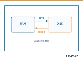 Save and reload prepared dataCache a windowed dataset to disk and reattach it without recompute.
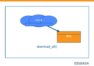 Download a dataset locallyPrefetch BIDS files to a local cache and validate the layout.
Swap any load_dataset(...) call for nm000394 to reproduce the tutorial on this dataset.
Citation
Dániel Fabó, Loránd Erőss, Zsigmond Benkő (20). Intracranial current source density (CSD) during epileptic seizures and interictal periods, one patient (Budapest). 10.82901/nemar.nm000394
Provenance
¹Contributed to nemar in BIDS format.
²Curated & ingested by the EEGDash catalog; see CITATION.cff for canonical reference.
³Persistent identifier: 10.82901/nemar.nm000394.
See Also#
eegdash.dataset.EEGDashDataseteegdash.dataset