


NM000373: ieeg dataset, 8 subjects#
Intracranial entrainment reveals statistical learning across levels of abstraction (Sherman et al., 2023): raw iEEG from 8 neurosurgical patients
Access recordings and metadata through EEGDash.
Citation: Brynn Sherman, Ayman Aljishi, Kathryn Graves, Imran Quraishi, Adithya Sivaraju, Eyiyemisi Damisah, Nicholas Turk-Browne (2023). Intracranial entrainment reveals statistical learning across levels of abstraction (Sherman et al., 2023): raw iEEG from 8 neurosurgical patients. 10.82901/nemar.nm000373
Modality: ieeg Subjects: 8 Recordings: 30 License: CC0-1.0 Source: nemar
Metadata: Complete (100%)
8-participant iEEG dataset — Intracranial entrainment reveals statistical learning across levels of abstraction (Sherman et al., 2023): raw iEEG from 8 neurosurgical patients.
Quickstart#
Install
pip install eegdash
Access the data
from eegdash.dataset import NM000373

dataset = NM000373(cache_dir="./data")
# Get the raw object of the first recording
raw = dataset.datasets[0].raw
print(raw.info)
Filter by subject
dataset = NM000373(cache_dir="./data", subject="01")
Advanced query
dataset = NM000373(
    cache_dir="./data",
    query={"subject": {"$in": ["01", "02"]}},
)
Iterate recordings
for rec in dataset:
    print(rec.subject, rec.raw.info['sfreq'])
If you use this dataset in your research, please cite the original authors.
BibTeX
@dataset{nm000373,
  title = {Intracranial entrainment reveals statistical learning across levels of abstraction (Sherman et al., 2023): raw iEEG from 8 neurosurgical patients},
  author = {Brynn Sherman and Ayman Aljishi and Kathryn Graves and Imran Quraishi and Adithya Sivaraju and Eyiyemisi Damisah and Nicholas Turk-Browne},
  doi = {10.82901/nemar.nm000373},
  url = {https://doi.org/10.82901/nemar.nm000373},
}
About This Dataset#
Raw intracranial EEG (iEEG) from 8 neurosurgical patients with electrodes implanted for seizure monitoring, recorded
while they viewed rapid streams of scene photographs with statistical structure at the category or exemplar level.
The study measured neural entrainment (phase coherence) at the image frequency (2 Hz) and at the pair frequency (1 Hz)
to track online statistical learning of exemplar-level and category-level regularities (Sherman et al., 2023).
Intracranial entrainment reveals statistical learning across levels of abstraction
Overview
Source
Dryad: Sherman B, Aljishi A, Graves K, Quraishi I, Sivaraju A, Damisah E, Turk-Browne N. Data for: Intracranial
View full README
Intracranial entrainment reveals statistical learning across levels of abstraction
Overview
Source
Dryad: Sherman B, Aljishi A, Graves K, Quraishi I, Sivaraju A, Damisah E, Turk-Browne N. Data for: Intracranial entrainment reveals statistical learning across levels of abstraction. doi:10.5061/dryad.q573n5tph (version 3, published 2023-07-07). License: CC0 1.0 (Dryad record
https://spdx.org/licenses/CC0-1.0.html; the Zenodo replica record 8018664 also states cc-zero).Article: Sherman et al. (2023) J Cogn Neurosci, doi:10.1162/jocn_a_02012. Preprint: doi:10.1101/2023.01.11.523605.
Acquired from the Zenodo replica of the Dryad record (community “dryad”, same DOI); every file matched the Dryad API sha-256 digest. The original files are kept unchanged in
sourcedata/dryad-q573n5tph/.
Participants
8 patients (7 male, 1 female; age range 21-61 years, mean 37.8 years) surgically implanted with intracranial electrodes for localization of the seizure onset zone, recruited through the Yale Comprehensive Epilepsy Center; all data were collected at Yale New Haven Hospital (paper, Methods, Participants).
Implants: 6 patients had depth electrodes combined with subdural grid/strip electrodes (“combined”), 2 had depth electrodes only; coverage was left, right, primarily left/right or bilateral depending on the patient. Electrode placement was determined solely by the clinical care team. Contacts were in the visual cortex ROI in 7 of 8 patients.
participants.tsv: age, sex, implant type, hemisphere and number of contacts are taken from Table 1 of the paper (age, sex and handedness are not given in the Dryad release itself; handedness is not reported in the paper either, so it stays n/a). Paper patient ID N corresponds to release participantsub-N(BIDSsub-0N): the hemisphere distribution of each participant’s contacts inallContacts_MNIcoords.csv, the presence/absence of grid (G*) electrode labels and the relative contact counts match Table 1 one-to-one (details in the NEMAR enrichment notes).Recording dates/years are not given (the source randomized the date information in the EDF metadata).
Task (from the release and the paper)
Category-level Structured (task-categorySL): trial-unique scene images whose categories were paired across repetitions (e.g., beach always followed by canyon). 6/8 participants completed two runs (run-1, run-2).
Exemplar-level Structured (task-exemplarSL): 6 repeating scene images presented in fixed pairs.
Random (task-random): the same kind of 6-image set in random order (baseline).
Each image 250 ms, followed by a 250 ms inter-stimulus interval.
Paper (Methods, Stimuli and Procedure): 720 unique outdoor scene images from 18 subcategories (40 per subcategory), 6 subcategories randomly assigned to each condition per participant; images 600 x 800 pixels, presented with MATLAB and the Psychophysics Toolbox on a laptop while the patient sat in the hospital bed. SOA fixed at 500 ms (fixation cross during the ISI); each run was 240 trials (2 min of viewing). Participants passively viewed the stream and were asked to pay attention to each image. Before category-level runs they were told the names of the six categories, but not that the sequence contained pairs. Condition order: category-level structured run(s) first (two back-to-back runs with the same sequence when possible), then one exemplar-level structured and one random run, counterbalanced. Each category pair / exemplar pair occurred 40 times per run.
Recording (from the Dryad methods and the paper)
Natus NeuroWorks EEG system, 4096 Hz. Reference: an electrode chosen by the clinical team to minimize noise.
Triggers: a custom DAQ converted signals from the research computer into 8-bit triggers inserted into an open EEG channel (TRIG). The authors state the iEEG files are the raw data, unprocessed except that the date information in the file metadata was randomized for anonymity (EDF start date 01.01.85, patient field anonymized).
Electrode localization (paper): post-operative CT and MRI, reconstruction in BioImage Suite, registration to the pre-operative MRI, conversion to native-space coordinates with FieldTrip, one-voxel-per-contact masks in FSL, linear registration (FLIRT, 12 dof) to the MNI T1 2 mm template.
Preprocessing
None in this dataset (raw data). For reference, the paper’s analysis (FieldTrip) applied a 60 Hz notch filter, no re-referencing, downsampling to 256 Hz and segmentation into trials; it excluded the first two trials of every run because of a computer-based timing error that shortened the first trial’s ISI in some runs.
Conversion (what changed and what did not)
EDF files are byte-identical copies of the source files (sha-256 listed in
code/source_to_bids_mapping.tsv). No filtering, resampling, re-referencing or channel removal.Renaming: sub-N -> sub-0N; task-categorySL1/2 -> task-categorySL run-1/2; task-exemplarSL1 -> task-exemplarSL run-1; task-random1 -> task-random run-1; datatype eeg -> ieeg.
channels.tsv: names, units, cut-offs and status from the source channels.tsv. Intracranial contacts keep the source type label
EEGbecause the release does not say which electrodes are grids, strips or depths. Channels the source README says to disregard (C*, DC*, OSAT, PR, Pleth) are typed MISC, ECG/EMG as such, TRIG as TRIG. Thegroupcolumn is the electrode prefix of the contact name.events.tsv: one row per TRIG pulse (onset = first sample away from the run’s resting TRIG level, duration = until it returns, value = the sequence of TRIG levels in the pulse), plus the rows of the source events.tsv where the release has one (sub-3).
Electrodes:
allContacts_MNIcoords.csvcopied verbatim into*_space-MNI152NLin6Asym_electrodes.tsv. The values are positive integers consistent with FSL MNI152 2 mm voxel indices, so units are declared n/a and the values are not converted (see coordsystem.json for the affine, an unverified assumption). For sub-01 the labels R1 and R2 each appear twice in the source file with different coordinates and are not among the recorded channels; these 4 ambiguous rows are left out of electrodes.tsv (the original file is in sourcedata).participants.tsv: age, sex and handedness are not given in the Dryad release (n/a); age, sex and implant details were added from Table 1 of the paper (2026-10-07, see CHANGES).
Files
sub-0N/ieeg/: EDF recordings,*_channels.tsv,*_events.tsv,*_ieeg.jsonper run; MNI electrode table and coordsystem per participant.sub-0N/sub-0N_scans.tsvlists the original Dryad file name of every recording.sourcedata/dryad-q573n5tph/: the unchanged Dryad files;code/: conversion script and reports.
Known caveats
Stimulus photographs are not part of the release and not included here.
The meanings of the TRIG codes are not documented in the release; source events.tsv files exist for sub-3 only.
Electrode coordinates are voxel indices (see Conversion); the electrode type of each contact (grid/strip/depth) is not given per contact in the release.
Contact counts in the release are slightly higher than the nContacts column of the paper’s Table 1 (kept as published in
n_contacts_paper).Two patients were tested a second time, 2 days later, because their first data set was unusable (eye irritation in one, a trigger error in the other); the paper does not say which patients (the release contains one set of runs per participant).
The first trial’s ISI was shorter than intended in some runs (paper); the authors dropped the first two trials.
How to load
from mne_bids import BIDSPath, read_raw_bids
bp = BIDSPath(root=".", subject="01", task="categorySL", run="1", datatype="ieeg", suffix="ieeg", extension=".edf")
raw = read_raw_bids(bp)
Citation
Sherman BE, Aljishi A, Graves KN, Quraishi IH, Sivaraju A, Damisah EC, Turk-Browne NB (2023). Intracranial Entrainment Reveals Statistical Learning across Levels of Abstraction. Journal of Cognitive Neuroscience 35(8):1312-1328. doi:10.1162/jocn_a_02012. Data: doi:10.5061/dryad.q573n5tph.
Provenance of the metadata
Dryad record and release README (sourcedata), and the article full text (Methods: Participants, Table 1, iEEG Recordings, iEEG Preprocessing, Electrode Localization, Stimuli, Procedure; Acknowledgments; Funding Information), read on direct.mit.edu on 2026-10-07.
Ethics approval
Verbatim from Sherman BE, Aljishi A, Graves KN, Quraishi IH, Sivaraju A, Damisah EC, Turk-Browne NB (2023). Intracranial Entrainment Reveals Statistical Learning across Levels of Abstraction. Journal of Cognitive Neuroscience 35(8):1312-1328. https://doi.org/10.1162/jocn_a_02012, Methods, “Participants”:
Patients were recruited through the Yale Comprehensive Epilepsy Center and provided informed consent in a manner approved by the Yale University Human Subjects Committee.
NEMAR Metadata#
[](https://doi.org/10.82901/nemar.nm000373) # Intracranial entrainment reveals statistical learning across levels of abstraction ## Overview Raw intracranial EEG (iEEG) from 8 neurosurgical patients with electrodes implanted for seizure monitoring, recorded while they viewed rapid streams of scene photographs with statistical structure at the category or exemplar level. The study measured neural entrainment (phase coherence) at the image frequency (2 Hz) and at the pair frequency (1 Hz) to track online statistical learning of exemplar-level and category-level regularities (Sherman et al., 2023). ## Source - Dryad: Sherman B, Aljishi A, Graves K, Quraishi I, Sivaraju A, Damisah E, Turk-Browne N. Data for: Intracranial
entrainment reveals statistical learning across levels of abstraction. doi:10.5061/dryad.q573n5tph (version 3, published 2023-07-07). License: CC0 1.0 (Dryad record https://spdx.org/licenses/CC0-1.0.html; the Zenodo replica record 8018664 also states cc-zero).
Article: Sherman et al. (2023) J Cogn Neurosci, doi:10.1162/jocn_a_02012. Preprint: doi:10.1101/2023.01.11.523605.
Acquired from the Zenodo replica of the Dryad record (community “dryad”, same DOI); every file matched the Dryad API sha-256 digest. The original files are kept unchanged in sourcedata/dryad-q573n5tph/.
## Participants - 8 patients (7 male, 1 female; age range 21-61 years, mean 37.8 years) surgically implanted with intracranial
electrodes for localization of the seizure onset zone, recruited through the Yale Comprehensive Epilepsy Center; all data were collected at Yale New Haven Hospital (paper, Methods, Participants).
Implants: 6 patients had depth electrodes combined with subdural grid/strip electrodes (“combined”), 2 had depth electrodes only; coverage was left, right, primarily left/right or bilateral depending on the patient. Electrode placement was determined solely by the clinical care team. Contacts were in the visual cortex ROI in 7 of 8 patients.
participants.tsv: age, sex, implant type, hemisphere and number of contacts are taken from Table 1 of the paper (age, sex and handedness are not given in the Dryad release itself; handedness is not reported in the paper either, so it stays n/a). Paper patient ID N corresponds to release participant sub-N (BIDS sub-0N): the hemisphere distribution of each participant’s contacts in allContacts_MNIcoords.csv, the presence/absence of grid (G*) electrode labels and the relative contact counts match Table 1 one-to-one (details in the NEMAR enrichment notes).
Recording dates/years are not given (the source randomized the date information in the EDF metadata).
## Task (from the release and the paper) - Category-level Structured (task-categorySL): trial-unique scene images whose categories were paired across
repetitions (e.g., beach always followed by canyon). 6/8 participants completed two runs (run-1, run-2).
Exemplar-level Structured (task-exemplarSL): 6 repeating scene images presented in fixed pairs.
Random (task-random): the same kind of 6-image set in random order (baseline).
Each image 250 ms, followed by a 250 ms inter-stimulus interval.
Paper (Methods, Stimuli and Procedure): 720 unique outdoor scene images from 18 subcategories (40 per subcategory), 6 subcategories randomly assigned to each condition per participant; images 600 x 800 pixels, presented with MATLAB and the Psychophysics Toolbox on a laptop while the patient sat in the hospital bed. SOA fixed at 500 ms (fixation cross during the ISI); each run was 240 trials (2 min of viewing). Participants passively viewed the stream and were asked to pay attention to each image. Before category-level runs they were told the names of the six categories, but not that the sequence contained pairs. Condition order: category-level structured run(s) first (two back-to-back runs with the same sequence when possible), then one exemplar-level structured and one random run, counterbalanced. Each category pair / exemplar pair occurred 40 times per run.
## Recording (from the Dryad methods and the paper) - Natus NeuroWorks EEG system, 4096 Hz. Reference: an electrode chosen by the clinical team to minimize noise. - Triggers: a custom DAQ converted signals from the research computer into 8-bit triggers inserted into an open EEG
channel (TRIG). The authors state the iEEG files are the raw data, unprocessed except that the date information in the file metadata was randomized for anonymity (EDF start date 01.01.85, patient field anonymized).
Electrode localization (paper): post-operative CT and MRI, reconstruction in BioImage Suite, registration to the pre-operative MRI, conversion to native-space coordinates with FieldTrip, one-voxel-per-contact masks in FSL, linear registration (FLIRT, 12 dof) to the MNI T1 2 mm template.
## Preprocessing - None in this dataset (raw data). For reference, the paper’s analysis (FieldTrip) applied a 60 Hz notch filter, no
re-referencing, downsampling to 256 Hz and segmentation into trials; it excluded the first two trials of every run because of a computer-based timing error that shortened the first trial’s ISI in some runs.
## Conversion (what changed and what did not) - EDF files are byte-identical copies of the source files (sha-256 listed in code/source_to_bids_mapping.tsv).
No filtering, resampling, re-referencing or channel removal.
Renaming: sub-N -> sub-0N; task-categorySL1/2 -> task-categorySL run-1/2; task-exemplarSL1 -> task-exemplarSL run-1; task-random1 -> task-random run-1; datatype eeg -> ieeg.
channels.tsv: names, units, cut-offs and status from the source channels.tsv. Intracranial contacts keep the source type label EEG because the release does not say which electrodes are grids, strips or depths. Channels the source README says to disregard (C*, DC*, OSAT, PR, Pleth) are typed MISC, ECG/EMG as such, TRIG as TRIG. The group column is the electrode prefix of the contact name.
events.tsv: one row per TRIG pulse (onset = first sample away from the run’s resting TRIG level, duration = until it returns, value = the sequence of TRIG levels in the pulse), plus the rows of the source events.tsv where the release has one (sub-3).
Electrodes: allContacts_MNIcoords.csv copied verbatim into *_space-MNI152NLin6Asym_electrodes.tsv. The values are positive integers consistent with FSL MNI152 2 mm voxel indices, so units are declared n/a and the values are not converted (see coordsystem.json for the affine, an unverified assumption). For sub-01 the labels R1 and R2 each appear twice in the source file with different coordinates and are not among the recorded channels; these 4 ambiguous rows are left out of electrodes.tsv (the original file is in sourcedata).
participants.tsv: age, sex and handedness are not given in the Dryad release (n/a); age, sex and implant details were added from Table 1 of the paper (2026-10-07, see CHANGES).
## Files - sub-0N/ieeg/: EDF recordings, *_channels.tsv, *_events.tsv, *_ieeg.json per run; MNI electrode table and
coordsystem per participant. sub-0N/sub-0N_scans.tsv lists the original Dryad file name of every recording.
sourcedata/dryad-q573n5tph/: the unchanged Dryad files; code/: conversion script and reports.
## Known caveats - Stimulus photographs are not part of the release and not included here. - The meanings of the TRIG codes are not documented in the release; source events.tsv files exist for sub-3 only. - Electrode coordinates are voxel indices (see Conversion); the electrode type of each contact (grid/strip/depth) is
not given per contact in the release.
Contact counts in the release are slightly higher than the nContacts column of the paper’s Table 1 (kept as published in n_contacts_paper).
Two patients were tested a second time, 2 days later, because their first data set was unusable (eye irritation in one, a trigger error in the other); the paper does not say which patients (the release contains one set of runs per participant).
The first trial’s ISI was shorter than intended in some runs (paper); the authors dropped the first two trials.
## How to load
`python
from mne_bids import BIDSPath, read_raw_bids
bp = BIDSPath(root=".", subject="01", task="categorySL", run="1", datatype="ieeg", suffix="ieeg", extension=".edf")
raw = read_raw_bids(bp)
`
## Citation
Sherman BE, Aljishi A, Graves KN, Quraishi IH, Sivaraju A, Damisah EC, Turk-Browne NB (2023). Intracranial Entrainment
Reveals Statistical Learning across Levels of Abstraction. Journal of Cognitive Neuroscience 35(8):1312-1328.
doi:10.1162/jocn_a_02012. Data: doi:10.5061/dryad.q573n5tph.
## Provenance of the metadata
- Dryad record and release README (sourcedata), and the article full text (Methods: Participants, Table 1, iEEG
Recordings, iEEG Preprocessing, Electrode Localization, Stimuli, Procedure; Acknowledgments; Funding Information), read on direct.mit.edu on 2026-10-07.
## Ethics approval Verbatim from Sherman BE, Aljishi A, Graves KN, Quraishi IH, Sivaraju A, Damisah EC, Turk-Browne NB (2023). Intracranial Entrainment Reveals Statistical Learning across Levels of Abstraction. Journal of Cognitive Neuroscience 35(8):1312-1328. https://doi.org/10.1162/jocn_a_02012, Methods, “Participants”: > Patients were recruited through the Yale Comprehensive Epilepsy Center and provided informed consent in a manner approved by the Yale University Human Subjects Committee.
License: CC0-1.0
Authors:
Brynn Sherman
Ayman Aljishi
Kathryn Graves
Imran Quraishi
Adithya Sivaraju
… and 2 more
Versions:
Version |
DOI |
Released |
|---|---|---|
|
Cohort#
Dataset Statistics#
Age distribution by gender (n=8, range 21–61 yr, mean 37.8 yr)
Sex composition
Channel counts (ch)
Sampling frequencies: 4096.0 Hz (n=30 recordings)
Total recording duration: 1 h 13 min
Signal · Electrodes & live trace#
Live trace viewer — sub-04 · task-random · run-1
Showing one representative recording out of
8 subjects and 30 recordings in this dataset.
Browse the full set on OpenNeuro;
drop any other _ieeg.{set,edf,bdf,vhdr} file onto the
viewer (or pass ?ieeg=<url>) to inspect it.
Electrode layout — iEEG · 122 sensors — 122 channels
NEMAR Processing Statistics#
The plots below are generated by NEMAR’s automated EEG pipeline. The histogram shows pipeline success for data cleaning and ICA decomposition, the percentage of data frames and EEG channels retained after artefact removal, line noise per channel (RMS, dB), and the age/gender distribution of participants.

HED event descriptors word cloud

Manifest#
File Explorer#
Browse the BIDS file structure of this dataset. Records are fetched on demand from the EEGDash catalog the first time you open the explorer.
Full dataset metadata table
Dataset ID |
|
Title |
Intracranial entrainment reveals statistical learning across levels of abstraction (Sherman et al., 2023): raw iEEG from 8 neurosurgical patients |
Author (year) |
— |
Canonical |
— |
Importable as |
|
Year |
2023 |
Authors |
Brynn Sherman, Ayman Aljishi, Kathryn Graves, Imran Quraishi, Adithya Sivaraju, Eyiyemisi Damisah, Nicholas Turk-Browne |
License |
CC0-1.0 |
Citation / DOI |
|
Source links |
OpenNeuro | NeMAR | Source URL |
Copy-paste BibTeX
@dataset{nm000373,
  title = {Intracranial entrainment reveals statistical learning across levels of abstraction (Sherman et al., 2023): raw iEEG from 8 neurosurgical patients},
  author = {Brynn Sherman and Ayman Aljishi and Kathryn Graves and Imran Quraishi and Adithya Sivaraju and Eyiyemisi Damisah and Nicholas Turk-Browne},
  doi = {10.82901/nemar.nm000373},
  url = {https://doi.org/10.82901/nemar.nm000373},
}
API Reference#
eegdash.datasetEEGDashDataset- class eegdash.dataset.NM000373(cache_dir: str, query: dict | None = None, s3_bucket: str | None = None, **kwargs)[source]#
Intracranial entrainment reveals statistical learning across levels of abstraction (Sherman et al., 2023): raw iEEG from 8 neurosurgical patients
- Study:
nm000373(NeMAR)- Author (year):
—
- Canonical:
—
Also importable as:
NM000373.Modality:
ieeg; Subject type:Unknown. Subjects: 8; recordings: 30; tasks: 3.- Parameters:
cache_dir (str | Path) – Directory where data are cached locally.
query (dict | None) – Additional MongoDB-style filters to AND with the dataset selection. Must not contain the key
dataset.s3_bucket (str | None) – Base S3 bucket used to locate the data.
**kwargs (dict) – Additional keyword arguments forwarded to
EEGDashDataset.
- data_dir#
Local dataset cache directory (
cache_dir / dataset_id).- Type:
Path
Notes
Each item is a recording; recording-level metadata are available via
dataset.description.querysupports MongoDB-style filters on fields inALLOWED_QUERY_FIELDSand is combined with the dataset filter. Dataset-specific caveats are not provided in the summary metadata.References
OpenNeuro dataset: https://openneuro.org/datasets/nm000373 NeMAR dataset: https://nemar.org/dataexplorer/detail?dataset_id=nm000373 DOI: https://doi.org/10.82901/nemar.nm000373
Examples
>>> from eegdash.dataset import NM000373
>>> dataset = NM000373(cache_dir="./data")
>>> recording = dataset[0]
>>> raw = recording.load()
- __init__(cache_dir: str, query: dict | None = None, s3_bucket: str | None = None, **kwargs)[source]#
- save(path: str, overwrite: bool = False, offset: int = 0)[source]#
Save datasets to files by creating one subdirectory for each dataset:
path/
    0/
        0-raw.fif | 0-epo.fif
        description.json
        raw_preproc_kwargs.json (if raws were preprocessed)
        window_kwargs.json (if this is a windowed dataset)
        window_preproc_kwargs.json  (if windows were preprocessed)
        target_name.json (if target_name is not None and dataset is raw)
    1/
        1-raw.fif | 1-epo.fif
        description.json
        raw_preproc_kwargs.json (if raws were preprocessed)
        window_kwargs.json (if this is a windowed dataset)
        window_preproc_kwargs.json  (if windows were preprocessed)
        target_name.json (if target_name is not None and dataset is raw)
- Parameters:
path (str) –
- Directory in which subdirectories are created to store
-raw.fif | -epo.fif and .json files to.
overwrite (bool) – Whether to delete old subdirectories that will be saved to in this call.
offset (int) – If provided, the integer is added to the id of the dataset in the concat. This is useful in the setting of very large datasets, where one dataset has to be processed and saved at a time to account for its original position.
BaseDataset from braindecode — windowed via create_windows_from_events.braindecodeDataLoader; supports parallel workers and on-the-fly augmentations.pytorch Find datasets with the EEGDash APIQuery the catalogue, filter by task or modality, list candidates.
 Load one EEG recordingResolve a single record to an MNE Raw with channels and events.
 EEG recording to PyTorch DataLoaderWrap braindecode windows in a DataLoader for model training.
 Preprocess EEG and create windowsFilter, resample, epoch — and persist the windowed dataset.
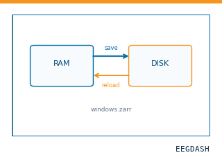 Save and reload prepared dataCache a windowed dataset to disk and reattach it without recompute.
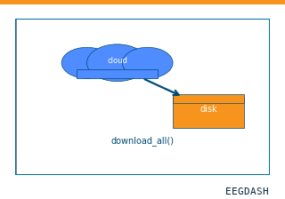 Download a dataset locallyPrefetch BIDS files to a local cache and validate the layout.
Swap any load_dataset(...) call for nm000373 to reproduce the tutorial on this dataset.
Citation
Brynn Sherman, Ayman Aljishi, Kathryn Graves, Imran Quraishi, Adithya Sivaraju, … (2023). Intracranial entrainment reveals statistical learning across levels of abstraction (Sherman et al., 2023): raw iEEG from 8 neurosurgical patients. 10.82901/nemar.nm000373
Provenance
¹Contributed to nemar in BIDS format.
²Curated & ingested by the EEGDash catalog; see CITATION.cff for canonical reference.
³Persistent identifier: 10.82901/nemar.nm000373.
See Also#
eegdash.dataset.EEGDashDataseteegdash.dataset