


NM000397: ieeg dataset, 4 subjects#
Executed and imagined grasp forces recorded with stereo-EEG (Murphy et al., 2016): 4 participants, power and pinch force-matching
Access recordings and metadata through EEGDash.
Citation: Brian A. Murphy, Jonathan P. Miller, Kabilar Gunalan, A. Bolu Ajiboye (2016). Executed and imagined grasp forces recorded with stereo-EEG (Murphy et al., 2016): 4 participants, power and pinch force-matching. 10.82901/nemar.nm000397
Modality: ieeg Subjects: 4 Recordings: 46 License: CC0-1.0 Source: nemar
Metadata: Complete (100%)
4-participant iEEG dataset — Executed and imagined grasp forces recorded with stereo-EEG (Murphy et al., 2016): 4 participants, power and pinch force-matching.
Quickstart#
Install
pip install eegdash
Access the data
from eegdash.dataset import NM000397

dataset = NM000397(cache_dir="./data")
# Get the raw object of the first recording
raw = dataset.datasets[0].raw
print(raw.info)
Filter by subject
dataset = NM000397(cache_dir="./data", subject="01")
Advanced query
dataset = NM000397(
    cache_dir="./data",
    query={"subject": {"$in": ["01", "02"]}},
)
Iterate recordings
for rec in dataset:
    print(rec.subject, rec.raw.info['sfreq'])
If you use this dataset in your research, please cite the original authors.
BibTeX
@dataset{nm000397,
  title = {Executed and imagined grasp forces recorded with stereo-EEG (Murphy et al., 2016): 4 participants, power and pinch force-matching},
  author = {Brian A. Murphy and Jonathan P. Miller and Kabilar Gunalan and A. Bolu Ajiboye},
  doi = {10.82901/nemar.nm000397},
  url = {https://doi.org/10.82901/nemar.nm000397},
}
About This Dataset#
Stereo-EEG from four adults with drug-resistant epilepsy (University Hospitals Case Medical Center, Cleveland, OH) who
performed force-matching tasks with a power grasp (dynamometer) and a lateral pinch (pinch gauge). Participants A and B also performed blocks in which they only imagined producing the force. Each block: 5 trials at each of 3 force targets (20, 30, 40% of maximum voluntary contraction), target shown 5 s with 5 s rest (article).
discrimination of executed and imagined grasp forces through stereoelectroencephalography. doi:10.5061/dryad.nq4fs
(version 1, 2016-04-21). License: CC0 1.0 (Dryad).
Executed and imagined grasp forces recorded with stereo-EEG (Murphy et al., 2016)
Article: PLoS One 11(3):e0150359 (2016), doi:10.1371/journal.pone.0150359 (open access, PMC4786254).
All 14 Dryad files were downloaded through the Dryad API and matched the Dryad md5 digests and sizes.
Contents
sub-<A-D>/ieeg/*_task-<power|pinch><executed|imagined>_run-<block>_ieeg.*: 46 blocks, 2.11 h in total, 2000 Hz.
View full README
Executed and imagined grasp forces recorded with stereo-EEG (Murphy et al., 2016)
Article: PLoS One 11(3):e0150359 (2016), doi:10.1371/journal.pone.0150359 (open access, PMC4786254).
All 14 Dryad files were downloaded through the Dryad API and matched the Dryad md5 digests and sizes.
Contents
sub-<A-D>/ieeg/*_task-<power|pinch><executed|imagined>_run-<block>_ieeg.*: 46 blocks, 2.11 h in total, 2000 Hz. Channelschan1..``chan128`` (participant C:chan49..``chan128``) are the Neuroport amplifier inputs, typed SEEG; the release does not say which inputs carried connected contacts, and gives no contact names, locations or anatomical labels (the article localised contacts with CT/MRI; those data are not released).dynamometeris the analog force signal (MISC, mV).*_events.tsv: force-target periods from the Simulink log (SLCdata). Target values are the game’s raw dynamometer units. Timing = SLCdata.NSPtime minus the NSx packet start; checked by correlating the SLCdata dynamometer log with the NSx dynamometer channel at those times: executed blocks r = 0.44-0.93 (median 0.83); imagined blocks r = -0.40-0.25 (no force produced, as expected). Logged target values: each block starts with one period at a lower value (e.g. 3475), then a value of 3660 alternates with the force targets (e.g. 4500, 5300, 6000); 3660 is most likely the rest/baseline target, but the release does not document the coding, so values are given as logged.sourcedata/dryad-nq4fs-deidentified/: every file of the 7 zips (NSx .ns3 and SLCData .mat) and the README files, de-identified as described below.DEIDENTIFICATION_MANIFEST.tsvlists original and new sha-256 per file.
Conversion
The int16 samples of each NSx file are written unchanged as BrainVision INT_16 (multiplexed); per-channel resolution from the NSx extended header ((max analog - min analog)/(max digital - min digital): 0.25 µV/bit for the amplifier inputs, 0.1526 mV/bit for the dynamometer). No filtering, resampling, re-referencing or channel removal. Read-back with MNE matches the source values. NSx data packets per file: [1]; packet gaps: 0.
Hardware filter settings are copied from the NSx extended header into
HardwareFilters.Hardware reference: a depth contact in a region not related to seizure generation (article). The article’s common average re-referencing was an analysis step and is not applied here.
Privacy
NSx headers store the recording date and time. In the BIDS
scans.tsvand in the sourcedata copies the day is set to 01 (year, month and time of day kept); the NSx weekday field is recomputed for the 1st. MAT-file text headers (‘Created on …’) of the SLCData files are reduced to month and year. No names or hospital identifiers were found in headers, file names or SLCdata fields.Age is given in the article only as a range (29-49 years); all participants were male.
Additional metadata and localisation (added 2026-10-08)
Compiled after the upload from the article, its supplement and the source deposit (each statement names its source). Text and sidecar metadata only; no data file was changed.
Sources: P = Murphy, Miller, Gunalan, Ajiboye 2016, PLoS ONE 11(3):e0150359, doi:10.1371/journal.pone.0150359 (PMC4786254), with S1 Text (supplemental methods), S2 Text and S1 Fig. R = deposit READMEs (README_for_Participant*.txt, identical). F = deposit files (.ns3 headers, probe on Voyager). Reference. Hardware reference was a depth-electrode contact in another anatomical region not involved in seizure generation. For analysis, signals were common-average referenced within groups of contacts on the same electrode passing through similar tissue, excluding noisy channels (P). The SLCdata CARchans field lists 1..128 (F). Electrodes. Integra electrodes have 12 Pt-Ir contacts (1.1 mm diameter, 2.3 mm long, 5 mm spacing). One participant had two PMT combination electrodes with 4 macro contacts (1.4 mm, 7 mm centre spacing) and 4x6 microwires. Only electrodes passing through sensorimotor areas were used in the study (P). Localisation. The post-operative CT (Philips Brilliance iCT or Siemens SOMATOM Sensation 16/Cardiac 64) was rigidly coregistered to the pre-operative 3T T1 (Siemens MAGNETOM Verio) with FSL FLIRT (mutual information, 6 DOF) (S1 Text). Each contact was shown as a sphere and coloured by the nearest structure from a FreeSurfer parcellation (motor, premotor, primary sensory, insula); SMA was drawn by hand in FSLView. Code: mcintyrelab (P). Per participant (P Results/Discussion): every participant had an electrode through the arm/hand area of motor cortex. A, B and C had motor electrodes near the central sulcus; D had two electrodes on the precentral gyrus (arm/hand and leg areas) and one through the SMA. A, B and C had contacts in anterior and posterior insula, D had none. C and D had contacts in primary sensory cortex; the contacts of A and B were too deep to record from sensory cortex. S1 Fig colour-codes every contact of every analysed electrode by region (counted in the table below). The mapping from deposit channel numbers (chanN) to these contacts and the coordinates are not published.
Regions per participant (as published; no coordinates exist)
| participant | region (as stated) | hemisphere | contacts | source |
|---|---|---|---|---|
| ParticipantA | AI (anterior insula electrode): premotor cortex | n/a | 1 | doi:10.1371/journal.pone.0150359, S1 Fig (pone.0150359.s003), visual count of colour-coded contact squares |
| ParticipantA | AI (anterior insula electrode): insular cortex | n/a | 4 | doi:10.1371/journal.pone.0150359, S1 Fig (pone.0150359.s003), visual count of colour-coded contact squares |
| ParticipantA | AI (anterior insula electrode): uncoloured: not grey matter (white matter or outside brain) | n/a | 7 | doi:10.1371/journal.pone.0150359, S1 Fig (pone.0150359.s003), visual count of colour-coded contact squares |
| ParticipantA | MI (middle insula electrode): motor cortex | n/a | 7 | doi:10.1371/journal.pone.0150359, S1 Fig (pone.0150359.s003), visual count of colour-coded contact squares |
| ParticipantA | MI (middle insula electrode): insular cortex | n/a | 1 | doi:10.1371/journal.pone.0150359, S1 Fig (pone.0150359.s003), visual count of colour-coded contact squares |
| ParticipantA | MI (middle insula electrode): uncoloured | n/a | 4 | doi:10.1371/journal.pone.0150359, S1 Fig (pone.0150359.s003), visual count of colour-coded contact squares |
| ParticipantA | PI (posterior insula electrode): insular cortex | n/a | 8 | doi:10.1371/journal.pone.0150359, S1 Fig (pone.0150359.s003), visual count of colour-coded contact squares |
| ParticipantA | PI (posterior insula electrode): uncoloured | n/a | 4 | doi:10.1371/journal.pone.0150359, S1 Fig (pone.0150359.s003), visual count of colour-coded contact squares |
| ParticipantB | AI: premotor cortex | n/a | 4 | doi:10.1371/journal.pone.0150359, S1 Fig (pone.0150359.s003), visual count of colour-coded contact squares |
| ParticipantB | AI: insular cortex | n/a | 5 | doi:10.1371/journal.pone.0150359, S1 Fig (pone.0150359.s003), visual count of colour-coded contact squares |
| ParticipantB | AI: uncoloured | n/a | 3 | doi:10.1371/journal.pone.0150359, S1 Fig (pone.0150359.s003), visual count of colour-coded contact squares |
| ParticipantB | MI: motor cortex | n/a | 7 | doi:10.1371/journal.pone.0150359, S1 Fig (pone.0150359.s003), visual count of colour-coded contact squares |
| ParticipantB | MI: insular cortex | n/a | 2 | doi:10.1371/journal.pone.0150359, S1 Fig (pone.0150359.s003), visual count of colour-coded contact squares |
| ParticipantB | MI: uncoloured | n/a | 3 | doi:10.1371/journal.pone.0150359, S1 Fig (pone.0150359.s003), visual count of colour-coded contact squares |
| ParticipantB | PI: insular cortex | n/a | 7 | doi:10.1371/journal.pone.0150359, S1 Fig (pone.0150359.s003), visual count of colour-coded contact squares |
| ParticipantB | PI: uncoloured | n/a | 5 | doi:10.1371/journal.pone.0150359, S1 Fig (pone.0150359.s003), visual count of colour-coded contact squares |
| ParticipantC | AI: premotor cortex | n/a | 2 | doi:10.1371/journal.pone.0150359, S1 Fig (pone.0150359.s003), visual count of colour-coded contact squares |
| ParticipantC | AI: insular cortex | n/a | 6 | doi:10.1371/journal.pone.0150359, S1 Fig (pone.0150359.s003), visual count of colour-coded contact squares |
| ParticipantC | AI: uncoloured | n/a | 4 | doi:10.1371/journal.pone.0150359, S1 Fig (pone.0150359.s003), visual count of colour-coded contact squares |
| ParticipantC | PS (precentral-sulcus side of primary motor): motor cortex | n/a | 3 | doi:10.1371/journal.pone.0150359, S1 Fig (pone.0150359.s003), visual count of colour-coded contact squares |
| ParticipantC | CS (central-sulcus side of primary motor): motor cortex | n/a | 3 | doi:10.1371/journal.pone.0150359, S1 Fig (pone.0150359.s003), visual count of colour-coded contact squares |
| ParticipantC | PI: primary sensory cortex | n/a | 5 | doi:10.1371/journal.pone.0150359, S1 Fig (pone.0150359.s003), visual count of colour-coded contact squares |
| ParticipantC | PI: insular cortex | n/a | 1 | doi:10.1371/journal.pone.0150359, S1 Fig (pone.0150359.s003), visual count of colour-coded contact squares |
| ParticipantC | PI: uncoloured | n/a | 6 | doi:10.1371/journal.pone.0150359, S1 Fig (pone.0150359.s003), visual count of colour-coded contact squares |
| ParticipantD | SMA: supplementary motor area | n/a | 3 | doi:10.1371/journal.pone.0150359, S1 Fig (pone.0150359.s003), visual count of colour-coded contact squares |
| ParticipantD | SMA: uncoloured | n/a | 9 | doi:10.1371/journal.pone.0150359, S1 Fig (pone.0150359.s003), visual count of colour-coded contact squares |
| ParticipantD | ML (primary motor, leg area): motor cortex | n/a | 4 | doi:10.1371/journal.pone.0150359, S1 Fig (pone.0150359.s003), visual count of colour-coded contact squares |
| ParticipantD | ML (primary motor, leg area): uncoloured | n/a | 8 | doi:10.1371/journal.pone.0150359, S1 Fig (pone.0150359.s003), visual count of colour-coded contact squares |
| ParticipantD | MA/H (primary motor, arm/hand area): motor cortex | n/a | 5 | doi:10.1371/journal.pone.0150359, S1 Fig (pone.0150359.s003), visual count of colour-coded contact squares |
| ParticipantD | MA/H (primary motor, arm/hand area): uncoloured | n/a | 7 | doi:10.1371/journal.pone.0150359, S1 Fig (pone.0150359.s003), visual count of colour-coded contact squares |
| ParticipantD | SL (sensory cortex, leg area): primary sensory cortex | n/a | 4 | doi:10.1371/journal.pone.0150359, S1 Fig (pone.0150359.s003), visual count of colour-coded contact squares |
| ParticipantD | SL (sensory cortex, leg area): uncoloured | n/a | 8 | doi:10.1371/journal.pone.0150359, S1 Fig (pone.0150359.s003), visual count of colour-coded contact squares |
| ParticipantD | SA/H (sensory cortex, arm/hand area): primary sensory cortex | n/a | 6 | doi:10.1371/journal.pone.0150359, S1 Fig (pone.0150359.s003), visual count of colour-coded contact squares |
| ParticipantD | SA/H (sensory cortex, arm/hand area): uncoloured | n/a | 6 | doi:10.1371/journal.pone.0150359, S1 Fig (pone.0150359.s003), visual count of colour-coded contact squares |
NEMAR Metadata#
[](https://doi.org/10.82901/nemar.nm000397) # Executed and imagined grasp forces recorded with stereo-EEG (Murphy et al., 2016) Stereo-EEG from four adults with drug-resistant epilepsy (University Hospitals Case Medical Center, Cleveland, OH) who performed force-matching tasks with a power grasp (dynamometer) and a lateral pinch (pinch gauge). Participants A and B also performed blocks in which they only imagined producing the force. Each block: 5 trials at each of 3 force targets (20, 30, 40% of maximum voluntary contraction), target shown 5 s with 5 s rest (article). ## Source - Dryad: Murphy BA, Miller JP, Gunalan K, Ajiboye AB. Data from: Contributions of subsurface cortical modulations to
discrimination of executed and imagined grasp forces through stereoelectroencephalography. doi:10.5061/dryad.nq4fs (version 1, 2016-04-21). License: CC0 1.0 (Dryad).
Article: PLoS One 11(3):e0150359 (2016), doi:10.1371/journal.pone.0150359 (open access, PMC4786254).
All 14 Dryad files were downloaded through the Dryad API and matched the Dryad md5 digests and sizes.
## Contents - sub-<A-D>/ieeg/*_task-<power|pinch><executed|imagined>_run-<block>_ieeg.*: 46 blocks, 2.11 h in total, 2000 Hz.
Channels chan1..`chan128` (participant C: chan49..`chan128`) are the Neuroport amplifier inputs, typed SEEG; the release does not say which inputs carried connected contacts, and gives no contact names, locations or anatomical labels (the article localised contacts with CT/MRI; those data are not released). dynamometer is the analog force signal (MISC, mV).
*_events.tsv: force-target periods from the Simulink log (SLCdata). Target values are the game’s raw dynamometer units. Timing = SLCdata.NSPtime minus the NSx packet start; checked by correlating the SLCdata dynamometer log with the NSx dynamometer channel at those times: executed blocks r = 0.44-0.93 (median 0.83); imagined blocks r = -0.40-0.25 (no force produced, as expected). Logged target values: each block starts with one period at a lower value (e.g. 3475), then a value of 3660 alternates with the force targets (e.g. 4500, 5300, 6000); 3660 is most likely the rest/baseline target, but the release does not document the coding, so values are given as logged.
sourcedata/dryad-nq4fs-deidentified/: every file of the 7 zips (NSx .ns3 and SLCData .mat) and the README files, de-identified as described below. DEIDENTIFICATION_MANIFEST.tsv lists original and new sha-256 per file.
## Conversion - The int16 samples of each NSx file are written unchanged as BrainVision INT_16 (multiplexed); per-channel resolution
from the NSx extended header ((max analog - min analog)/(max digital - min digital): 0.25 µV/bit for the amplifier inputs, 0.1526 mV/bit for the dynamometer). No filtering, resampling, re-referencing or channel removal. Read-back with MNE matches the source values. NSx data packets per file: [1]; packet gaps: 0.
Hardware filter settings are copied from the NSx extended header into HardwareFilters.
Hardware reference: a depth contact in a region not related to seizure generation (article). The article’s common average re-referencing was an analysis step and is not applied here.
## Privacy - NSx headers store the recording date and time. In the BIDS scans.tsv and in the sourcedata copies the day is set
to 01 (year, month and time of day kept); the NSx weekday field is recomputed for the 1st. MAT-file text headers (‘Created on …’) of the SLCData files are reduced to month and year. No names or hospital identifiers were found in headers, file names or SLCdata fields.
Age is given in the article only as a range (29-49 years); all participants were male.
## Additional metadata and localisation (added 2026-10-08) Compiled after the upload from the article, its supplement and the source deposit (each statement names its source). Text and sidecar metadata only; no data file was changed. Sources: P = Murphy, Miller, Gunalan, Ajiboye 2016, PLoS ONE 11(3):e0150359, doi:10.1371/journal.pone.0150359 (PMC4786254), with S1 Text (supplemental methods), S2 Text and S1 Fig. R = deposit READMEs (README_for_Participant*.txt, identical). F = deposit files (.ns3 headers, probe on Voyager). Reference. Hardware reference was a depth-electrode contact in another anatomical region not involved in seizure generation. For analysis, signals were common-average referenced within groups of contacts on the same electrode passing through similar tissue, excluding noisy channels (P). The SLCdata CARchans field lists 1..128 (F). Electrodes. Integra electrodes have 12 Pt-Ir contacts (1.1 mm diameter, 2.3 mm long, 5 mm spacing). One participant had two PMT combination electrodes with 4 macro contacts (1.4 mm, 7 mm centre spacing) and 4x6 microwires. Only electrodes passing through sensorimotor areas were used in the study (P). Localisation. The post-operative CT (Philips Brilliance iCT or Siemens SOMATOM Sensation 16/Cardiac 64) was rigidly coregistered to the pre-operative 3T T1 (Siemens MAGNETOM Verio) with FSL FLIRT (mutual information, 6 DOF) (S1 Text). Each contact was shown as a sphere and coloured by the nearest structure from a FreeSurfer parcellation (motor, premotor, primary sensory, insula); SMA was drawn by hand in FSLView. Code: mcintyrelab (P). Per participant (P Results/Discussion): every participant had an electrode through the arm/hand area of motor cortex. A, B and C had motor electrodes near the central sulcus; D had two electrodes on the precentral gyrus (arm/hand and leg areas) and one through the SMA. A, B and C had contacts in anterior and posterior insula, D had none. C and D had contacts in primary sensory cortex; the contacts of A and B were too deep to record from sensory cortex. S1 Fig colour-codes every contact of every analysed electrode by region (counted in the table below). The mapping from deposit channel numbers (chanN) to these contacts and the coordinates are not published. ### Regions per participant (as published; no coordinates exist) | participant | region (as stated) | hemisphere | contacts | source | |---|—|---|—|---| | ParticipantA | AI (anterior insula electrode): premotor cortex | n/a | 1 | doi:10.1371/journal.pone.0150359, S1 Fig (pone.0150359.s003), visual count of colour-coded contact squares | | ParticipantA | AI (anterior insula electrode): insular cortex | n/a | 4 | doi:10.1371/journal.pone.0150359, S1 Fig (pone.0150359.s003), visual count of colour-coded contact squares | | ParticipantA | AI (anterior insula electrode): uncoloured: not grey matter (white matter or outside brain) | n/a | 7 | doi:10.1371/journal.pone.0150359, S1 Fig (pone.0150359.s003), visual count of colour-coded contact squares | | ParticipantA | MI (middle insula electrode): motor cortex | n/a | 7 | doi:10.1371/journal.pone.0150359, S1 Fig (pone.0150359.s003), visual count of colour-coded contact squares | | ParticipantA | MI (middle insula electrode): insular cortex | n/a | 1 | doi:10.1371/journal.pone.0150359, S1 Fig (pone.0150359.s003), visual count of colour-coded contact squares | | ParticipantA | MI (middle insula electrode): uncoloured | n/a | 4 | doi:10.1371/journal.pone.0150359, S1 Fig (pone.0150359.s003), visual count of colour-coded contact squares | | ParticipantA | PI (posterior insula electrode): insular cortex | n/a | 8 | doi:10.1371/journal.pone.0150359, S1 Fig (pone.0150359.s003), visual count of colour-coded contact squares | | ParticipantA | PI (posterior insula electrode): uncoloured | n/a | 4 | doi:10.1371/journal.pone.0150359, S1 Fig (pone.0150359.s003), visual count of colour-coded contact squares | | ParticipantB | AI: premotor cortex | n/a | 4 | doi:10.1371/journal.pone.0150359, S1 Fig (pone.0150359.s003), visual count of colour-coded contact squares | | ParticipantB | AI: insular cortex | n/a | 5 | doi:10.1371/journal.pone.0150359, S1 Fig (pone.0150359.s003), visual count of colour-coded contact squares | | ParticipantB | AI: uncoloured | n/a | 3 | doi:10.1371/journal.pone.0150359, S1 Fig (pone.0150359.s003), visual count of colour-coded contact squares | | ParticipantB | MI: motor cortex | n/a | 7 | doi:10.1371/journal.pone.0150359, S1 Fig (pone.0150359.s003), visual count of colour-coded contact squares | | ParticipantB | MI: insular cortex | n/a | 2 | doi:10.1371/journal.pone.0150359, S1 Fig (pone.0150359.s003), visual count of colour-coded contact squares | | ParticipantB | MI: uncoloured | n/a | 3 | doi:10.1371/journal.pone.0150359, S1 Fig (pone.0150359.s003), visual count of colour-coded contact squares | | ParticipantB | PI: insular cortex | n/a | 7 | doi:10.1371/journal.pone.0150359, S1 Fig (pone.0150359.s003), visual count of colour-coded contact squares | | ParticipantB | PI: uncoloured | n/a | 5 | doi:10.1371/journal.pone.0150359, S1 Fig (pone.0150359.s003), visual count of colour-coded contact squares | | ParticipantC | AI: premotor cortex | n/a | 2 | doi:10.1371/journal.pone.0150359, S1 Fig (pone.0150359.s003), visual count of colour-coded contact squares | | ParticipantC | AI: insular cortex | n/a | 6 | doi:10.1371/journal.pone.0150359, S1 Fig (pone.0150359.s003), visual count of colour-coded contact squares | | ParticipantC | AI: uncoloured | n/a | 4 | doi:10.1371/journal.pone.0150359, S1 Fig (pone.0150359.s003), visual count of colour-coded contact squares | | ParticipantC | PS (precentral-sulcus side of primary motor): motor cortex | n/a | 3 | doi:10.1371/journal.pone.0150359, S1 Fig (pone.0150359.s003), visual count of colour-coded contact squares | | ParticipantC | CS (central-sulcus side of primary motor): motor cortex | n/a | 3 | doi:10.1371/journal.pone.0150359, S1 Fig (pone.0150359.s003), visual count of colour-coded contact squares | | ParticipantC | PI: primary sensory cortex | n/a | 5 | doi:10.1371/journal.pone.0150359, S1 Fig (pone.0150359.s003), visual count of colour-coded contact squares | | ParticipantC | PI: insular cortex | n/a | 1 | doi:10.1371/journal.pone.0150359, S1 Fig (pone.0150359.s003), visual count of colour-coded contact squares | | ParticipantC | PI: uncoloured | n/a | 6 | doi:10.1371/journal.pone.0150359, S1 Fig (pone.0150359.s003), visual count of colour-coded contact squares | | ParticipantD | SMA: supplementary motor area | n/a | 3 | doi:10.1371/journal.pone.0150359, S1 Fig (pone.0150359.s003), visual count of colour-coded contact squares | | ParticipantD | SMA: uncoloured | n/a | 9 | doi:10.1371/journal.pone.0150359, S1 Fig (pone.0150359.s003), visual count of colour-coded contact squares | | ParticipantD | ML (primary motor, leg area): motor cortex | n/a | 4 | doi:10.1371/journal.pone.0150359, S1 Fig (pone.0150359.s003), visual count of colour-coded contact squares | | ParticipantD | ML (primary motor, leg area): uncoloured | n/a | 8 | doi:10.1371/journal.pone.0150359, S1 Fig (pone.0150359.s003), visual count of colour-coded contact squares | | ParticipantD | MA/H (primary motor, arm/hand area): motor cortex | n/a | 5 | doi:10.1371/journal.pone.0150359, S1 Fig (pone.0150359.s003), visual count of colour-coded contact squares | | ParticipantD | MA/H (primary motor, arm/hand area): uncoloured | n/a | 7 | doi:10.1371/journal.pone.0150359, S1 Fig (pone.0150359.s003), visual count of colour-coded contact squares | | ParticipantD | SL (sensory cortex, leg area): primary sensory cortex | n/a | 4 | doi:10.1371/journal.pone.0150359, S1 Fig (pone.0150359.s003), visual count of colour-coded contact squares | | ParticipantD | SL (sensory cortex, leg area): uncoloured | n/a | 8 | doi:10.1371/journal.pone.0150359, S1 Fig (pone.0150359.s003), visual count of colour-coded contact squares | | ParticipantD | SA/H (sensory cortex, arm/hand area): primary sensory cortex | n/a | 6 | doi:10.1371/journal.pone.0150359, S1 Fig (pone.0150359.s003), visual count of colour-coded contact squares | | ParticipantD | SA/H (sensory cortex, arm/hand area): uncoloured | n/a | 6 | doi:10.1371/journal.pone.0150359, S1 Fig (pone.0150359.s003), visual count of colour-coded contact squares |
License: CC0-1.0
Authors:
Brian A. Murphy
Jonathan P. Miller
Kabilar Gunalan
Bolu Ajiboye
Versions:
Version |
DOI |
Released |
|---|---|---|
|
Cohort#
Dataset Statistics#
Sex composition
Channel counts (ch)
Sampling frequencies: 2000.0 Hz (n=46 recordings)
Total recording duration: 2 h 6 min
Signal · Electrodes & live trace#
Live trace viewer — sub-A · task-pinchimagined · run-3
Showing one representative recording out of
4 subjects and 46 recordings in this dataset.
Browse the full set on OpenNeuro;
drop any other _ieeg.{set,edf,bdf,vhdr} file onto the
viewer (or pass ?ieeg=<url>) to inspect it.
No scalp electrode layout is currently indexed for this dataset. Once the eegdash montage registry ingests it, the interactive viewer will appear here automatically.
NEMAR Processing Statistics#
The plots below are generated by NEMAR’s automated EEG pipeline. The histogram shows pipeline success for data cleaning and ICA decomposition, the percentage of data frames and EEG channels retained after artefact removal, line noise per channel (RMS, dB), and the age/gender distribution of participants.

HED event descriptors word cloud

Manifest#
File Explorer#
Browse the BIDS file structure of this dataset. Records are fetched on demand from the EEGDash catalog the first time you open the explorer.
Full dataset metadata table
Dataset ID |
|
Title |
Executed and imagined grasp forces recorded with stereo-EEG (Murphy et al., 2016): 4 participants, power and pinch force-matching |
Author (year) |
— |
Canonical |
— |
Importable as |
|
Year |
2016 |
Authors |
Brian A. Murphy, Jonathan P. Miller, Kabilar Gunalan, A. Bolu Ajiboye |
License |
CC0-1.0 |
Citation / DOI |
|
Source links |
OpenNeuro | NeMAR | Source URL |
Copy-paste BibTeX
@dataset{nm000397,
  title = {Executed and imagined grasp forces recorded with stereo-EEG (Murphy et al., 2016): 4 participants, power and pinch force-matching},
  author = {Brian A. Murphy and Jonathan P. Miller and Kabilar Gunalan and A. Bolu Ajiboye},
  doi = {10.82901/nemar.nm000397},
  url = {https://doi.org/10.82901/nemar.nm000397},
}
API Reference#
eegdash.datasetEEGDashDataset- class eegdash.dataset.NM000397(cache_dir: str, query: dict | None = None, s3_bucket: str | None = None, **kwargs)[source]#
Executed and imagined grasp forces recorded with stereo-EEG (Murphy et al., 2016): 4 participants, power and pinch force-matching
- Study:
nm000397(NeMAR)- Author (year):
—
- Canonical:
—
Also importable as:
NM000397.Modality:
ieeg; Subject type:Unknown. Subjects: 4; recordings: 46; tasks: 4.- Parameters:
cache_dir (str | Path) – Directory where data are cached locally.
query (dict | None) – Additional MongoDB-style filters to AND with the dataset selection. Must not contain the key
dataset.s3_bucket (str | None) – Base S3 bucket used to locate the data.
**kwargs (dict) – Additional keyword arguments forwarded to
EEGDashDataset.
- data_dir#
Local dataset cache directory (
cache_dir / dataset_id).- Type:
Path
Notes
Each item is a recording; recording-level metadata are available via
dataset.description.querysupports MongoDB-style filters on fields inALLOWED_QUERY_FIELDSand is combined with the dataset filter. Dataset-specific caveats are not provided in the summary metadata.References
OpenNeuro dataset: https://openneuro.org/datasets/nm000397 NeMAR dataset: https://nemar.org/dataexplorer/detail?dataset_id=nm000397 DOI: https://doi.org/10.82901/nemar.nm000397
Examples
>>> from eegdash.dataset import NM000397
>>> dataset = NM000397(cache_dir="./data")
>>> recording = dataset[0]
>>> raw = recording.load()
- __init__(cache_dir: str, query: dict | None = None, s3_bucket: str | None = None, **kwargs)[source]#
- save(path: str, overwrite: bool = False, offset: int = 0)[source]#
Save datasets to files by creating one subdirectory for each dataset:
path/
    0/
        0-raw.fif | 0-epo.fif
        description.json
        raw_preproc_kwargs.json (if raws were preprocessed)
        window_kwargs.json (if this is a windowed dataset)
        window_preproc_kwargs.json  (if windows were preprocessed)
        target_name.json (if target_name is not None and dataset is raw)
    1/
        1-raw.fif | 1-epo.fif
        description.json
        raw_preproc_kwargs.json (if raws were preprocessed)
        window_kwargs.json (if this is a windowed dataset)
        window_preproc_kwargs.json  (if windows were preprocessed)
        target_name.json (if target_name is not None and dataset is raw)
- Parameters:
path (str) –
- Directory in which subdirectories are created to store
-raw.fif | -epo.fif and .json files to.
overwrite (bool) – Whether to delete old subdirectories that will be saved to in this call.
offset (int) – If provided, the integer is added to the id of the dataset in the concat. This is useful in the setting of very large datasets, where one dataset has to be processed and saved at a time to account for its original position.
BaseDataset from braindecode — windowed via create_windows_from_events.braindecodeDataLoader; supports parallel workers and on-the-fly augmentations.pytorch Find datasets with the EEGDash APIQuery the catalogue, filter by task or modality, list candidates.
 Load one EEG recordingResolve a single record to an MNE Raw with channels and events.
 EEG recording to PyTorch DataLoaderWrap braindecode windows in a DataLoader for model training.
 Preprocess EEG and create windowsFilter, resample, epoch — and persist the windowed dataset.
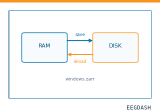 Save and reload prepared dataCache a windowed dataset to disk and reattach it without recompute.
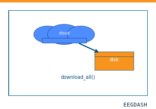 Download a dataset locallyPrefetch BIDS files to a local cache and validate the layout.
Swap any load_dataset(...) call for nm000397 to reproduce the tutorial on this dataset.
Citation
Brian A. Murphy, Jonathan P. Miller, Kabilar Gunalan, A. Bolu Ajiboye (2016). Executed and imagined grasp forces recorded with stereo-EEG (Murphy et al., 2016): 4 participants, power and pinch force-matching. 10.82901/nemar.nm000397
Provenance
¹Contributed to nemar in BIDS format.
²Curated & ingested by the EEGDash catalog; see CITATION.cff for canonical reference.
³Persistent identifier: 10.82901/nemar.nm000397.
See Also#
eegdash.dataset.EEGDashDataseteegdash.dataset