


NM000355: ieeg dataset, 1 subjects#
Stolk et al. 2018 FieldTrip iEEG protocol dataset (SubjectUCI29): authors’ preprocessed ECoG+SEEG epochs
Access recordings and metadata through EEGDash.
Citation: Arjen Stolk, Sandon Griffin, Roemer van der Meij, Callum Dewar, Ignacio Saez, Jack J. Lin, Giovanni Piantoni, Jan-Mathijs Schoffelen, Robert T. Knight, Robert Oostenveld (6502). Stolk et al. 2018 FieldTrip iEEG protocol dataset (SubjectUCI29): authors’ preprocessed ECoG+SEEG epochs. 10.82901/nemar.nm000355
Modality: ieeg Subjects: 1 Recordings: 1 License: CC-BY-SA-4.0 Source: nemar
Metadata: Complete (100%)
1-participant iEEG dataset — Stolk et al. 2018 FieldTrip iEEG protocol dataset (SubjectUCI29): authors' preprocessed ECoG+SEEG epochs.
Quickstart#
Install
pip install eegdash
Access the data
from eegdash.dataset import NM000355

dataset = NM000355(cache_dir="./data")
# Get the raw object of the first recording
raw = dataset.datasets[0].raw
print(raw.info)
Filter by subject
dataset = NM000355(cache_dir="./data", subject="01")
Advanced query
dataset = NM000355(
    cache_dir="./data",
    query={"subject": {"$in": ["01", "02"]}},
)
Iterate recordings
for rec in dataset:
    print(rec.subject, rec.raw.info['sfreq'])
If you use this dataset in your research, please cite the original authors.
BibTeX
@dataset{nm000355,
  title = {Stolk et al. 2018 FieldTrip iEEG protocol dataset (SubjectUCI29): authors' preprocessed ECoG+SEEG epochs},
  author = {Arjen Stolk and Sandon Griffin and Roemer van der Meij and Callum Dewar and Ignacio Saez and Jack J. Lin and Giovanni Piantoni and Jan-Mathijs Schoffelen and Robert T. Knight and Robert Oostenveld},
  doi = {10.82901/nemar.nm000355},
  url = {https://doi.org/10.82901/nemar.nm000355},
}
About This Dataset#
**These are not raw recordings.**This dataset is a BIDS packaging of the*preprocessed* intracranial EEG released
with the FieldTrip human intracranial analysis protocol (Stolk et al., 2018, Nature Protocols). The authors did not share the
raw recording: “Raw recording files are not shared, in order to protect the subject’s identity.” What they released, and
what is packaged here, is the output of protocol steps 35-36: 26 trials of 152 channels at 5000 Hz, cut from -0.4 to
0.9002 s around tone onset, demeaned, low-pass filtered at 200 Hz and band-stop filtered at 59-61, 119-121 and
179-181 Hz. dataset_description.json therefore declares DatasetType: derivative, with GeneratedBy and
SourceDatasets pointing at the authors’ Zenodo record (doi:10.5281/zenodo.1201560, v1.0, CC-BY-SA-4.0).
University of California, Irvine Medical Center. Study approved by the Office for the Protection of Human Subjects of
the University of California, Berkeley; the subject gave informed consent (as stated by the authors). Age, sex and handedness are not given in the release.
Stolk et al. 2018 FieldTrip iEEG protocol dataset (SubjectUCI29): the authors’ preprocessed epochs
Overview
Electrodes: 56 SEEG contacts on 7 depth leads (RAM/LAM amygdala, RHH/LHH hippocampal head, RTH/LTH hippocampal tail, ROC right occipital) and 96 ECoG contacts (LPG 64-contact left parietal grid, LTG 32-contact left temporal grid).
SubjectUCI29_grids.pnginsourcedata/is the authors’ schematic of the grid numbering.
View full README
Stolk et al. 2018 FieldTrip iEEG protocol dataset (SubjectUCI29): the authors’ preprocessed epochs
Overview
Electrodes: 56 SEEG contacts on 7 depth leads (RAM/LAM amygdala, RHH/LHH hippocampal head, RTH/LTH hippocampal tail, ROC right occipital) and 96 ECoG contacts (LPG 64-contact left parietal grid, LTG 32-contact left temporal grid).
SubjectUCI29_grids.pnginsourcedata/is the authors’ schematic of the grid numbering.Task: the patient pressed a button with the right hand on hearing a target tone. Trials are aligned to tone onset (trigger value 4).
Additional acquisition details (Stolk et al. 2018, “Materials” and “Experimental design”; PMC6548463): - Implant: grids LPG (64 contacts, 8 x 8, left parietal) and LTG (32 contacts, 4 x 8, left temporal), Integra,
10 mm inter-electrode spacing; depth leads LAM, LHH, LTH, RAM, RHH, RTH and ROC, 8 contacts each, Ad-Tech, 5 mm inter-electrode spacing. Implanted “as part of the preparation for the epilepsy surgery”.
Amplifier: “All neural recordings were acquired using a Nihon Kohden recording system with a JE-120A amplifier (Nihon Kohden Corporation, Tokyo, Japan), analogfiltered above 0.01 Hz, and digitally sampled at 5 KHz”.
Reference: the acquisition reference electrode is not stated for this recording (see
iEEGReference).Anatomical imaging used by the authors (not redistributed here): pre-implant T1 MRI (Siemens 3T TrioTim), post-implant CT (Philips iCT 256), post-implant T1 MRI (Siemens 1.5T Avanto).
Task / paradigm and timing
“The neural data were recorded in the context of an experiment that required the patient to press a button with
the right hand when hearing a target tone” (Stolk et al. 2018). The authors defined trials from trigger value 4
(tone onset) in the recording’s trigger channel, from 400 ms before to 900 ms after the tone (protocol step 34:
cfg.trialdef.eventvalue = 4; prestim = 0.4; poststim = 0.9), giving the “experiment’s twenty-six trials” (step 36).
Button-press times are not in the release as events; the meaning of trialinfo is not documented (see
_events.json).
Files
sub-UCI29/ieeg/sub-UCI29_task-tonedetection_ieeg.vhdr/.vmrk/.eeg: BrainVision, 152 channels, 5000 Hz, IEEE float32. The 26 epochs (6502 samples each) are stored **back to back**. The sidecar saysRecordingType: epochedandEpochLength: 1.3004, and the marker file has aNew Segmentat each epoch start. The file’s time axis is not the original recording’s time axis. Do not filter across epoch boundaries as if the data were continuous. To get epochs, split the file every 6502 samples. Inside each epoch, sample 2000 (0-based) is tone onset (t = 0).mne_bids.read_raw_bidsrefuses epoched recordings. You can read the file withmne.io.read_raw_brainvisionand then reshape it...._events.tsv: oneepochrow and onetonerow per trial. The rows also carry the authors’data.trialinfovalue, which the release does not explain, anddata.sampleinfo, the epoch’s begin and end sample in the original unshared recording. Usesampleinfoto recover the original spacing between trials...._channels.tsv: channel type (SEEG/ECOG), lead group and filter settings. Units: the FieldTrip structure has no unit field. The stored amplitudes (median |x| ≈ 49, max |x| ≈ 1398) are only plausible as microvolts, so µV is used here. The numbers are the authors’ values.sub-UCI29_space-ACPC_electrodes.tsv+_coordsystem.json: x/y/z areelec_acpc_fr, the subject-ACPC positions the authors attached to the data (CT-MRI fusion, then brain-shift compensation of the grids). Further columns giveelec_acpc_f(before brain-shift compensation),elec_mni_frv(volume-based MNI normalisation; the template variant is not stated by the authors) andelec_fsavg_frs(fsaverage, grids only). All are in mm, as stored.sourcedata/zenodo-1201560/: the authors’ non-imaging files, byte-identical, with SHA-256 insourcedata/sourcedata_provenance.json. These are the FieldTrip data (_data.mat, float64), header, the four electrode structures, the electrode table, the time-frequency result_freq.mat(a further derivative computed after re-montage) and the left cortical hull mesh.
Preprocessing already applied by the source
Protocol step 35 (Stolk et al. 2018), applied by the authors before release with FieldTrip ft_preprocessing:
\`cfg.demean = ‘yes’; cfg.baselinewindow = ‘all’; cfg.lpfilter = ‘yes’; cfg.lpfreq = 200; cfg.padding = 2;
cfg.padtype = ‘data’; cfg.bsfilter = ‘yes’; cfg.bsfiltord = 3; cfg.bsfreq = [59 61; 119 121; 179 181]\`.
The later protocol steps (bad-segment rejection, re-montage to common average for grids and bipolar for depths,
time-frequency analysis) were not applied to the packaged signals; SubjectUCI29_freq.mat in sourcedata/ is the
authors’ time-frequency result from those later steps. Hardware filtering: analog high-pass above 0.01 Hz
(low_cutoff in _channels.tsv).
Known caveats
Not raw data: authors’ preprocessed epochs only (see Overview).
Epochs are stored back to back; the file time axis is not the recording time axis (see Files).
Units (µV) are inferred from amplitudes, not stated by the source (see Files).
trialinfois undocumented by the authors.Head imaging is not redistributed (see “What is not included, and why”).
Precision
The source stores float64 and BrainVision float32. The largest absolute difference between the BrainVision samples and
the source values is reported in the conversion summary. It is below 1e-4 µV, against a signal of tens of µV. For
bit-exact values, use sourcedata/zenodo-1201560/SubjectUCI29_data.mat.
What is not included, and why
The record’s head imaging is not redistributed here. That covers the pre-implant MRI, the post-implant MRI, the CT, the
CT/MRI overlay figure and freesurfer.zip, whose subject directory contains whole-head T1/orig/rawavg volumes. The
authors state the imaging was defaced with ft_defacevolume, and our low-resolution renders show rectangular defacing
masks. We did not run an independent full-resolution identifiability review. These files remain available from the
authors at https://zenodo.org/records/1201560. The same data are also archived at the Donders Repository
(hdl:11633/di.dccn.DSC_3015000.00_734; Zenodo marks it as identical).
How to load
mne_bids.read_raw_bids refuses epoched recordings, so read the BrainVision file directly and cut it into the
26 epochs of 6502 samples (tone onset at sample 2000 of each epoch):
import mne
import numpy as np
raw = mne.io.read_raw_brainvision(
    "sub-UCI29/ieeg/sub-UCI29_task-tonedetection_ieeg.vhdr", preload=True)

data = raw.get_data()                      # (152, 26 * 6502)
epochs = data.reshape(152, 26, 6502).transpose(1, 0, 2)
times = (np.arange(6502) - 2000) / raw.info["sfreq"]   # -0.4 ... 0.9002 s
The signal files are stored with git-annex on NEMAR; fetch them first (for example git annex get sub-UCI29).
Electrode positions are in sub-UCI29_space-ACPC_electrodes.tsv (subject ACPC, mm).
Licence and citation
CC-BY-SA-4.0 (from the Zenodo record), so adaptations must be shared under the same licence. Please cite Stolk et al. (2018), doi:10.1038/s41596-018-0009-6, and the data record doi:10.5281/zenodo.1201560.
Source and provenance
Data record: Zenodo doi:10.5281/zenodo.1201560 (v1.0, published 2018-03-21; identical copy at the Donders Repository, hdl:11633/di.dccn.DSC_3015000.00_734).
Article: Stolk et al. (2018) Nature Protocols 13:1699-1723, doi:10.1038/s41596-018-0009-6 (author manuscript PMC6548463; preprint doi:10.1101/230912). Acquisition, implant and funding details above were read from the PMC full text on 2026-10-06.
Packaging:
laneD_convert.py(iEEG-NEMAR campaign); seeGeneratedByindataset_description.jsonand SHA-256 checksums insourcedata/sourcedata_provenance.json.
NEMAR Metadata#
[](https://doi.org/10.82901/nemar.nm000355) # Stolk et al. 2018 FieldTrip iEEG protocol dataset (SubjectUCI29): the authors’ preprocessed epochs ## Overview These are not raw recordings. This dataset is a BIDS packaging of the preprocessed intracranial EEG released with the FieldTrip human intracranial analysis protocol (Stolk et al., 2018, Nature Protocols). The authors did not share the raw recording: “Raw recording files are not shared, in order to protect the subject’s identity.” What they released, and what is packaged here, is the output of protocol steps 35-36: 26 trials of 152 channels at 5000 Hz, cut from -0.4 to 0.9002 s around tone onset, demeaned, low-pass filtered at 200 Hz and band-stop filtered at 59-61, 119-121 and 179-181 Hz. dataset_description.json therefore declares DatasetType: derivative, with GeneratedBy and SourceDatasets pointing at the authors’ Zenodo record (doi:10.5281/zenodo.1201560, v1.0, CC-BY-SA-4.0). ## Cohort and recording - One adult patient with medication-refractory epilepsy (sub-UCI29, source ID SubjectUCI29), recorded at the
University of California, Irvine Medical Center. Study approved by the Office for the Protection of Human Subjects of the University of California, Berkeley; the subject gave informed consent (as stated by the authors). Age, sex and handedness are not given in the release.
Electrodes: 56 SEEG contacts on 7 depth leads (RAM/LAM amygdala, RHH/LHH hippocampal head, RTH/LTH hippocampal tail, ROC right occipital) and 96 ECoG contacts (LPG 64-contact left parietal grid, LTG 32-contact left temporal grid). SubjectUCI29_grids.png in sourcedata/ is the authors’ schematic of the grid numbering.
Task: the patient pressed a button with the right hand on hearing a target tone. Trials are aligned to tone onset (trigger value 4).
Additional acquisition details (Stolk et al. 2018, “Materials” and “Experimental design”; PMC6548463): - Implant: grids LPG (64 contacts, 8 x 8, left parietal) and LTG (32 contacts, 4 x 8, left temporal), Integra,
10 mm inter-electrode spacing; depth leads LAM, LHH, LTH, RAM, RHH, RTH and ROC, 8 contacts each, Ad-Tech, 5 mm inter-electrode spacing. Implanted “as part of the preparation for the epilepsy surgery”.
Amplifier: “All neural recordings were acquired using a Nihon Kohden recording system with a JE-120A amplifier (Nihon Kohden Corporation, Tokyo, Japan), analogfiltered above 0.01 Hz, and digitally sampled at 5 KHz”.
Reference: the acquisition reference electrode is not stated for this recording (see iEEGReference).
Anatomical imaging used by the authors (not redistributed here): pre-implant T1 MRI (Siemens 3T TrioTim), post-implant CT (Philips iCT 256), post-implant T1 MRI (Siemens 1.5T Avanto).
## Task / paradigm and timing “The neural data were recorded in the context of an experiment that required the patient to press a button with the right hand when hearing a target tone” (Stolk et al. 2018). The authors defined trials from trigger value 4 (tone onset) in the recording’s trigger channel, from 400 ms before to 900 ms after the tone (protocol step 34: cfg.trialdef.eventvalue = 4; prestim = 0.4; poststim = 0.9), giving the “experiment’s twenty-six trials” (step 36). Button-press times are not in the release as events; the meaning of trialinfo is not documented (see _events.json). ## Files - sub-UCI29/ieeg/sub-UCI29_task-tonedetection_ieeg.vhdr/.vmrk/.eeg: BrainVision, 152 channels, 5000 Hz, IEEE float32.
The 26 epochs (6502 samples each) are stored back to back. The sidecar says RecordingType: epoched and EpochLength: 1.3004, and the marker file has a New Segment at each epoch start. The file’s time axis is not the original recording’s time axis. Do not filter across epoch boundaries as if the data were continuous. To get epochs, split the file every 6502 samples. Inside each epoch, sample 2000 (0-based) is tone onset (t = 0). mne_bids.read_raw_bids refuses epoched recordings. You can read the file with mne.io.read_raw_brainvision and then reshape it.
…_events.tsv: one epoch row and one tone row per trial. The rows also carry the authors’ data.trialinfo value, which the release does not explain, and data.sampleinfo, the epoch’s begin and end sample in the original unshared recording. Use sampleinfo to recover the original spacing between trials.
…_channels.tsv: channel type (SEEG/ECOG), lead group and filter settings. Units: the FieldTrip structure has no unit field. The stored amplitudes (median |x| ≈ 49, max |x| ≈ 1398) are only plausible as microvolts, so µV is used here. The numbers are the authors’ values.
sub-UCI29_space-ACPC_electrodes.tsv + _coordsystem.json: x/y/z are elec_acpc_fr, the subject-ACPC positions the authors attached to the data (CT-MRI fusion, then brain-shift compensation of the grids). Further columns give elec_acpc_f (before brain-shift compensation), elec_mni_frv (volume-based MNI normalisation; the template variant is not stated by the authors) and elec_fsavg_frs (fsaverage, grids only). All are in mm, as stored.
sourcedata/zenodo-1201560/: the authors’ non-imaging files, byte-identical, with SHA-256 in sourcedata/sourcedata_provenance.json. These are the FieldTrip data (_data.mat, float64), header, the four electrode structures, the electrode table, the time-frequency result _freq.mat (a further derivative computed after re-montage) and the left cortical hull mesh.
## Preprocessing already applied by the source Protocol step 35 (Stolk et al. 2018), applied by the authors before release with FieldTrip ft_preprocessing: cfg.demean = ‘yes’; cfg.baselinewindow = ‘all’; cfg.lpfilter = ‘yes’; cfg.lpfreq = 200; cfg.padding = 2; cfg.padtype = ‘data’; cfg.bsfilter = ‘yes’; cfg.bsfiltord = 3; cfg.bsfreq = [59 61; 119 121; 179 181]. The later protocol steps (bad-segment rejection, re-montage to common average for grids and bipolar for depths, time-frequency analysis) were not applied to the packaged signals; SubjectUCI29_freq.mat in sourcedata/ is the authors’ time-frequency result from those later steps. Hardware filtering: analog high-pass above 0.01 Hz (low_cutoff in _channels.tsv). ## Known caveats - Not raw data: authors’ preprocessed epochs only (see Overview). - Epochs are stored back to back; the file time axis is not the recording time axis (see Files). - Units (µV) are inferred from amplitudes, not stated by the source (see Files). - trialinfo is undocumented by the authors. - Head imaging is not redistributed (see “What is not included, and why”). ### Precision The source stores float64 and BrainVision float32. The largest absolute difference between the BrainVision samples and the source values is reported in the conversion summary. It is below 1e-4 µV, against a signal of tens of µV. For bit-exact values, use sourcedata/zenodo-1201560/SubjectUCI29_data.mat. ### What is not included, and why The record’s head imaging is not redistributed here. That covers the pre-implant MRI, the post-implant MRI, the CT, the CT/MRI overlay figure and freesurfer.zip, whose subject directory contains whole-head T1/orig/rawavg volumes. The authors state the imaging was defaced with ft_defacevolume, and our low-resolution renders show rectangular defacing masks. We did not run an independent full-resolution identifiability review. These files remain available from the authors at https://zenodo.org/records/1201560. The same data are also archived at the Donders Repository (hdl:11633/di.dccn.DSC_3015000.00_734; Zenodo marks it as identical). ## How to load mne_bids.read_raw_bids refuses epoched recordings, so read the BrainVision file directly and cut it into the 26 epochs of 6502 samples (tone onset at sample 2000 of each epoch): ```python import mne import numpy as np raw = mne.io.read_raw_brainvision(
“sub-UCI29/ieeg/sub-UCI29_task-tonedetection_ieeg.vhdr”, preload=True)
data = raw.get_data() # (152, 26 * 6502) epochs = data.reshape(152, 26, 6502).transpose(1, 0, 2) times = (np.arange(6502) - 2000) / raw.info[“sfreq”] # -0.4 … 0.9002 s ``` The signal files are stored with git-annex on NEMAR; fetch them first (for example git annex get sub-UCI29). Electrode positions are in sub-UCI29_space-ACPC_electrodes.tsv (subject ACPC, mm). ## Licence and citation CC-BY-SA-4.0 (from the Zenodo record), so adaptations must be shared under the same licence. Please cite Stolk et al. (2018), doi:10.1038/s41596-018-0009-6, and the data record doi:10.5281/zenodo.1201560. ## Source and provenance - Data record: Zenodo doi:10.5281/zenodo.1201560 (v1.0, published 2018-03-21; identical copy at the Donders
Repository, hdl:11633/di.dccn.DSC_3015000.00_734).
Article: Stolk et al. (2018) Nature Protocols 13:1699-1723, doi:10.1038/s41596-018-0009-6 (author manuscript PMC6548463; preprint doi:10.1101/230912). Acquisition, implant and funding details above were read from the PMC full text on 2026-10-06.
Packaging: laneD_convert.py (iEEG-NEMAR campaign); see GeneratedBy in dataset_description.json and SHA-256 checksums in sourcedata/sourcedata_provenance.json.
License: CC-BY-SA-4.0
Authors:
Arjen Stolk
Sandon Griffin
Roemer van der Meij
Callum Dewar
Ignacio Saez
… and 5 more
Versions:
Version |
DOI |
Released |
|---|---|---|
|
Cohort#
Dataset Statistics#
Channel counts: 152 ch (n=1 recordings)
Sampling frequencies: 5000.0 Hz (n=1 recordings)
Total recording duration: 0 min
Signal · Electrodes & live trace#
Live trace viewer — sub-UCI29 · task-tonedetection
Showing one representative recording out of
1 subjects and 1 recordings in this dataset.
Browse the full set on OpenNeuro;
drop any other _ieeg.{set,edf,bdf,vhdr} file onto the
viewer (or pass ?ieeg=<url>) to inspect it.
Electrode layout — iEEG · 152 sensors — 152 channels
NEMAR Processing Statistics#
The plots below are generated by NEMAR’s automated EEG pipeline. The histogram shows pipeline success for data cleaning and ICA decomposition, the percentage of data frames and EEG channels retained after artefact removal, line noise per channel (RMS, dB), and the age/gender distribution of participants.

HED event descriptors word cloud

Manifest#
File Explorer#
Browse the BIDS file structure of this dataset. Records are fetched on demand from the EEGDash catalog the first time you open the explorer.
Full dataset metadata table
Dataset ID |
|
Title |
Stolk et al. 2018 FieldTrip iEEG protocol dataset (SubjectUCI29): authors’ preprocessed ECoG+SEEG epochs |
Author (year) |
— |
Canonical |
— |
Importable as |
|
Year |
6502 |
Authors |
Arjen Stolk, Sandon Griffin, Roemer van der Meij, Callum Dewar, Ignacio Saez, Jack J. Lin, Giovanni Piantoni, Jan-Mathijs Schoffelen, Robert T. Knight, Robert Oostenveld |
License |
CC-BY-SA-4.0 |
Citation / DOI |
|
Source links |
OpenNeuro | NeMAR | Source URL |
Copy-paste BibTeX
@dataset{nm000355,
  title = {Stolk et al. 2018 FieldTrip iEEG protocol dataset (SubjectUCI29): authors' preprocessed ECoG+SEEG epochs},
  author = {Arjen Stolk and Sandon Griffin and Roemer van der Meij and Callum Dewar and Ignacio Saez and Jack J. Lin and Giovanni Piantoni and Jan-Mathijs Schoffelen and Robert T. Knight and Robert Oostenveld},
  doi = {10.82901/nemar.nm000355},
  url = {https://doi.org/10.82901/nemar.nm000355},
}
API Reference#
eegdash.datasetEEGDashDataset- class eegdash.dataset.NM000355(cache_dir: str, query: dict | None = None, s3_bucket: str | None = None, **kwargs)[source]#
Stolk et al. 2018 FieldTrip iEEG protocol dataset (SubjectUCI29): authors’ preprocessed ECoG+SEEG epochs
- Study:
nm000355(NeMAR)- Author (year):
—
- Canonical:
—
Also importable as:
NM000355.Modality:
ieeg; Subject type:Unknown. Subjects: 1; recordings: 1; tasks: 1.- Parameters:
cache_dir (str | Path) – Directory where data are cached locally.
query (dict | None) – Additional MongoDB-style filters to AND with the dataset selection. Must not contain the key
dataset.s3_bucket (str | None) – Base S3 bucket used to locate the data.
**kwargs (dict) – Additional keyword arguments forwarded to
EEGDashDataset.
- data_dir#
Local dataset cache directory (
cache_dir / dataset_id).- Type:
Path
Notes
Each item is a recording; recording-level metadata are available via
dataset.description.querysupports MongoDB-style filters on fields inALLOWED_QUERY_FIELDSand is combined with the dataset filter. Dataset-specific caveats are not provided in the summary metadata.References
OpenNeuro dataset: https://openneuro.org/datasets/nm000355 NeMAR dataset: https://nemar.org/dataexplorer/detail?dataset_id=nm000355 DOI: https://doi.org/10.82901/nemar.nm000355
Examples
>>> from eegdash.dataset import NM000355
>>> dataset = NM000355(cache_dir="./data")
>>> recording = dataset[0]
>>> raw = recording.load()
- __init__(cache_dir: str, query: dict | None = None, s3_bucket: str | None = None, **kwargs)[source]#
- save(path: str, overwrite: bool = False, offset: int = 0)[source]#
Save datasets to files by creating one subdirectory for each dataset:
path/
    0/
        0-raw.fif | 0-epo.fif
        description.json
        raw_preproc_kwargs.json (if raws were preprocessed)
        window_kwargs.json (if this is a windowed dataset)
        window_preproc_kwargs.json  (if windows were preprocessed)
        target_name.json (if target_name is not None and dataset is raw)
    1/
        1-raw.fif | 1-epo.fif
        description.json
        raw_preproc_kwargs.json (if raws were preprocessed)
        window_kwargs.json (if this is a windowed dataset)
        window_preproc_kwargs.json  (if windows were preprocessed)
        target_name.json (if target_name is not None and dataset is raw)
- Parameters:
path (str) –
- Directory in which subdirectories are created to store
-raw.fif | -epo.fif and .json files to.
overwrite (bool) – Whether to delete old subdirectories that will be saved to in this call.
offset (int) – If provided, the integer is added to the id of the dataset in the concat. This is useful in the setting of very large datasets, where one dataset has to be processed and saved at a time to account for its original position.
BaseDataset from braindecode — windowed via create_windows_from_events.braindecodeDataLoader; supports parallel workers and on-the-fly augmentations.pytorch Find datasets with the EEGDash APIQuery the catalogue, filter by task or modality, list candidates.
 Load one EEG recordingResolve a single record to an MNE Raw with channels and events.
 EEG recording to PyTorch DataLoaderWrap braindecode windows in a DataLoader for model training.
 Preprocess EEG and create windowsFilter, resample, epoch — and persist the windowed dataset.
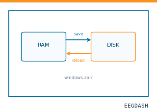 Save and reload prepared dataCache a windowed dataset to disk and reattach it without recompute.
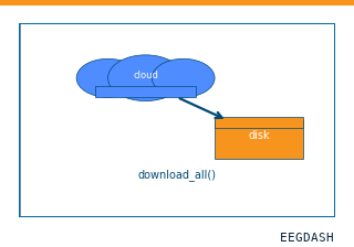 Download a dataset locallyPrefetch BIDS files to a local cache and validate the layout.
Swap any load_dataset(...) call for nm000355 to reproduce the tutorial on this dataset.
Citation
Arjen Stolk, Sandon Griffin, Roemer van der Meij, Callum Dewar, Ignacio Saez, … (6502). Stolk et al. 2018 FieldTrip iEEG protocol dataset (SubjectUCI29): authors' preprocessed ECoG+SEEG epochs. 10.82901/nemar.nm000355
Provenance
¹Contributed to nemar in BIDS format.
²Curated & ingested by the EEGDash catalog; see CITATION.cff for canonical reference.
³Persistent identifier: 10.82901/nemar.nm000355.
See Also#
eegdash.dataset.EEGDashDataseteegdash.dataset