


NM000361: ieeg dataset, 71 subjects#
MNI Open iEEG Atlas: high-sampling-rate NREM sleep sEEG (normal high-frequency oscillations)
Access recordings and metadata through EEGDash.
Citation: Birgit Frauscher, Nicolás von Ellenrieder, Rina Zelmann, Christine Rogers, Dang Khoa Nguyen, Philippe Kahane, François Dubeau, Jean Gotman (1152). MNI Open iEEG Atlas: high-sampling-rate NREM sleep sEEG (normal high-frequency oscillations). 10.82901/nemar.nm000361
Modality: ieeg Subjects: 71 Recordings: 71 License: CC-BY-NC-4.0 Source: nemar
Metadata: Complete (100%)
71-participant iEEG dataset — MNI Open iEEG Atlas: high-sampling-rate NREM sleep sEEG (normal high-frequency oscillations).
Quickstart#
Install
pip install eegdash
Access the data
from eegdash.dataset import NM000361

dataset = NM000361(cache_dir="./data")
# Get the raw object of the first recording
raw = dataset.datasets[0].raw
print(raw.info)
Filter by subject
dataset = NM000361(cache_dir="./data", subject="01")
Advanced query
dataset = NM000361(
    cache_dir="./data",
    query={"subject": {"$in": ["01", "02"]}},
)
Iterate recordings
for rec in dataset:
    print(rec.subject, rec.raw.info['sfreq'])
If you use this dataset in your research, please cite the original authors.
BibTeX
@dataset{nm000361,
  title = {MNI Open iEEG Atlas: high-sampling-rate NREM sleep sEEG (normal high-frequency oscillations)},
  author = {Birgit Frauscher and Nicolás von Ellenrieder and Rina Zelmann and Christine Rogers and Dang Khoa Nguyen and Philippe Kahane and François Dubeau and Jean Gotman},
  doi = {10.82901/nemar.nm000361},
  url = {https://doi.org/10.82901/nemar.nm000361},
}
About This Dataset#
Stereo-EEG during non-REM sleep (N2/N3) at the original sampling rates (512, 1024 and 2000 Hz) from
71 patients, as published in the MNI Open iEEG Atlas for the normative high-frequency oscillation (HFO) study (Frauscher et al. 2018, Ann Neurol). The source calls these “the raw data of physiological sEEG recordings used to analyze HFOs”. The processed 200 Hz atlas clips (wakefulness, N2, N3, REM) are deposited separately as a derivative dataset (“MNI Open iEEG Atlas: normal intracranial EEG in wakefulness and sleep”).
The HFO paper (Frauscher et al. 2018, Ann Neurol 84:374, abstract) used “Intracerebral stereo-encephalographic
recordings with channels displaying normal physiological activity from nonlesional tissue […] from 2 tertiary epilepsy centers. Twenty-minute sections from N2/N3 sleep were selected for automatic detection of ripples (80-250Hz), fast ripples (>250Hz), and HFA defined as long-lasting activity > 80Hz. Normative values are provided for 17 brain regions.” and reports “A total of 1,171 bipolar channels with normal physiological activity from 71 patients”. This dataset contains 1152 bipolar channels from 71 patients (575 + 35 + 542, see Files), i.e. the channel set released by the authors.
MNI Open iEEG Atlas: high-sampling-rate NREM sleep sEEG (normal high-frequency oscillations)
Overview
License
CC-BY-NC-4.0
View full README
MNI Open iEEG Atlas: high-sampling-rate NREM sleep sEEG (normal high-frequency oscillations)
Overview
License
CC-BY-NC-4.0
Cohort
Patients with drug-resistant focal epilepsy investigated with intracranial EEG at three tertiary epilepsy
centres (Frauscher et al. 2018). Only channels judged by the authors to record normal activity (outside the
epileptic zone and lesions, in grey matter) are included. participants.tsv holds only what the source
publishes: the source patient number (1-110; participant label sub-<3-digit number>), sex and age at time
of study (PatientInformation.csv), and the recording-centre code (first character of the source channel
names: G, M or N; the source does not name the centres). No other clinical information exists in the source
and none was added.
- Patients: 71 (35 with centre code G, 36 with centre code M; participants.tsv). The HFO paper abstract states
that the recordings come from 2 tertiary epilepsy centres.
Centres of the MNI Open iEEG Atlas (Frauscher et al. 2018, Brain 141:1130, Methods, “Selection of intracranial EEG recordings”): Montreal Neurological Institute and Hospital (MNI), Centre Hospitalier de l’Université de Montréal (CHUM) and Grenoble-Alpes University Hospital (CHUGA). The source does not state which centre code (G, M, N) corresponds to which centre, so no mapping is given here.
Implant: stereo-EEG depth electrodes only in this dataset (source groups “DIXI electrodes or MNI Home Made (HM) electrodes”, readme_HighSamplingRate.txt).
Selection criteria of the atlas (Frauscher et al. 2018, Brain, Methods): “A channel with normal activity is defined as a channel localized in normal tissue as assessed by MRI, is located outside the seizure onset zone, does not show at any time of the circadian cycle interictal epileptic discharges (according to the clinical report of the complete implantation and to a careful investigation of one night of sleep by a board-certified electrophysiologist), and shows the absence of overt slow-wave anomaly”; contacts in white matter were excluded; recordings had to be obtained “after a minimum of 72 h after insertion of stereo-EEG electrodes or 1 week after placement of subdural grids or strips […] and at least 12 h after a generalized tonic-clonic seizure, 6 h in case of focal clinical seizures, or 2 h in case of purely electrographic seizures, and not after electrical stimulation”. Patients with large cortical malformations were excluded.
Recording and task
All task-sleepNREM (N2/N3 sleep; the paper used twenty-minute sections of N2/N3 sleep). There is no task or
stimulus; the recordings are spontaneous sleep during clinical monitoring.
- Sampling rates (original, not resampled): 512 Hz (32 patients), 1024 Hz (3 patients), 2000 Hz (36 patients).
- Reference: every channel is a bipolar derivation between adjacent contacts of one electrode (source: “one
bipolar channel per column”).
Power-line frequency per the atlas readme (readme_MNI_Open_iEEG_Atlas.txt): “50 Hz for channels with name beginning with ‘G’, and 60 Hz for channels beginning with ‘M’ or ‘N’” (
PowerLineFrequencyin*_ieeg.json).Amplifier/acquisition system and hardware filters: not stated in the released readmes; the full text of the HFO paper was not accessible to us, so these fields are left
n/a.
Files
| acq | Source file (in HFOsInTheNormalHumanBrain.zip) | Electrodes | Subjects | Channels | Samples per file |
|---|---|---|---|---|---|
| hfo512Hz | DIXI-512Hz.mat | DIXI | 32 | 575 | 616448 (1204 s) |
| hfo1024Hz | DIXI-1024Hz.mat | DIXI | 3 | 35 | 1230848 (1202 s) |
| hfo2000Hz | DIXI-2000Hz.mat and HM-2000Hz.mat | DIXI (18 subjects) / MNI homemade (18 subjects) | 36 | 542 | 2406001 / 2406000 |
Per recording:
- *_ieeg.vhdr/.vmrk/.eeg: BrainVision, INT_16 samples with resolution = source Gain (see Units below).
- *_ieeg.json: recording metadata (sampling rate, power-line frequency, reference, electrode manufacturer,
source file and scaling).
*_channels.tsv: one row per bipolar channel (source names), type SEEG, units µV.*_space-Talairach_electrodes.tsv+*_coordsystem.json: one row per bipolar channel at the midpoint of its two contacts, with hemisphere, region number/name (17 regions) and electrode type.*_events.tsv/.json: artifact-free sections, zero buffers and end padding (see Known caveats).sub-*_scans.tsv: list of recordings (no acquisition times exist).sourcedata/document_repository/: the original release files, byte-identical, with SHA-256 checksums.
Units: values are the source int16 samples stored as BrainVision INT_16 with resolution = the source Gain
(0.0977 µV per unit for the DIXI 512/1024 Hz files, 0.1526 µV per unit for the 2000 Hz files): readme: “In order
to obtain the recordings in uV this matrix should be multiplied by the Gain variable.” Nothing was filtered or
resampled by us. The readme does not describe any filtering by the source.
Coordinates: Talairach, readme: “Position: Talairach coordinates of the channels (midpoint of bipolar
channels), in mm.” (space-Talairach). Regions: 17 regions (RegionList in each .mat file, the
labels.nii/template.nii volumes in the source zip). The readme states that the NifTi files “are modified
versions of an existing ATLAS (Landman BA, Warfield SK, editors. MICCAI 2012 Workshop on Multi-Atlas Labeling
[…]). They were non-linearly registered to the ICBM152 2009c nonlinear symmetric brain model”. The HFO detector
used in the paper (HFODetector.m) is in the source zip; a later version is on Zenodo (doi:10.5281/zenodo.7191089,
CC BY 4.0).
Channels and coordinates
Every channel is a bipolar derivation between adjacent contacts of one electrode. electrodes.tsv has one
row per bipolar channel, positioned at the midpoint of its two contacts, coordinates copied unchanged from
the source. Channel names are the source names. Channel type SEEG for depth electrodes (source types D = DIXI,
M = MNI homemade, A = AdTech), ECOG for subdural strips/grids (type G = AdTech).
Preprocessing applied by the source
The release readme for this set (readme_HighSamplingRate.txt) describes the data as raw sEEG recordings stored as int16 with a gain and does not describe filtering, resampling or re-referencing beyond the bipolar derivation. The selection of artifact-free sections and their concatenation with zero buffers is visible in the samples (see Known caveats). The 200 Hz preprocessing described in readme_MNI_Open_iEEG_Atlas.txt (anti-alias filter, resampling, power-line removal, mean removal) applies to the separate atlas dataset, not to these files.
Known caveats
Segments: the files contain all-channel exact-zero runs of exactly one second between sections and zero
padding at the end. This is not described in readme_HighSamplingRate.txt; it is detected from the samples and
listed in *_events.tsv (artifact_free_segment, zero_buffer_between_segments, zero_padding_end). Files
with more than one section set RecordingType: discontinuous; consecutive sections were not necessarily
contiguous in the original recording. Onsets are relative to the file start; no acquisition times exist.
Patient numbers are shared with the atlas (see participants.json). - The sleep stage of each section is given only as “N2/N3”: the source does not separate N2 from N3 in these files. - The channel count (1152) differs from the 1,171 channels analysed in the paper abstract; the source does not
explain the difference.
How to load
from mne_bids import BIDSPath, read_raw_bids
bp = BIDSPath(root=".", subject="002", task="sleepNREM", acquisition="hfo512Hz",
              datatype="ieeg", suffix="ieeg", extension=".vhdr")

raw = read_raw_bids(bp)          # bipolar SEEG channels (MNE stores volts)
events = raw.annotations         # artifact-free sections, zero buffers, end padding
Exclude zero_buffer_between_segments and zero_padding_end intervals before analysis (for example by cropping
to the artifact_free_segment rows of *_events.tsv).
How to cite
Cite all papers that describe the data you use: - Frauscher B, von Ellenrieder N, Zelmann R, Doležalová I, Minotti L, Olivier A, Hall J, Hoffmann D, Nguyen DK, Kahane P, Dubeau F, Gotman J. Atlas of the normal intracranial electroencephalogram: neurophysiological awake activity in different cortical areas. Brain 2018;141(4):1130-1144. doi:10.1093/brain/awy035 - Frauscher B, von Ellenrieder N, Zelmann R, Rogers C, Nguyen DK, Kahane P, Dubeau F, Gotman J. High-Frequency Oscillations in the Normal Human Brain. Ann Neurol 2018;84(3):374-385. doi:10.1002/ana.25304 - von Ellenrieder N, Gotman J, Zelmann R, Rogers C, Nguyen DK, Kahane P, Dubeau F, Frauscher B. How the Human Brain Sleeps: Direct Cortical Recordings of Normal Brain Activity. Ann Neurol 2020;87(2):289-301. doi:10.1002/ana.25651
Source: MNI Open iEEG Atlas, Montreal Neurological Institute, https://mni-open-ieegatlas.research.mcgill.ca/
Ethics
Frauscher et al. 2018 (Brain 141:1130), Methods: “Ethical approval was granted at the MNI as lead ethics organization (REB vote: MUHC-15-950).” The data were recorded during clinical presurgical evaluation of drug-resistant focal epilepsy at three tertiary epilepsy centres. The HFO patients are patients of the same atlas (shared patient numbers).
Funding
Frauscher et al. 2018 (Brain 141:1130), Funding: “This work was supported by the Savoy Epilepsy Foundation (project grant to B.F. and post-doctoral fellowship to R.Z.), the Botterell Powell’s Foundation (grant to B.F.), and the Canadian Institute of Health Research (grant FDN-143208 to J.G.).” The Crossref funder record of the HFO paper (doi:10.1002/ana.25304) lists the same funders (CIHR award FDN-143208) plus the Fonds de la Recherche en Santé du Québec.
Privacy
The source files were already anonymised by the authors (EDF headers carry “X” placeholders and the date
01-JAN-1970; no names, birth dates or recording dates). A byte-level review of every converted file and of
sourcedata/ found no participant identifiers. The only personal names are author attributions (the
HFODetector.m author line and the PDF author field). The NIfTI volumes are brain templates, not participant
images.
Conversion and provenance
Conversion (iEEG-NEMAR campaign, lane F, 2026-10-06) is a lossless repack: every sample in the BrainVision
.eeg files is bit-identical to the source arrays (606/606 recordings verified, raw bytes compared). No
filtering, resampling, re-referencing, rescaling or channel selection was done. Channels absent in a stage
(NaN columns in the source) are omitted from that recording, never filled. All original files are kept
byte-identical under sourcedata/ with SHA-256 checksums.
Source: MNI Open iEEG Atlas document repository (https://mni-open-ieegatlas.research.mcgill.ca/),
HFOsInTheNormalHumanBrain.zip with readme_HighSamplingRate.txt, downloaded 2026-10-06.
NEMAR Metadata#
[](https://doi.org/10.82901/nemar.nm000361) # MNI Open iEEG Atlas: high-sampling-rate NREM sleep sEEG (normal high-frequency oscillations) ## Overview Stereo-EEG during non-REM sleep (N2/N3) at the original sampling rates (512, 1024 and 2000 Hz) from 71 patients, as published in the MNI Open iEEG Atlas for the normative high-frequency oscillation (HFO) study (Frauscher et al. 2018, Ann Neurol). The source calls these “the raw data of physiological sEEG recordings used to analyze HFOs”. The processed 200 Hz atlas clips (wakefulness, N2, N3, REM) are deposited separately as a derivative dataset (“MNI Open iEEG Atlas: normal intracranial EEG in wakefulness and sleep”). The HFO paper (Frauscher et al. 2018, Ann Neurol 84:374, abstract) used “Intracerebral stereo-encephalographic recordings with channels displaying normal physiological activity from nonlesional tissue […] from 2 tertiary epilepsy centers. Twenty-minute sections from N2/N3 sleep were selected for automatic detection of ripples (80-250Hz), fast ripples (>250Hz), and HFA defined as long-lasting activity > 80Hz. Normative values are provided for 17 brain regions.” and reports “A total of 1,171 bipolar channels with normal physiological activity from 71 patients”. This dataset contains 1152 bipolar channels from 71 patients (575 + 35 + 542, see Files), i.e. the channel set released by the authors. ## License CC-BY-NC-4.0 ## Cohort Patients with drug-resistant focal epilepsy investigated with intracranial EEG at three tertiary epilepsy centres (Frauscher et al. 2018). Only channels judged by the authors to record normal activity (outside the epileptic zone and lesions, in grey matter) are included. participants.tsv holds only what the source publishes: the source patient number (1-110; participant label sub-<3-digit number>), sex and age at time of study (PatientInformation.csv), and the recording-centre code (first character of the source channel names: G, M or N; the source does not name the centres). No other clinical information exists in the source and none was added. - Patients: 71 (35 with centre code G, 36 with centre code M; participants.tsv). The HFO paper abstract states
that the recordings come from 2 tertiary epilepsy centres.
Centres of the MNI Open iEEG Atlas (Frauscher et al. 2018, Brain 141:1130, Methods, “Selection of intracranial EEG recordings”): Montreal Neurological Institute and Hospital (MNI), Centre Hospitalier de l’Université de Montréal (CHUM) and Grenoble-Alpes University Hospital (CHUGA). The source does not state which centre code (G, M, N) corresponds to which centre, so no mapping is given here.
Implant: stereo-EEG depth electrodes only in this dataset (source groups “DIXI electrodes or MNI Home Made (HM) electrodes”, readme_HighSamplingRate.txt).
Selection criteria of the atlas (Frauscher et al. 2018, Brain, Methods): “A channel with normal activity is defined as a channel localized in normal tissue as assessed by MRI, is located outside the seizure onset zone, does not show at any time of the circadian cycle interictal epileptic discharges (according to the clinical report of the complete implantation and to a careful investigation of one night of sleep by a board-certified electrophysiologist), and shows the absence of overt slow-wave anomaly”; contacts in white matter were excluded; recordings had to be obtained “after a minimum of 72 h after insertion of stereo-EEG electrodes or 1 week after placement of subdural grids or strips […] and at least 12 h after a generalized tonic-clonic seizure, 6 h in case of focal clinical seizures, or 2 h in case of purely electrographic seizures, and not after electrical stimulation”. Patients with large cortical malformations were excluded.
## Recording and task All task-sleepNREM (N2/N3 sleep; the paper used twenty-minute sections of N2/N3 sleep). There is no task or stimulus; the recordings are spontaneous sleep during clinical monitoring. - Sampling rates (original, not resampled): 512 Hz (32 patients), 1024 Hz (3 patients), 2000 Hz (36 patients). - Reference: every channel is a bipolar derivation between adjacent contacts of one electrode (source: “one
bipolar channel per column”).
Power-line frequency per the atlas readme (readme_MNI_Open_iEEG_Atlas.txt): “50 Hz for channels with name beginning with ‘G’, and 60 Hz for channels beginning with ‘M’ or ‘N’” (PowerLineFrequency in *_ieeg.json).
Amplifier/acquisition system and hardware filters: not stated in the released readmes; the full text of the HFO paper was not accessible to us, so these fields are left n/a.
## Files | acq | Source file (in HFOsInTheNormalHumanBrain.zip) | Electrodes | Subjects | Channels | Samples per file | |---|—|---|—|---|—| | hfo512Hz | DIXI-512Hz.mat | DIXI | 32 | 575 | 616448 (1204 s) | | hfo1024Hz | DIXI-1024Hz.mat | DIXI | 3 | 35 | 1230848 (1202 s) | | hfo2000Hz | DIXI-2000Hz.mat and HM-2000Hz.mat | DIXI (18 subjects) / MNI homemade (18 subjects) | 36 | 542 | 2406001 / 2406000 | Per recording: - *_ieeg.vhdr/.vmrk/.eeg: BrainVision, INT_16 samples with resolution = source Gain (see Units below). - *_ieeg.json: recording metadata (sampling rate, power-line frequency, reference, electrode manufacturer,
source file and scaling).
*_channels.tsv: one row per bipolar channel (source names), type SEEG, units µV.
*_space-Talairach_electrodes.tsv + *_coordsystem.json: one row per bipolar channel at the midpoint of its two contacts, with hemisphere, region number/name (17 regions) and electrode type.
*_events.tsv/.json: artifact-free sections, zero buffers and end padding (see Known caveats).
sub-*_scans.tsv: list of recordings (no acquisition times exist).
sourcedata/document_repository/: the original release files, byte-identical, with SHA-256 checksums.
Units: values are the source int16 samples stored as BrainVision INT_16 with resolution = the source Gain (0.0977 µV per unit for the DIXI 512/1024 Hz files, 0.1526 µV per unit for the 2000 Hz files): readme: “In order to obtain the recordings in uV this matrix should be multiplied by the Gain variable.” Nothing was filtered or resampled by us. The readme does not describe any filtering by the source. Coordinates: Talairach, readme: “Position: Talairach coordinates of the channels (midpoint of bipolar channels), in mm.” (space-Talairach). Regions: 17 regions (RegionList in each .mat file, the labels.nii/template.nii volumes in the source zip). The readme states that the NifTi files “are modified versions of an existing ATLAS (Landman BA, Warfield SK, editors. MICCAI 2012 Workshop on Multi-Atlas Labeling […]). They were non-linearly registered to the ICBM152 2009c nonlinear symmetric brain model”. The HFO detector used in the paper (HFODetector.m) is in the source zip; a later version is on Zenodo (doi:10.5281/zenodo.7191089, CC BY 4.0). ## Channels and coordinates Every channel is a bipolar derivation between adjacent contacts of one electrode. electrodes.tsv has one row per bipolar channel, positioned at the midpoint of its two contacts, coordinates copied unchanged from the source. Channel names are the source names. Channel type SEEG for depth electrodes (source types D = DIXI, M = MNI homemade, A = AdTech), ECOG for subdural strips/grids (type G = AdTech). ## Preprocessing applied by the source The release readme for this set (readme_HighSamplingRate.txt) describes the data as raw sEEG recordings stored as int16 with a gain and does not describe filtering, resampling or re-referencing beyond the bipolar derivation. The selection of artifact-free sections and their concatenation with zero buffers is visible in the samples (see Known caveats). The 200 Hz preprocessing described in readme_MNI_Open_iEEG_Atlas.txt (anti-alias filter, resampling, power-line removal, mean removal) applies to the separate atlas dataset, not to these files. ## Known caveats Segments: the files contain all-channel exact-zero runs of exactly one second between sections and zero padding at the end. This is not described in readme_HighSamplingRate.txt; it is detected from the samples and listed in *_events.tsv (artifact_free_segment, zero_buffer_between_segments, zero_padding_end). Files with more than one section set RecordingType: discontinuous; consecutive sections were not necessarily contiguous in the original recording. Onsets are relative to the file start; no acquisition times exist. Patient numbers are shared with the atlas (see participants.json). - The sleep stage of each section is given only as “N2/N3”: the source does not separate N2 from N3 in these files. - The channel count (1152) differs from the 1,171 channels analysed in the paper abstract; the source does not
explain the difference.
## How to load ```python from mne_bids import BIDSPath, read_raw_bids bp = BIDSPath(root=”.”, subject=”002”, task=”sleepNREM”, acquisition=”hfo512Hz”,
datatype=”ieeg”, suffix=”ieeg”, extension=”.vhdr”)
raw = read_raw_bids(bp) # bipolar SEEG channels (MNE stores volts) events = raw.annotations # artifact-free sections, zero buffers, end padding ``` Exclude zero_buffer_between_segments and zero_padding_end intervals before analysis (for example by cropping to the artifact_free_segment rows of *_events.tsv). ## How to cite Cite all papers that describe the data you use: - Frauscher B, von Ellenrieder N, Zelmann R, Doležalová I, Minotti L, Olivier A, Hall J, Hoffmann D, Nguyen DK, Kahane P, Dubeau F, Gotman J. Atlas of the normal intracranial electroencephalogram: neurophysiological awake activity in different cortical areas. Brain 2018;141(4):1130-1144. doi:10.1093/brain/awy035 - Frauscher B, von Ellenrieder N, Zelmann R, Rogers C, Nguyen DK, Kahane P, Dubeau F, Gotman J. High-Frequency Oscillations in the Normal Human Brain. Ann Neurol 2018;84(3):374-385. doi:10.1002/ana.25304 - von Ellenrieder N, Gotman J, Zelmann R, Rogers C, Nguyen DK, Kahane P, Dubeau F, Frauscher B. How the Human Brain Sleeps: Direct Cortical Recordings of Normal Brain Activity. Ann Neurol 2020;87(2):289-301. doi:10.1002/ana.25651 Source: MNI Open iEEG Atlas, Montreal Neurological Institute, https://mni-open-ieegatlas.research.mcgill.ca/ ## Ethics Frauscher et al. 2018 (Brain 141:1130), Methods: “Ethical approval was granted at the MNI as lead ethics organization (REB vote: MUHC-15-950).” The data were recorded during clinical presurgical evaluation of drug-resistant focal epilepsy at three tertiary epilepsy centres. The HFO patients are patients of the same atlas (shared patient numbers). ## Funding Frauscher et al. 2018 (Brain 141:1130), Funding: “This work was supported by the Savoy Epilepsy Foundation (project grant to B.F. and post-doctoral fellowship to R.Z.), the Botterell Powell’s Foundation (grant to B.F.), and the Canadian Institute of Health Research (grant FDN-143208 to J.G.).” The Crossref funder record of the HFO paper (doi:10.1002/ana.25304) lists the same funders (CIHR award FDN-143208) plus the Fonds de la Recherche en Santé du Québec. ## Privacy The source files were already anonymised by the authors (EDF headers carry “X” placeholders and the date 01-JAN-1970; no names, birth dates or recording dates). A byte-level review of every converted file and of sourcedata/ found no participant identifiers. The only personal names are author attributions (the HFODetector.m author line and the PDF author field). The NIfTI volumes are brain templates, not participant images. ## Conversion and provenance Conversion (iEEG-NEMAR campaign, lane F, 2026-10-06) is a lossless repack: every sample in the BrainVision .eeg files is bit-identical to the source arrays (606/606 recordings verified, raw bytes compared). No filtering, resampling, re-referencing, rescaling or channel selection was done. Channels absent in a stage (NaN columns in the source) are omitted from that recording, never filled. All original files are kept byte-identical under sourcedata/ with SHA-256 checksums. Source: MNI Open iEEG Atlas document repository (https://mni-open-ieegatlas.research.mcgill.ca/), HFOsInTheNormalHumanBrain.zip with readme_HighSamplingRate.txt, downloaded 2026-10-06.
License: CC-BY-NC-4.0
Authors:
Birgit Frauscher
Nicolás von Ellenrieder
Rina Zelmann
Christine Rogers
Dang Khoa Nguyen
… and 3 more
Versions:
Version |
DOI |
Released |
|---|---|---|
|
Cohort#
Dataset Statistics#
Age distribution by gender (n=71, range 14–57 yr, mean 34.0 yr)
Sex composition
Channel counts (ch)
Sampling frequencies (Hz)
Total recording duration: 23 h 44 min
Signal · Electrodes & live trace#
Live trace viewer — sub-031 · task-sleepNREM
Showing one representative recording out of
71 subjects and 71 recordings in this dataset.
Browse the full set on OpenNeuro;
drop any other _ieeg.{set,edf,bdf,vhdr} file onto the
viewer (or pass ?ieeg=<url>) to inspect it.
Electrode layout — iEEG · 18 sensors — 18 channels
NEMAR Processing Statistics#
The plots below are generated by NEMAR’s automated EEG pipeline. The histogram shows pipeline success for data cleaning and ICA decomposition, the percentage of data frames and EEG channels retained after artefact removal, line noise per channel (RMS, dB), and the age/gender distribution of participants.

HED event descriptors word cloud

Manifest#
File Explorer#
Browse the BIDS file structure of this dataset. Records are fetched on demand from the EEGDash catalog the first time you open the explorer.
Full dataset metadata table
Dataset ID |
|
Title |
MNI Open iEEG Atlas: high-sampling-rate NREM sleep sEEG (normal high-frequency oscillations) |
Author (year) |
— |
Canonical |
— |
Importable as |
|
Year |
1152 |
Authors |
Birgit Frauscher, Nicolás von Ellenrieder, Rina Zelmann, Christine Rogers, Dang Khoa Nguyen, Philippe Kahane, François Dubeau, Jean Gotman |
License |
CC-BY-NC-4.0 |
Citation / DOI |
|
Source links |
OpenNeuro | NeMAR | Source URL |
Copy-paste BibTeX
@dataset{nm000361,
  title = {MNI Open iEEG Atlas: high-sampling-rate NREM sleep sEEG (normal high-frequency oscillations)},
  author = {Birgit Frauscher and Nicolás von Ellenrieder and Rina Zelmann and Christine Rogers and Dang Khoa Nguyen and Philippe Kahane and François Dubeau and Jean Gotman},
  doi = {10.82901/nemar.nm000361},
  url = {https://doi.org/10.82901/nemar.nm000361},
}
API Reference#
eegdash.datasetEEGDashDataset- class eegdash.dataset.NM000361(cache_dir: str, query: dict | None = None, s3_bucket: str | None = None, **kwargs)[source]#
MNI Open iEEG Atlas: high-sampling-rate NREM sleep sEEG (normal high-frequency oscillations)
- Study:
nm000361(NeMAR)- Author (year):
—
- Canonical:
—
Also importable as:
NM000361.Modality:
ieeg; Subject type:Unknown. Subjects: 71; recordings: 71; tasks: 1.- Parameters:
cache_dir (str | Path) – Directory where data are cached locally.
query (dict | None) – Additional MongoDB-style filters to AND with the dataset selection. Must not contain the key
dataset.s3_bucket (str | None) – Base S3 bucket used to locate the data.
**kwargs (dict) – Additional keyword arguments forwarded to
EEGDashDataset.
- data_dir#
Local dataset cache directory (
cache_dir / dataset_id).- Type:
Path
Notes
Each item is a recording; recording-level metadata are available via
dataset.description.querysupports MongoDB-style filters on fields inALLOWED_QUERY_FIELDSand is combined with the dataset filter. Dataset-specific caveats are not provided in the summary metadata.References
OpenNeuro dataset: https://openneuro.org/datasets/nm000361 NeMAR dataset: https://nemar.org/dataexplorer/detail?dataset_id=nm000361 DOI: https://doi.org/10.82901/nemar.nm000361
Examples
>>> from eegdash.dataset import NM000361
>>> dataset = NM000361(cache_dir="./data")
>>> recording = dataset[0]
>>> raw = recording.load()
- __init__(cache_dir: str, query: dict | None = None, s3_bucket: str | None = None, **kwargs)[source]#
- save(path: str, overwrite: bool = False, offset: int = 0)[source]#
Save datasets to files by creating one subdirectory for each dataset:
path/
    0/
        0-raw.fif | 0-epo.fif
        description.json
        raw_preproc_kwargs.json (if raws were preprocessed)
        window_kwargs.json (if this is a windowed dataset)
        window_preproc_kwargs.json  (if windows were preprocessed)
        target_name.json (if target_name is not None and dataset is raw)
    1/
        1-raw.fif | 1-epo.fif
        description.json
        raw_preproc_kwargs.json (if raws were preprocessed)
        window_kwargs.json (if this is a windowed dataset)
        window_preproc_kwargs.json  (if windows were preprocessed)
        target_name.json (if target_name is not None and dataset is raw)
- Parameters:
path (str) –
- Directory in which subdirectories are created to store
-raw.fif | -epo.fif and .json files to.
overwrite (bool) – Whether to delete old subdirectories that will be saved to in this call.
offset (int) – If provided, the integer is added to the id of the dataset in the concat. This is useful in the setting of very large datasets, where one dataset has to be processed and saved at a time to account for its original position.
BaseDataset from braindecode — windowed via create_windows_from_events.braindecodeDataLoader; supports parallel workers and on-the-fly augmentations.pytorch Find datasets with the EEGDash APIQuery the catalogue, filter by task or modality, list candidates.
 Load one EEG recordingResolve a single record to an MNE Raw with channels and events.
 EEG recording to PyTorch DataLoaderWrap braindecode windows in a DataLoader for model training.
 Preprocess EEG and create windowsFilter, resample, epoch — and persist the windowed dataset.
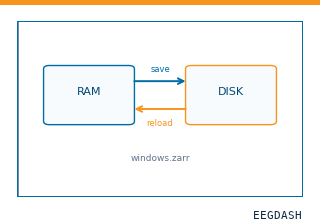 Save and reload prepared dataCache a windowed dataset to disk and reattach it without recompute.
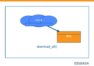 Download a dataset locallyPrefetch BIDS files to a local cache and validate the layout.
Swap any load_dataset(...) call for nm000361 to reproduce the tutorial on this dataset.
Citation
Birgit Frauscher, Nicolás von Ellenrieder, Rina Zelmann, Christine Rogers, Dang Khoa Nguyen, … (1152). MNI Open iEEG Atlas: high-sampling-rate NREM sleep sEEG (normal high-frequency oscillations). 10.82901/nemar.nm000361
Provenance
¹Contributed to nemar in BIDS format.
²Curated & ingested by the EEGDash catalog; see CITATION.cff for canonical reference.
³Persistent identifier: 10.82901/nemar.nm000361.
See Also#
eegdash.dataset.EEGDashDataseteegdash.dataset