


NM000412: ieeg dataset, 6 subjects#
ECoG hemispheric asymmetry during reaching (Merrick et al. 2022): authors’ 1000 Hz intracranial exports with reach kinematics
Access recordings and metadata through EEGDash.
Citation: Christina M. Merrick, Tanner C. Dixon, Assaf Breska, Jack J. Lin, Edward F. Chang, David King-Stephens, Kenneth D. Laxer, Peter B. Weber, Jose M. Carmena, Robert T. Knight, Richard B. Ivry (2022). ECoG hemispheric asymmetry during reaching (Merrick et al. 2022): authors’ 1000 Hz intracranial exports with reach kinematics. 10.82901/nemar.nm000412
Modality: ieeg Subjects: 6 Recordings: 24 License: CC-BY-4.0 Source: nemar
Metadata: Complete (100%)
6-participant iEEG dataset — ECoG hemispheric asymmetry during reaching (Merrick et al. 2022): authors' 1000 Hz intracranial exports with reach kinematics.
Quickstart#
Install
pip install eegdash
Access the data
from eegdash.dataset import NM000412

dataset = NM000412(cache_dir="./data")
# Get the raw object of the first recording
raw = dataset.datasets[0].raw
print(raw.info)
Filter by subject
dataset = NM000412(cache_dir="./data", subject="01")
Advanced query
dataset = NM000412(
    cache_dir="./data",
    query={"subject": {"$in": ["01", "02"]}},
)
Iterate recordings
for rec in dataset:
    print(rec.subject, rec.raw.info['sfreq'])
If you use this dataset in your research, please cite the original authors.
BibTeX
@dataset{nm000412,
  title = {ECoG hemispheric asymmetry during reaching (Merrick et al. 2022): authors' 1000 Hz intracranial exports with reach kinematics},
  author = {Christina M. Merrick and Tanner C. Dixon and Assaf Breska and Jack J. Lin and Edward F. Chang and David King-Stephens and Kenneth D. Laxer and Peter B. Weber and Jose M. Carmena and Robert T. Knight and Richard B. Ivry},
  doi = {10.82901/nemar.nm000412},
  url = {https://doi.org/10.82901/nemar.nm000412},
}
About This Dataset#
Intracranial recordings from six patients with intractable epilepsy implanted with subdural grids, three with
left-hemisphere implants (L1-L3) and three with right-hemisphere implants (R4-R6). They were recorded at the University of California, Irvine, the University of California, San Francisco Medical Center and California Pacific Medical Center, San Francisco, two patients per site. Patients performed an instructed-delay reaching task on a touchscreen, with two blocks of 40 trials for each arm, in counterbalanced order. Re-packaged from the authors’ public release (Zenodo record 4761390, “ECoG_Hemispheric_Asymmetry_dataset”, CC-BY-4.0).
Reference article: Merrick CM, Dixon TC, Breska A, Lin J, Chang EF, King-Stephens D, Laxer KD, Weber PB, Carmena J,
Knight RT, Ivry RB. Left hemisphere dominance for bilateral kinematic encoding in the human brain. eLife 11:e69977 (2022). https://doi.org/10.7554/eLife.69977. Code: cmerrick15/Asymmetry_ECoG_dataset
ECoG hemispheric asymmetry during reaching (Merrick et al. 2022): authors’ 1000 Hz exports
This is a derivative dataset
The neural arrays in the release are the authors’ exports at **1000 Hz**. The paper says UCI recorded at 5000 Hz
(Nihon Kohden), UCSF at 3052 Hz (Tucker Davis Technologies) and CPMC at 1000 Hz (Nihon Kohden). The UCI and UCSF data
were low-pass filtered at 500 Hz and downsampled to 1000 Hz. Channels that the release metadata lists as bad
(SDATA.metadata.bad_electrodes) are already absent from the arrays. The release does not say whether its exports also
View full README
ECoG hemispheric asymmetry during reaching (Merrick et al. 2022): authors’ 1000 Hz exports
This is a derivative dataset
The neural arrays in the release are the authors’ exports at **1000 Hz**. The paper says UCI recorded at 5000 Hz
(Nihon Kohden), UCSF at 3052 Hz (Tucker Davis Technologies) and CPMC at 1000 Hz (Nihon Kohden). The UCI and UCSF data
were low-pass filtered at 500 Hz and downsampled to 1000 Hz. Channels that the release metadata lists as bad
(SDATA.metadata.bad_electrodes) are already absent from the arrays. The release does not say whether its exports also
include the paper’s common-average reference and 60/120/180 Hz notch filters. The dataset is therefore labelled
DatasetType: derivative. The packaging itself changes no values.
Layout
sub-L1…``sub-R6`` = release patient IDs.task-reach,acq-lefthand/acq-righthand= arm used,run-1/run-2= block.*_ieeg.vhdr:SDATA.data(float32 BrainVision; values are the stored float64 values rounded to float32). Channel names are as inSDATA.info.channel_labels. The release does not give units; the magnitudes differ by site (seetask-reach_channels.json).*_recording-kinematics_physio.tsv.gz: the releasekin_mat(position, speed, x, y, z, theta, phi) at 100 Hz. The paper estimated the movement profiles from the button and touch times.StartTime0 assumes that the kinematics start with the neural export: the lengths agree at 10:1 to within one sample, but the release does not state the alignment.The release holds no trial events: the
SDATA.eventsstructures are empty, and R5’s is an undocumented MATLAB table object. The photodiode and microswitch analog channels (SDATA.analog, 4 channels at 24414 Hz for UCSF, 5000 Hz for UCI L3 and 1000 Hz for CPMC; absent for R4) remain only in the source files insourcedata/.participants.tsv: age and handedness from Table 1 for L1-L3. Table 1 names the right-hemisphere patients R1-R3 (ages 26, 32 and 34, all right-handed), but the release uses R4-R6 and does not state the correspondence, so R4-R6 aren/a. Sex is given only at group level (two of six female).recording_siteis inferred from the release session codes (EC = UCSF, IR = UCI, CP = CPMC) and the analog sampling rates, which matches the paper’s two patients per site.Channel types: the release types electrodes only for L1 and L2 (grid / strip). Elsewhere all channels are typed ECOG (see
task-reach_channels.json). There are no electrode coordinates in the release.electrodes.tsvlists names withn/acoordinates.
Source files
sourcedata/zenodo-4761390/ holds all 48 release .mat files: the neural SDATA structures, including the analog
channels, the empty events, info.id session codes and, for R4 right-hand block 1, an extra data_ref array. It also
holds the kinematics files and the Zenodo record metadata. The only change is that the MATLAB header text “Created on”
(file export date, 2019-2021) has its weekday and day masked. The provenance JSON gives the SHA-256 of each file before
and after masking.
License
CC-BY-4.0
Additional metadata and localisation (added 2026-10-08)
Compiled after the upload from the article, its supplement and the source deposit (each statement names its source). Text and sidecar metadata only; no data file was changed. Recording system. Nihon Kohden at UCI (128 ch, 5000 Hz) and CPMC (128 ch, 1000 Hz); Tucker Davis Technologies at UCSF (128 ch, 3052 Hz); signals not at 1000 Hz were low-pass filtered at 500 Hz and downsampled to 1000 Hz (paper Methods). Deposit SDATA.info.sampling_rate = 1000 for all six; L1/L2 also carry analog_sr 24414.0625 (TDT analog rate). Reference scheme. Common average reference applied in the paper’s preprocessing; notch 60/120/180 Hz (paper Methods). Whether the deposited SDATA.data is pre- or post-CAR is not stated (n/a). Electrode types. Subdural grids and strips; spacing 1 cm in four patients and 4 mm in the other two (paper “Electrode localization”). 752 electrodes in total, 82 removed for noise and 5 for epileptic activity -> 665 analysed (paper Methods). Localisation. Electrode locations visualised on each patient’s 3D cortical reconstruction by co-registering post-operative CT to pre-operative MRI (Stolk et al. 2018 pipeline); each electrode was categorised with the Desikan-Killiany parcellation; pooled proportions are shown per hemisphere in Figure 2-figure supplement 1 (premotor: superior frontal, caudal middle frontal, pars opercularis; sensorimotor: pre-/post-central; parietal: supramarginal, superior parietal, inferior parietal; 70% of electrodes in these parcels). Per-electrode coordinates/labels are not deposited.
Regions per participant (as published; no coordinates exist)
| participant | region (as stated) | hemisphere | contacts | source |
|---|---|---|---|---|
| cohort-left(L1-L3) | Superior frontal; Caudal middle frontal; Opercularis | left | n/a | doi:10.7554/eLife.69977, Figure 2-figure supplement 1 (premotor group |
| cohort-left(L1-L3) | Pre-central; Post-central | left | n/a | doi:10.7554/eLife.69977, Figure 2-figure supplement 1 (sensorimotor) |
| cohort-left(L1-L3) | Supra marginal; Superior parietal; Inferior parietal | left | n/a | doi:10.7554/eLife.69977, Figure 2-figure supplement 1 (parietal) |
| cohort-right(R4-R6) | Superior frontal; Caudal middle frontal; Opercularis | right | n/a | doi:10.7554/eLife.69977, Figure 2-figure supplement 1 |
| cohort-right(R4-R6) | Pre-central; Post-central | right | n/a | doi:10.7554/eLife.69977, Figure 2-figure supplement 1 |
| cohort-right(R4-R6) | Supra marginal; Superior parietal; Inferior parietal | right | n/a | doi:10.7554/eLife.69977, Figure 2-figure supplement 1 |
NEMAR Metadata#
[](https://doi.org/10.82901/nemar.nm000412) # ECoG hemispheric asymmetry during reaching (Merrick et al. 2022): authors’ 1000 Hz exports Intracranial recordings from six patients with intractable epilepsy implanted with subdural grids, three with left-hemisphere implants (L1-L3) and three with right-hemisphere implants (R4-R6). They were recorded at the University of California, Irvine, the University of California, San Francisco Medical Center and California Pacific Medical Center, San Francisco, two patients per site. Patients performed an instructed-delay reaching task on a touchscreen, with two blocks of 40 trials for each arm, in counterbalanced order. Re-packaged from the authors’ public release (Zenodo record [4761390](https://doi.org/10.5281/zenodo.4761390), “ECoG_Hemispheric_Asymmetry_dataset”, CC-BY-4.0). Reference article: Merrick CM, Dixon TC, Breska A, Lin J, Chang EF, King-Stephens D, Laxer KD, Weber PB, Carmena J, Knight RT, Ivry RB. Left hemisphere dominance for bilateral kinematic encoding in the human brain. eLife 11:e69977 (2022). https://doi.org/10.7554/eLife.69977. Code: cmerrick15/Asymmetry_ECoG_dataset ## This is a derivative dataset The neural arrays in the release are the authors’ exports at 1000 Hz. The paper says UCI recorded at 5000 Hz (Nihon Kohden), UCSF at 3052 Hz (Tucker Davis Technologies) and CPMC at 1000 Hz (Nihon Kohden). The UCI and UCSF data were low-pass filtered at 500 Hz and downsampled to 1000 Hz. Channels that the release metadata lists as bad (SDATA.metadata.bad_electrodes) are already absent from the arrays. The release does not say whether its exports also include the paper’s common-average reference and 60/120/180 Hz notch filters. The dataset is therefore labelled DatasetType: derivative. The packaging itself changes no values. ## Layout - sub-L1…`sub-R6` = release patient IDs. task-reach, acq-lefthand / acq-righthand = arm used,
run-1 / run-2 = block.
*_ieeg.vhdr: SDATA.data (float32 BrainVision; values are the stored float64 values rounded to float32). Channel names are as in SDATA.info.channel_labels. The release does not give units; the magnitudes differ by site (see task-reach_channels.json).
*_recording-kinematics_physio.tsv.gz: the release kin_mat (position, speed, x, y, z, theta, phi) at 100 Hz. The paper estimated the movement profiles from the button and touch times. StartTime 0 assumes that the kinematics start with the neural export: the lengths agree at 10:1 to within one sample, but the release does not state the alignment.
The release holds no trial events: the SDATA.events structures are empty, and R5’s is an undocumented MATLAB table object. The photodiode and microswitch analog channels (SDATA.analog, 4 channels at 24414 Hz for UCSF, 5000 Hz for UCI L3 and 1000 Hz for CPMC; absent for R4) remain only in the source files in sourcedata/.
participants.tsv: age and handedness from Table 1 for L1-L3. Table 1 names the right-hemisphere patients R1-R3 (ages 26, 32 and 34, all right-handed), but the release uses R4-R6 and does not state the correspondence, so R4-R6 are n/a. Sex is given only at group level (two of six female). recording_site is inferred from the release session codes (EC = UCSF, IR = UCI, CP = CPMC) and the analog sampling rates, which matches the paper’s two patients per site.
Channel types: the release types electrodes only for L1 and L2 (grid / strip). Elsewhere all channels are typed ECOG (see task-reach_channels.json). There are no electrode coordinates in the release. electrodes.tsv lists names with n/a coordinates.
## Source files sourcedata/zenodo-4761390/ holds all 48 release .mat files: the neural SDATA structures, including the analog channels, the empty events, info.id session codes and, for R4 right-hand block 1, an extra data_ref array. It also holds the kinematics files and the Zenodo record metadata. The only change is that the MATLAB header text “Created on” (file export date, 2019-2021) has its weekday and day masked. The provenance JSON gives the SHA-256 of each file before and after masking. ## License CC-BY-4.0 ## Additional metadata and localisation (added 2026-10-08) Compiled after the upload from the article, its supplement and the source deposit (each statement names its source). Text and sidecar metadata only; no data file was changed. Recording system. Nihon Kohden at UCI (128 ch, 5000 Hz) and CPMC (128 ch, 1000 Hz); Tucker Davis Technologies at UCSF (128 ch, 3052 Hz); signals not at 1000 Hz were low-pass filtered at 500 Hz and downsampled to 1000 Hz (paper Methods). Deposit SDATA.info.sampling_rate = 1000 for all six; L1/L2 also carry analog_sr 24414.0625 (TDT analog rate). Reference scheme. Common average reference applied in the paper’s preprocessing; notch 60/120/180 Hz (paper Methods). Whether the deposited SDATA.data is pre- or post-CAR is not stated (n/a). Electrode types. Subdural grids and strips; spacing 1 cm in four patients and 4 mm in the other two (paper “Electrode localization”). 752 electrodes in total, 82 removed for noise and 5 for epileptic activity -> 665 analysed (paper Methods). Localisation. Electrode locations visualised on each patient’s 3D cortical reconstruction by co-registering post-operative CT to pre-operative MRI (Stolk et al. 2018 pipeline); each electrode was categorised with the Desikan-Killiany parcellation; pooled proportions are shown per hemisphere in Figure 2-figure supplement 1 (premotor: superior frontal, caudal middle frontal, pars opercularis; sensorimotor: pre-/post-central; parietal: supramarginal, superior parietal, inferior parietal; 70% of electrodes in these parcels). Per-electrode coordinates/labels are not deposited. ### Regions per participant (as published; no coordinates exist) | participant | region (as stated) | hemisphere | contacts | source | |---|—|---|—|---| | cohort-left(L1-L3) | Superior frontal; Caudal middle frontal; Opercularis | left | n/a | doi:10.7554/eLife.69977, Figure 2-figure supplement 1 (premotor group | | cohort-left(L1-L3) | Pre-central; Post-central | left | n/a | doi:10.7554/eLife.69977, Figure 2-figure supplement 1 (sensorimotor) | | cohort-left(L1-L3) | Supra marginal; Superior parietal; Inferior parietal | left | n/a | doi:10.7554/eLife.69977, Figure 2-figure supplement 1 (parietal) | | cohort-right(R4-R6) | Superior frontal; Caudal middle frontal; Opercularis | right | n/a | doi:10.7554/eLife.69977, Figure 2-figure supplement 1 | | cohort-right(R4-R6) | Pre-central; Post-central | right | n/a | doi:10.7554/eLife.69977, Figure 2-figure supplement 1 | | cohort-right(R4-R6) | Supra marginal; Superior parietal; Inferior parietal | right | n/a | doi:10.7554/eLife.69977, Figure 2-figure supplement 1 |
License: CC-BY-4.0
Authors:
Christina M. Merrick
Tanner C. Dixon
Assaf Breska
Jack J. Lin
Edward F. Chang
… and 6 more
Versions:
Version |
DOI |
Released |
|---|---|---|
|
Cohort#
Dataset Statistics#
Age distribution by gender (n=3, range 26–55 yr, mean 39.0 yr)
Channel counts (ch)
Sampling frequencies: 1000.0 Hz (n=24 recordings)
Total recording duration: 2 h 7 min
Signal · Electrodes & live trace#
Live trace viewer — sub-L1 · task-reach · run-2
Showing one representative recording out of
6 subjects and 24 recordings in this dataset.
Browse the full set on OpenNeuro;
drop any other _ieeg.{set,edf,bdf,vhdr} file onto the
viewer (or pass ?ieeg=<url>) to inspect it.
No scalp electrode layout is currently indexed for this dataset. Once the eegdash montage registry ingests it, the interactive viewer will appear here automatically.
NEMAR Processing Statistics#
The plots below are generated by NEMAR’s automated EEG pipeline. The histogram shows pipeline success for data cleaning and ICA decomposition, the percentage of data frames and EEG channels retained after artefact removal, line noise per channel (RMS, dB), and the age/gender distribution of participants.

HED event descriptors word cloud

Manifest#
File Explorer#
Browse the BIDS file structure of this dataset. Records are fetched on demand from the EEGDash catalog the first time you open the explorer.
Full dataset metadata table
Dataset ID |
|
Title |
ECoG hemispheric asymmetry during reaching (Merrick et al. 2022): authors’ 1000 Hz intracranial exports with reach kinematics |
Author (year) |
— |
Canonical |
— |
Importable as |
|
Year |
2022 |
Authors |
Christina M. Merrick, Tanner C. Dixon, Assaf Breska, Jack J. Lin, Edward F. Chang, David King-Stephens, Kenneth D. Laxer, Peter B. Weber, Jose M. Carmena, Robert T. Knight, Richard B. Ivry |
License |
CC-BY-4.0 |
Citation / DOI |
|
Source links |
OpenNeuro | NeMAR | Source URL |
Copy-paste BibTeX
@dataset{nm000412,
  title = {ECoG hemispheric asymmetry during reaching (Merrick et al. 2022): authors' 1000 Hz intracranial exports with reach kinematics},
  author = {Christina M. Merrick and Tanner C. Dixon and Assaf Breska and Jack J. Lin and Edward F. Chang and David King-Stephens and Kenneth D. Laxer and Peter B. Weber and Jose M. Carmena and Robert T. Knight and Richard B. Ivry},
  doi = {10.82901/nemar.nm000412},
  url = {https://doi.org/10.82901/nemar.nm000412},
}
API Reference#
eegdash.datasetEEGDashDataset- class eegdash.dataset.NM000412(cache_dir: str, query: dict | None = None, s3_bucket: str | None = None, **kwargs)[source]#
ECoG hemispheric asymmetry during reaching (Merrick et al. 2022): authors’ 1000 Hz intracranial exports with reach kinematics
- Study:
nm000412(NeMAR)- Author (year):
—
- Canonical:
—
Also importable as:
NM000412.Modality:
ieeg; Subject type:Unknown. Subjects: 6; recordings: 24; tasks: 1.- Parameters:
cache_dir (str | Path) – Directory where data are cached locally.
query (dict | None) – Additional MongoDB-style filters to AND with the dataset selection. Must not contain the key
dataset.s3_bucket (str | None) – Base S3 bucket used to locate the data.
**kwargs (dict) – Additional keyword arguments forwarded to
EEGDashDataset.
- data_dir#
Local dataset cache directory (
cache_dir / dataset_id).- Type:
Path
Notes
Each item is a recording; recording-level metadata are available via
dataset.description.querysupports MongoDB-style filters on fields inALLOWED_QUERY_FIELDSand is combined with the dataset filter. Dataset-specific caveats are not provided in the summary metadata.References
OpenNeuro dataset: https://openneuro.org/datasets/nm000412 NeMAR dataset: https://nemar.org/dataexplorer/detail?dataset_id=nm000412 DOI: https://doi.org/10.82901/nemar.nm000412
Examples
>>> from eegdash.dataset import NM000412
>>> dataset = NM000412(cache_dir="./data")
>>> recording = dataset[0]
>>> raw = recording.load()
- __init__(cache_dir: str, query: dict | None = None, s3_bucket: str | None = None, **kwargs)[source]#
- save(path: str, overwrite: bool = False, offset: int = 0)[source]#
Save datasets to files by creating one subdirectory for each dataset:
path/
    0/
        0-raw.fif | 0-epo.fif
        description.json
        raw_preproc_kwargs.json (if raws were preprocessed)
        window_kwargs.json (if this is a windowed dataset)
        window_preproc_kwargs.json  (if windows were preprocessed)
        target_name.json (if target_name is not None and dataset is raw)
    1/
        1-raw.fif | 1-epo.fif
        description.json
        raw_preproc_kwargs.json (if raws were preprocessed)
        window_kwargs.json (if this is a windowed dataset)
        window_preproc_kwargs.json  (if windows were preprocessed)
        target_name.json (if target_name is not None and dataset is raw)
- Parameters:
path (str) –
- Directory in which subdirectories are created to store
-raw.fif | -epo.fif and .json files to.
overwrite (bool) – Whether to delete old subdirectories that will be saved to in this call.
offset (int) – If provided, the integer is added to the id of the dataset in the concat. This is useful in the setting of very large datasets, where one dataset has to be processed and saved at a time to account for its original position.
BaseDataset from braindecode — windowed via create_windows_from_events.braindecodeDataLoader; supports parallel workers and on-the-fly augmentations.pytorch Find datasets with the EEGDash APIQuery the catalogue, filter by task or modality, list candidates.
 Load one EEG recordingResolve a single record to an MNE Raw with channels and events.
 EEG recording to PyTorch DataLoaderWrap braindecode windows in a DataLoader for model training.
 Preprocess EEG and create windowsFilter, resample, epoch — and persist the windowed dataset.
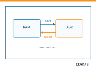 Save and reload prepared dataCache a windowed dataset to disk and reattach it without recompute.
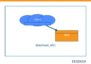 Download a dataset locallyPrefetch BIDS files to a local cache and validate the layout.
Swap any load_dataset(...) call for nm000412 to reproduce the tutorial on this dataset.
Citation
Christina M. Merrick, Tanner C. Dixon, Assaf Breska, Jack J. Lin, Edward F. Chang, … (2022). ECoG hemispheric asymmetry during reaching (Merrick et al. 2022): authors' 1000 Hz intracranial exports with reach kinematics. 10.82901/nemar.nm000412
Provenance
¹Contributed to nemar in BIDS format.
²Curated & ingested by the EEGDash catalog; see CITATION.cff for canonical reference.
³Persistent identifier: 10.82901/nemar.nm000412.
See Also#
eegdash.dataset.EEGDashDataseteegdash.dataset