


NM000377: ieeg dataset, 3 subjects#
Human Responses to Visually Evoked Threat: intracranial EEG during virtual-reality heights, no-heights and shark stimuli (3 patients)
Access recordings and metadata through EEGDash.
Citation: Melis Yilmaz Balban, Erin Cafaro, Lauren Saue-Fletcher, Marlon Joseph Washington, Maryam Bijanzadeh, Andrew Moses Lee, Edward Chang, Andrew Huberman (2021). Human Responses to Visually Evoked Threat: intracranial EEG during virtual-reality heights, no-heights and shark stimuli (3 patients). 10.82901/nemar.nm000377
Modality: ieeg Subjects: 3 Recordings: 8 License: CC0-1.0 Source: nemar
Metadata: Complete (100%)
3-participant iEEG dataset — Human Responses to Visually Evoked Threat: intracranial EEG during virtual-reality heights, no-heights and shark stimuli (3 patients).
Quickstart#
Install
pip install eegdash
Access the data
from eegdash.dataset import NM000377

dataset = NM000377(cache_dir="./data")
# Get the raw object of the first recording
raw = dataset.datasets[0].raw
print(raw.info)
Filter by subject
dataset = NM000377(cache_dir="./data", subject="01")
Advanced query
dataset = NM000377(
    cache_dir="./data",
    query={"subject": {"$in": ["01", "02"]}},
)
Iterate recordings
for rec in dataset:
    print(rec.subject, rec.raw.info['sfreq'])
If you use this dataset in your research, please cite the original authors.
BibTeX
@dataset{nm000377,
  title = {Human Responses to Visually Evoked Threat: intracranial EEG during virtual-reality heights, no-heights and shark stimuli (3 patients)},
  author = {Melis Yilmaz Balban and Erin Cafaro and Lauren Saue-Fletcher and Marlon Joseph Washington and Maryam Bijanzadeh and Andrew Moses Lee and Edward Chang and Andrew Huberman},
  doi = {10.82901/nemar.nm000377},
  url = {https://doi.org/10.82901/nemar.nm000377},
}
About This Dataset#
Intracranial EEG of 3 patients with treatment-resistant epilepsy (UCSF, under the care of Dr. Edward Chang) who
experienced immersive virtual-reality (VR) stimuli: a modified virtual-heights stimulus, a “no heights” baseline in VR and (two patients) a 360° shark movie, with simultaneous skin conductance and eye/visual-scanning measures.
This is the iEEG part of the release “Human Responses to Visually Evoked Threat” (Dryad doi:10.5061/dryad.wdbrv15mq,
mirrored on Zenodo as record 4283021; licence CC0 1.0).
Human Responses to Visually Evoked Threat — intracranial EEG (3 patients)
Overview
Reference article: Yilmaz Balban M, Cafaro E, Saue-Fletcher L, Washington MJ, Bijanzadeh M, Lee AM, Chang EF, Huberman AD (2021). Human Responses to Visually Evoked Threat. Current Biology 31(3):601–612.e3. https://doi.org/10.1016/j.cub.2020.11.035 (author manuscript: PMC8407368). Data and analysis code: Dryad doi:10.5061/dryad.wdbrv15mq.
View full README
Human Responses to Visually Evoked Threat — intracranial EEG (3 patients)
Overview
Reference article: Yilmaz Balban M, Cafaro E, Saue-Fletcher L, Washington MJ, Bijanzadeh M, Lee AM, Chang EF, Huberman AD (2021). Human Responses to Visually Evoked Threat. Current Biology 31(3):601–612.e3. https://doi.org/10.1016/j.cub.2020.11.035 (author manuscript: PMC8407368). Data and analysis code: Dryad doi:10.5061/dryad.wdbrv15mq.
Participants / cohort
Three patients with treatment-resistant epilepsy implanted with intracranial electrodes for localisation of seizure foci,
recorded at the UCSF Hospital (under the care of Dr. Edward Chang); they were included if they had electrodes in the regions
of interest (insula, orbitofrontal cortex) and were willing to do the VR task; implantation was guided solely by clinical
decision (paper, STAR Methods). participants.tsv gives, per patient, age, gender, STAI state/trait and GAD-7 from Table S1
of the paper (EC192: M, 33 y; EC200: M, 23 y; EC205: F, 44 y); the patient labels (EC 192, EC 200, EC 205) are identical in
the release folders and in the paper. Electrodes: EC192 and EC205 depth electrodes; EC200 a 64-contact grid and strips.
Electrodes in insula (EC192, EC205) and orbitofrontal cortex (EC200, EC205) were the focus of the paper. Further columns: implant type, number of electrode leads and implanted hemisphere (right for all three; derived from the release electrode tables), the paper’s regions of interest, the number of visual scanning episodes during heights reported in the paper (EC192: 3, EC200: 0, EC205: 2), and the year-month of the TDT iEEG exports (MAT-file headers: 2019-02, 2019-05, 2019-08; the recordings took place on or before these months, exact dates are not documented).
The healthy and anxious VR participants of the paper (no iEEG) are not part of this dataset.
Tasks
task-heights: modified heights stimulus, adapted to the patients’ mobility constraints: seated in a chair in a virtual copy of their hospital room, playing the ‘lights-out’ task (4 × 4 light grid; clicking a light toggles it and its neighbours, goal: all lights off) in front of them; 1 min into the task the walls and the floor of the virtual room fall away, leaving the subject seated on a platform of a building 50 stories high. iEEG covers 5 min before the stimulus (“No Visual Stim”) and the stimulus. (The healthy-participant version with a narrow plank ~150 ft above ground that had to be crossed is described in the paper but is not the patients’ version.)task-noheights: “No Heights” baseline: 5 min in the virtual hospital room without the heights stimulus, measured before the heights stimulus (paper, Modified Heights Stimulus); release fileTDTData_B*_baseline(EC205:baselineinVR).task-sharks: 360° movie of swimming with great white sharks (filmed at the Guadalupe Island field station), presented on the inner surface of a virtual sphere (EC192, EC205; Figure S5).
VR: Unity-programmed stimuli shown in an HTC Vive headset (secured with a custom Velcro cap); skin conductance was recorded simultaneously with the iEEG (paper).
Acquisition
Tucker-Davis Technologies PZ2 (256-ch) or PZ5 (512-ch) amplifier with an RZ2 acquisition system, 3051.7578 Hz. The VR headset was secured with a custom Velcro cap. Reference and ground not documented in the release. Electrode localisation in the paper: pre-operative 3T T1 MRI co-registered with post-operative CT (SPM12), confirmed by a neurologist; pial surfaces reconstructed with FreeSurfer.
Preprocessing already applied by the source
None: the release holds the raw TDT exports. The paper’s analysis steps (downsampling to 400 Hz, notch at 60/120/180 Hz, common average reference per lead, exclusion of noisy channels/epochs) were NOT applied to these data.
What was converted, and how
Each
TDTData_B<block>_<condition>.mat(MATLAB 7.3) holds the iEEG array (rawDataorData; where both exist they were verified identical), its sampling rate, andANIN(4 analog inputs at 24414.0625 Hz). The iEEG array (channels × samples, float64, volts) was written as BrainVision IEEE float32 in V (file value = stored value rounded to float32). No filtering, resampling, re-referencing or channel removal; all 128 (EC192, EC200) / 64 (EC205) inputs kept.Channel names: the release gives no channel labels; channels are
ch001…in array order. The electrode tables of the release (<EC n>_TDT_elecs_all_warped.mat: 94 / 100 / 52 rows, name, long name, type, anatomical label, xyz) are provided aselectrodes.tsv(space-Other) with the row number, but the mapping of TDT inputs to electrode rows is not documented, so no link between channels and electrodes is asserted. Channeltypeis the patient’s implant type. Rows whose name and type areNaNin the release (EC192 rows 31, 32, 63, 64; EC205 rows 31, 32; all with the same dummy position) are listed asunlabeled_row<k>with n/a coordinates. (Observation only: such placeholders at positions 31–32 and 63–64 are consistent with rows following amplifier-input order, but the release does not state it.)Events: the release holds per-condition
syncOnTimeandgsrshift/GSRsynctimevalues used by the authors’AACorrGSR_stanford.mto time-lock iEEG and skin conductance, and visual-scanning vectors (VS_heights, 1 value per second). Their time origin is not documented precisely enough to place them on the iEEG time axis, so noevents.tsvis generated; the files are insourcedata/.sourcedata/zenodo-4283021/: the iEEG folder (TDT files incl. ANIN, skin-conductance arrays, sync times, visual scans, analysis code), the electrode-location folder and the release read-me, extracted from the zips (macOS__MACOSXand.DS_Storeentries dropped). In every.mat, only the day in the MAT-file header text “Created on: …” was masked to01(weekday---); SHA-256 of original and public bytes are insourcedata/b2zen_provenance_IEEG044.json.Not included: the healthy/anxious VR participants’ data (gaze, game, physiology, survey; no iEEG) — their session logs contain real session date-times. They are available from the CC0 source record.
Known caveats (summary)
Channel-to-electrode mapping is not documented (see above); channel names are
ch001….No
events.tsv(time origin of the sync values not documented).Units: volts as stored in the TDT export.
In
electrodes.tsvof EC205 thegroupvalueOFCcovers three 10-contact leads (OFC11–OFC110, OFC21–OFC210, OFC31–OFC310);iEEGElectrodeGroupsandn_electrode_leadscount them separately.The paper’s Results text refers to EC 200 as “her”, while Table S1 lists EC 200 as M;
participants.tsvkeeps Table S1.Earlier versions of the
*_ieeg.jsonTaskDescription described the heights task with the healthy-participant plank and the no-heights run as the healthy-participant control; both were corrected on 2026-10-07 per the paper’s “Modified Heights Stimulus” section.
How to load
from mne_bids import BIDSPath, read_raw_bids
bp = BIDSPath(root=".", subject="EC192", task="heights", datatype="ieeg")
raw = read_raw_bids(bp)   # 128 channels ch001..ch128, volts, 3051.7578 Hz
Citation
Yilmaz Balban M, et al. (2021) Human Responses to Visually Evoked Threat. Curr Biol 31(3):601–612.e3. doi:10.1016/j.cub.2020.11.035; data: doi:10.5061/dryad.wdbrv15mq.
Provenance / sources
Dryad doi:10.5061/dryad.wdbrv15mq / Zenodo record 4283021 (record JSON, “Read me for STARS.docx”, electrode tables, MAT headers); Yilmaz Balban et al. 2021 (author manuscript PMC8407368: STAR Methods, Results, Acknowledgments) and its Supplemental Information (Table S1, Figures S3–S5). Metadata enrichment 2026-10-07 (see CHANGES).
Licence
CC0 1.0 (Zenodo metadata license id cc-zero; Dryad publishes all data under CC0).
NEMAR Metadata#
[](https://doi.org/10.82901/nemar.nm000377) # Human Responses to Visually Evoked Threat — intracranial EEG (3 patients) ## Overview Intracranial EEG of 3 patients with treatment-resistant epilepsy (UCSF, under the care of Dr. Edward Chang) who experienced immersive virtual-reality (VR) stimuli: a modified virtual-heights stimulus, a “no heights” baseline in VR and (two patients) a 360° shark movie, with simultaneous skin conductance and eye/visual-scanning measures. This is the iEEG part of the release “Human Responses to Visually Evoked Threat” (Dryad doi:10.5061/dryad.wdbrv15mq, mirrored on Zenodo as record [4283021](https://zenodo.org/records/4283021); licence CC0 1.0). Reference article: Yilmaz Balban M, Cafaro E, Saue-Fletcher L, Washington MJ, Bijanzadeh M, Lee AM, Chang EF, Huberman AD (2021). Human Responses to Visually Evoked Threat. Current Biology 31(3):601–612.e3. https://doi.org/10.1016/j.cub.2020.11.035 (author manuscript: PMC8407368). Data and analysis code: Dryad doi:10.5061/dryad.wdbrv15mq. ## Participants / cohort Three patients with treatment-resistant epilepsy implanted with intracranial electrodes for localisation of seizure foci, recorded at the UCSF Hospital (under the care of Dr. Edward Chang); they were included if they had electrodes in the regions of interest (insula, orbitofrontal cortex) and were willing to do the VR task; implantation was guided solely by clinical decision (paper, STAR Methods). participants.tsv gives, per patient, age, gender, STAI state/trait and GAD-7 from Table S1 of the paper (EC192: M, 33 y; EC200: M, 23 y; EC205: F, 44 y); the patient labels (EC 192, EC 200, EC 205) are identical in the release folders and in the paper. Electrodes: EC192 and EC205 depth electrodes; EC200 a 64-contact grid and strips. Electrodes in insula (EC192, EC205) and orbitofrontal cortex (EC200, EC205) were the focus of the paper. Further columns: implant type, number of electrode leads and implanted hemisphere (right for all three; derived from the release electrode tables), the paper’s regions of interest, the number of visual scanning episodes during heights reported in the paper (EC192: 3, EC200: 0, EC205: 2), and the year-month of the TDT iEEG exports (MAT-file headers: 2019-02, 2019-05, 2019-08; the recordings took place on or before these months, exact dates are not documented). The healthy and anxious VR participants of the paper (no iEEG) are not part of this dataset. ## Tasks - task-heights: modified heights stimulus, adapted to the patients’ mobility constraints: seated in a chair in a virtual
copy of their hospital room, playing the ‘lights-out’ task (4 × 4 light grid; clicking a light toggles it and its neighbours, goal: all lights off) in front of them; 1 min into the task the walls and the floor of the virtual room fall away, leaving the subject seated on a platform of a building 50 stories high. iEEG covers 5 min before the stimulus (“No Visual Stim”) and the stimulus. (The healthy-participant version with a narrow plank ~150 ft above ground that had to be crossed is described in the paper but is not the patients’ version.)
task-noheights: “No Heights” baseline: 5 min in the virtual hospital room without the heights stimulus, measured before the heights stimulus (paper, Modified Heights Stimulus); release file TDTData_B*_baseline (EC205: baselineinVR).
task-sharks: 360° movie of swimming with great white sharks (filmed at the Guadalupe Island field station), presented on the inner surface of a virtual sphere (EC192, EC205; Figure S5).
VR: Unity-programmed stimuli shown in an HTC Vive headset (secured with a custom Velcro cap); skin conductance was recorded simultaneously with the iEEG (paper). ## Acquisition Tucker-Davis Technologies PZ2 (256-ch) or PZ5 (512-ch) amplifier with an RZ2 acquisition system, 3051.7578 Hz. The VR headset was secured with a custom Velcro cap. Reference and ground not documented in the release. Electrode localisation in the paper: pre-operative 3T T1 MRI co-registered with post-operative CT (SPM12), confirmed by a neurologist; pial surfaces reconstructed with FreeSurfer. ## Preprocessing already applied by the source None: the release holds the raw TDT exports. The paper’s analysis steps (downsampling to 400 Hz, notch at 60/120/180 Hz, common average reference per lead, exclusion of noisy channels/epochs) were NOT applied to these data. ## What was converted, and how - Each TDTData_B<block>_<condition>.mat (MATLAB 7.3) holds the iEEG array (rawData or Data; where both exist they
were verified identical), its sampling rate, and ANIN (4 analog inputs at 24414.0625 Hz). The iEEG array (channels × samples, float64, volts) was written as BrainVision IEEE float32 in V (file value = stored value rounded to float32). No filtering, resampling, re-referencing or channel removal; all 128 (EC192, EC200) / 64 (EC205) inputs kept.
Channel names: the release gives no channel labels; channels are ch001… in array order. The electrode tables of the release (<EC n>_TDT_elecs_all_warped.mat: 94 / 100 / 52 rows, name, long name, type, anatomical label, xyz) are provided as electrodes.tsv (space-Other) with the row number, but the mapping of TDT inputs to electrode rows is not documented, so no link between channels and electrodes is asserted. Channel type is the patient’s implant type. Rows whose name and type are NaN in the release (EC192 rows 31, 32, 63, 64; EC205 rows 31, 32; all with the same dummy position) are listed as unlabeled_row<k> with n/a coordinates. (Observation only: such placeholders at positions 31–32 and 63–64 are consistent with rows following amplifier-input order, but the release does not state it.)
Events: the release holds per-condition syncOnTime and gsrshift/GSRsynctime values used by the authors’ AACorrGSR_stanford.m to time-lock iEEG and skin conductance, and visual-scanning vectors (VS_heights, 1 value per second). Their time origin is not documented precisely enough to place them on the iEEG time axis, so no events.tsv is generated; the files are in sourcedata/.
sourcedata/zenodo-4283021/: the iEEG folder (TDT files incl. ANIN, skin-conductance arrays, sync times, visual scans, analysis code), the electrode-location folder and the release read-me, extracted from the zips (macOS __MACOSX and .DS_Store entries dropped). In every .mat, only the day in the MAT-file header text “Created on: …” was masked to 01 (weekday —); SHA-256 of original and public bytes are in sourcedata/b2zen_provenance_IEEG044.json.
Not included: the healthy/anxious VR participants’ data (gaze, game, physiology, survey; no iEEG) — their session logs contain real session date-times. They are available from the CC0 source record.
## Known caveats (summary) - Channel-to-electrode mapping is not documented (see above); channel names are ch001…. - No events.tsv (time origin of the sync values not documented). - Units: volts as stored in the TDT export. - In electrodes.tsv of EC205 the group value OFC covers three 10-contact leads (OFC11–OFC110, OFC21–OFC210,
OFC31–OFC310); iEEGElectrodeGroups and n_electrode_leads count them separately.
The paper’s Results text refers to EC 200 as “her”, while Table S1 lists EC 200 as M; participants.tsv keeps Table S1.
Earlier versions of the *_ieeg.json TaskDescription described the heights task with the healthy-participant plank and the no-heights run as the healthy-participant control; both were corrected on 2026-10-07 per the paper’s “Modified Heights Stimulus” section.
## How to load
`python
from mne_bids import BIDSPath, read_raw_bids
bp = BIDSPath(root=".", subject="EC192", task="heights", datatype="ieeg")
raw = read_raw_bids(bp)   # 128 channels ch001..ch128, volts, 3051.7578 Hz
`
## Citation
Yilmaz Balban M, et al. (2021) Human Responses to Visually Evoked Threat. Curr Biol 31(3):601–612.e3.
doi:10.1016/j.cub.2020.11.035; data: doi:10.5061/dryad.wdbrv15mq.
## Provenance / sources
Dryad doi:10.5061/dryad.wdbrv15mq / Zenodo record 4283021 (record JSON, “Read me for STARS.docx”, electrode tables, MAT
headers); Yilmaz Balban et al. 2021 (author manuscript PMC8407368: STAR Methods, Results, Acknowledgments) and its
Supplemental Information (Table S1, Figures S3–S5). Metadata enrichment 2026-10-07 (see CHANGES).
## Licence
CC0 1.0 (Zenodo metadata license id cc-zero; Dryad publishes all data under CC0).
License: CC0-1.0
Authors:
Melis Yilmaz Balban
Erin Cafaro
Lauren Saue-Fletcher
Marlon Joseph Washington
Maryam Bijanzadeh
… and 3 more
Versions:
Version |
DOI |
Released |
|---|---|---|
|
Cohort#
Dataset Statistics#
Age distribution by gender (n=3, range 23–44 yr, mean 33.3 yr)
Sex composition
Channel counts (ch)
Sampling frequencies: 3051.7578 Hz (n=8 recordings)
Total recording duration: 54 min
Signal · Electrodes & live trace#
Live trace viewer — sub-EC205 · task-heights
Showing one representative recording out of
3 subjects and 8 recordings in this dataset.
Browse the full set on OpenNeuro;
drop any other _ieeg.{set,edf,bdf,vhdr} file onto the
viewer (or pass ?ieeg=<url>) to inspect it.
Electrode layout — iEEG · 90 sensors — 90 channels
NEMAR Processing Statistics#
The plots below are generated by NEMAR’s automated EEG pipeline. The histogram shows pipeline success for data cleaning and ICA decomposition, the percentage of data frames and EEG channels retained after artefact removal, line noise per channel (RMS, dB), and the age/gender distribution of participants.

HED event descriptors word cloud

Manifest#
File Explorer#
Browse the BIDS file structure of this dataset. Records are fetched on demand from the EEGDash catalog the first time you open the explorer.
Full dataset metadata table
Dataset ID |
|
Title |
Human Responses to Visually Evoked Threat: intracranial EEG during virtual-reality heights, no-heights and shark stimuli (3 patients) |
Author (year) |
— |
Canonical |
— |
Importable as |
|
Year |
2021 |
Authors |
Melis Yilmaz Balban, Erin Cafaro, Lauren Saue-Fletcher, Marlon Joseph Washington, Maryam Bijanzadeh, Andrew Moses Lee, Edward Chang, Andrew Huberman |
License |
CC0-1.0 |
Citation / DOI |
|
Source links |
OpenNeuro | NeMAR | Source URL |
Copy-paste BibTeX
@dataset{nm000377,
  title = {Human Responses to Visually Evoked Threat: intracranial EEG during virtual-reality heights, no-heights and shark stimuli (3 patients)},
  author = {Melis Yilmaz Balban and Erin Cafaro and Lauren Saue-Fletcher and Marlon Joseph Washington and Maryam Bijanzadeh and Andrew Moses Lee and Edward Chang and Andrew Huberman},
  doi = {10.82901/nemar.nm000377},
  url = {https://doi.org/10.82901/nemar.nm000377},
}
API Reference#
eegdash.datasetEEGDashDataset- class eegdash.dataset.NM000377(cache_dir: str, query: dict | None = None, s3_bucket: str | None = None, **kwargs)[source]#
Human Responses to Visually Evoked Threat: intracranial EEG during virtual-reality heights, no-heights and shark stimuli (3 patients)
- Study:
nm000377(NeMAR)- Author (year):
—
- Canonical:
—
Also importable as:
NM000377.Modality:
ieeg; Subject type:Unknown. Subjects: 3; recordings: 8; tasks: 3.- Parameters:
cache_dir (str | Path) – Directory where data are cached locally.
query (dict | None) – Additional MongoDB-style filters to AND with the dataset selection. Must not contain the key
dataset.s3_bucket (str | None) – Base S3 bucket used to locate the data.
**kwargs (dict) – Additional keyword arguments forwarded to
EEGDashDataset.
- data_dir#
Local dataset cache directory (
cache_dir / dataset_id).- Type:
Path
Notes
Each item is a recording; recording-level metadata are available via
dataset.description.querysupports MongoDB-style filters on fields inALLOWED_QUERY_FIELDSand is combined with the dataset filter. Dataset-specific caveats are not provided in the summary metadata.References
OpenNeuro dataset: https://openneuro.org/datasets/nm000377 NeMAR dataset: https://nemar.org/dataexplorer/detail?dataset_id=nm000377 DOI: https://doi.org/10.82901/nemar.nm000377
Examples
>>> from eegdash.dataset import NM000377
>>> dataset = NM000377(cache_dir="./data")
>>> recording = dataset[0]
>>> raw = recording.load()
- __init__(cache_dir: str, query: dict | None = None, s3_bucket: str | None = None, **kwargs)[source]#
- save(path: str, overwrite: bool = False, offset: int = 0)[source]#
Save datasets to files by creating one subdirectory for each dataset:
path/
    0/
        0-raw.fif | 0-epo.fif
        description.json
        raw_preproc_kwargs.json (if raws were preprocessed)
        window_kwargs.json (if this is a windowed dataset)
        window_preproc_kwargs.json  (if windows were preprocessed)
        target_name.json (if target_name is not None and dataset is raw)
    1/
        1-raw.fif | 1-epo.fif
        description.json
        raw_preproc_kwargs.json (if raws were preprocessed)
        window_kwargs.json (if this is a windowed dataset)
        window_preproc_kwargs.json  (if windows were preprocessed)
        target_name.json (if target_name is not None and dataset is raw)
- Parameters:
path (str) –
- Directory in which subdirectories are created to store
-raw.fif | -epo.fif and .json files to.
overwrite (bool) – Whether to delete old subdirectories that will be saved to in this call.
offset (int) – If provided, the integer is added to the id of the dataset in the concat. This is useful in the setting of very large datasets, where one dataset has to be processed and saved at a time to account for its original position.
BaseDataset from braindecode — windowed via create_windows_from_events.braindecodeDataLoader; supports parallel workers and on-the-fly augmentations.pytorch Find datasets with the EEGDash APIQuery the catalogue, filter by task or modality, list candidates.
 Load one EEG recordingResolve a single record to an MNE Raw with channels and events.
 EEG recording to PyTorch DataLoaderWrap braindecode windows in a DataLoader for model training.
 Preprocess EEG and create windowsFilter, resample, epoch — and persist the windowed dataset.
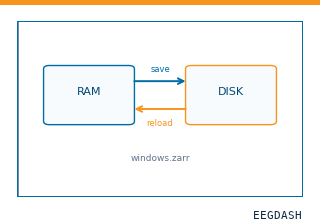 Save and reload prepared dataCache a windowed dataset to disk and reattach it without recompute.
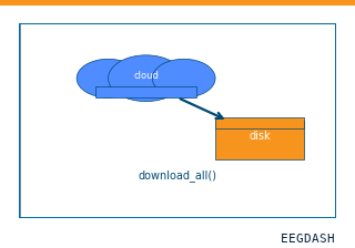 Download a dataset locallyPrefetch BIDS files to a local cache and validate the layout.
Swap any load_dataset(...) call for nm000377 to reproduce the tutorial on this dataset.
Citation
Melis Yilmaz Balban, Erin Cafaro, Lauren Saue-Fletcher, Marlon Joseph Washington, Maryam Bijanzadeh, … (2021). Human Responses to Visually Evoked Threat: intracranial EEG during virtual-reality heights, no-heights and shark stimuli (3 patients). 10.82901/nemar.nm000377
Provenance
¹Contributed to nemar in BIDS format.
²Curated & ingested by the EEGDash catalog; see CITATION.cff for canonical reference.
³Persistent identifier: 10.82901/nemar.nm000377.
See Also#
eegdash.dataset.EEGDashDataseteegdash.dataset