
DS004194: ieeg dataset, 14 subjects#
Visual ECoG dataset
Access recordings and metadata through EEGDash.
Citation: Iris Groen, Kenichi Yuasa, Amber Brands, Giovanni Piantoni, Stephanie Montenegro, Adeen Flinker, Sasha Devore, Orrin Devinsky, Werner Doyle, Patricia Dugan, Daniel Friedman, Nick Ramsey, Natalia Petridou, Jonathan Winawer (2022). Visual ECoG dataset. 10.18112/openneuro.ds004194.v3.0.0
Modality: ieeg Subjects: 14 Recordings: 209 License: CC0 Source: openneuro Citations: 4.0
Metadata: Complete (100%)
14-participant iEEG dataset — Visual ECoG dataset.
Quickstart#
Install
pip install eegdash
Access the data
from eegdash.dataset import DS004194

dataset = DS004194(cache_dir="./data")
# Get the raw object of the first recording
raw = dataset.datasets[0].raw
print(raw.info)
Filter by subject
dataset = DS004194(cache_dir="./data", subject="01")
Advanced query
dataset = DS004194(
    cache_dir="./data",
    query={"subject": {"$in": ["01", "02"]}},
)
Iterate recordings
for rec in dataset:
    print(rec.subject, rec.raw.info['sfreq'])
If you use this dataset in your research, please cite the original authors.
BibTeX
@dataset{ds004194,
  title = {Visual ECoG dataset},
  author = {Iris Groen and Kenichi Yuasa and Amber Brands and Giovanni Piantoni and Stephanie Montenegro and Adeen Flinker and Sasha Devore and Orrin Devinsky and Werner Doyle and Patricia Dugan and Daniel Friedman and Nick Ramsey and Natalia Petridou and Jonathan Winawer},
  doi = {10.18112/openneuro.ds004194.v3.0.0},
  url = {https://doi.org/10.18112/openneuro.ds004194.v3.0.0},
}
About This Dataset#
Please contact Iris Groen (i.i.a.groen@uva.nl, https://orcid.org/0000-0002-5536-6128) for more information.
Please see the following papers for more details on the data collection and preprocessing:
Details related to access to the data
Contact person
Groen IIA, Piantoni G, Montenegro S, Flinker A, Devore S, Devinsky O, Doyle W, Dugan P, Friedman D, Ramsey N, Petridou N, Winawer JA (2022) Temporal dynamics of neural responses in human visual cortex. The Journal of Neuroscience 42(40):7562-7580 (https://doi.org/10.1523/JNEUROSCI.1812-21.2022) Yuasa K, Groen IIA, Piantoni G, Montenegro S, Flinker A, Devore S, Devinsky O, Doyle W, Dugan P, Friedman D, Ramsey N, Petridou N, Winawer JA. Precise Spatial Tuning of Visually Driven Alpha Oscillations in Human Visual Cortex. eLife12:RP90387 https://doi.org/10.7554/eLife.90387.1 Brands AM, Devore S, Devinsky O, Doyle W, Flinker A, Friedman D, Dugan P, Winawer JA, Groen IIA (2024). Temporal dynamics of short-term neural adaptation in human visual cortex. https://doi.org/10.1101/2023.09.13.557378 - Practical information to access the data
Processed data and model fits reported in Groen et al., (2022) are available in derivatives/Groenetal2022TemporalDynamicsECoG as matlab .mat files. Matlab code to load, process and plot these data (including 3D renderings of the participant’s surface reconstructions and electrode positions) is available in WinawerLab/ECoG_utils and irisgroen/temporalECoG. These repositories have dependencies on other Matlab toolboxes (e.g., FieldTrip). See instructions on Github for relevant links and guidelines.
View full README
Details related to access to the data
Contact person
Groen IIA, Piantoni G, Montenegro S, Flinker A, Devore S, Devinsky O, Doyle W, Dugan P, Friedman D, Ramsey N, Petridou N, Winawer JA (2022) Temporal dynamics of neural responses in human visual cortex. The Journal of Neuroscience 42(40):7562-7580 (https://doi.org/10.1523/JNEUROSCI.1812-21.2022) Yuasa K, Groen IIA, Piantoni G, Montenegro S, Flinker A, Devore S, Devinsky O, Doyle W, Dugan P, Friedman D, Ramsey N, Petridou N, Winawer JA. Precise Spatial Tuning of Visually Driven Alpha Oscillations in Human Visual Cortex. eLife12:RP90387 https://doi.org/10.7554/eLife.90387.1 Brands AM, Devore S, Devinsky O, Doyle W, Flinker A, Friedman D, Dugan P, Winawer JA, Groen IIA (2024). Temporal dynamics of short-term neural adaptation in human visual cortex. https://doi.org/10.1101/2023.09.13.557378 - Practical information to access the data
Processed data and model fits reported in Groen et al., (2022) are available in derivatives/Groenetal2022TemporalDynamicsECoG as matlab .mat files. Matlab code to load, process and plot these data (including 3D renderings of the participant’s surface reconstructions and electrode positions) is available in WinawerLab/ECoG_utils and irisgroen/temporalECoG. These repositories have dependencies on other Matlab toolboxes (e.g., FieldTrip). See instructions on Github for relevant links and guidelines.
Processed data and model fits reported in Yuasa et al., (2023) are available in the Github repositories described in the paper. Processed data and model fits reported in Brands et al., (2024) are available in derivatives/Brandsetal2024TemporalAdaptationECoGCategories as python .py files. Python code to process and analyze these data is available in the Github repositories described in the paper.
Overview
Project name
Visual ECoG dataset - Years that the project ran
Data were collected between 2017-2020. Exact recording dates have been scrubbed for anonymization purposes. - Brief overview of the tasks in the experiment
Participants sub-p01 to sub-p11 viewed grayscale visual pattern stimuli that were varied in temporal or spatial properties. Participans sub-p11 to sub-p14 additionally saw color images of different image classes (faces, bodies, buildings, objects, scenes, and scrambled) that were varied in temporal properties. See ‘Independent Variables’ below for more details.
In all tasks, participants were instructed to fixate a cross or point in the center of the screen and monitor it for a color change, i.e. to perform a stimulus-orthogonal task (see the task-specific _events.json files, e.g., task-prf_events.json, for further details). - Description of the contents of the dataset
The data consists of cortical iEEG recordings in 14 epilepsy patients in response to visual stimulation. Patients were implanted with standard clinical surface (grid) and depth electrodes. Two patients were additionally implanted with a high-density research grid. In addition to the ieeg recordings, pre-implantation MRI T1 scans are provided for the purpose of localizing electrodes. Participants performed a varying number of tasks and runs. - Independent variables
The data are divided in 6 different sets of stimulus types or events: 1. prf: grayscale, oriented bar stimuli consisting of curved, band-pass filtered lines that were swept across the screen (up to (~16 degree of visual angle) in a fixed order for the purpose of estimating spatial population receptive fields (pRFs). 2. spatialpattern: grayscale, centrally presented pattern stimuli (~16 degree of visual angle diameter) consisting of curved, band-pass filtered lines that were systematically varied in level of contrast and density, as well as various oriented grating stimuli. 3. temporalpattern: grayscale, centrally presented pattern stimuli (~16 degree of visual angle diameter) consisting of curved, band-pass filtered lines that were systematically varied in temporal duration and interval. 4. soc: combination of the spatialpattern and temporalpattern stimuli. 5. sixcatloctemporal: color images of six stimulus classes: faces, bodies (hands/feet only), buildings, objects, scenes and scrambled, systematically varied in temporal duration and interval, whereby interval stimuli consisted of direct repeats of the identical image. 6. sixcatlocisidiff/sixcatlocdiffisi: color images of six stimulus classes: faces, bodies (hands/feet only), buildings, objects, scenes and scrambled, systematically varied in temporal duration and interval, whereby the first interval stimulus was followed by images from either the same or a different category (but not the identical image).
Participant-, task- and run-specific stimuli are provided in the /stimuli folder as matlab .mat files. - Dependent variables
The main BIDS folder contains the raw voltage data, split up in individual task runs.
The /derivatives/ECoGCAR folder contains common-average-referenced version of the data. The /derivatives/ECoGBroadband folder contains time-varying broadband responses estimated by band-pass filtering the common-average-referenced voltage data and taking the average power envelope.
The /derivatives/ECoGPreprocessed folder contains epoched trials used in Brands et al., (2024). The /derivatives/freesurfer folder contains surface reconstructions of each participant’s T1, along with retinotopic atlas files.
The /derivatives/Groen2022TemporalDynamicsECoG contains preprocessed data and model fits that can be used to reproduce the results reported in Groen et al., (2022). The /derivatives/Brands2024TemporalAdaptationECoG contains preprocessed data and model fits that can be used to reproduce the results reported in Brands et al., (2024). - Quality assessment of the data
Data quality and number of trials per subjects varies considerably across patients, for various reasons.
First, for each recording session, attempts were made to optimize the environment for running visual experiments; e.g. room illumination was stabilized as much as possible by closing blinds when available, the visual display was calibrated (for most patients), and interference from medical staff or visitors was minimized. However, it was not possible to equate this with great precision across patients and sessions/runs. Second, implantations were determined based on clinical needs and electrode locations therefore vary across participants. The strength and robustness of the neural responses varies greatly with the electrode location (e.g. early vs higher-level visual cortex), as well as with uncontrolled factors such as how well the electrode made contact with the cortex and whether it was primarily situated on grey matter (surface/grid electrodes) or could be located in white matter (some depth electrodes). Electrodes that were marked as containing epileptic activity by clinicians, or that did not have good signal based on visual inspection of the raw data, are marked as ‘bad’ in the channels.tsv files.
Third, patients varied greatly in their cognitive abilities and mental/medical state, which affected their ability to follow task instructions, e.g. to remain alert and fixation. Some patients were able to perform repeated runs of multiple tasks across multiple sessions, while others only managed to do a few runs. All patients included in this dataset have sufficiently good responses in some electrodes/tasks as judged by Groen et al., (2022) and Brands et al., (2024). However, when using this dataset to address further research questions, it is advisable to set stringent requirements on electrode and trial selection. See Groen et al., (2022) and associated code repository for an example preprocessing pipeline that selected for robust visual responses to temporally- and contrast-varying stimuli.
Methods
Subjects
All participants were intractable epilepsy patients who were undergoing ECoG for the purpose of monitoring seizures. Participants were included if their implantation covered parts of visual cortex and if they consented to participate in research. - Apparatus
Data were collected in a clinical setting, i.e. at bedside in the patient’s hospital room. Information about iEEG recording apparatus is provided the meta data for each patient. Information about the visual stimulation equipment and behavioral response recordings are provided in Groen et al., (2022), Yuasa et al., (2023) and Brands et al., (2024). - Experimental location
Data were collected at NYU University Langone Hospital (New York, USA) or at University Medical Center Utrecht (The Netherlands). - Missing data
Stimulus files are missing for a few runs of sub-02. These are marked as N/A in the associated event files.
Notes
Further participant-specific notes: - For sub-03 and sub-04 the spatial pattern and temporal pattern stimuli are combined in the soc task runs, for the remaining participants these are split across the spatialpattern and temporalpattern task runs. - The pRF task from sub-04 has different prf parameters (bar duration and gap). - The first two runs of the pRF task from sub-05 are not of good quality (participant repeatedly broke fixation). In addition, the triggers in all pRF runs from sub-05 are not correct due to a stimulus coding problem and will need to be re-interpolated if one wishes to use these data. - Participants sub-10 and sub-11 have high density grids in addition to clinical grids. - Note that all stimuli and stimulus parameters can be found in the participant-specific stimulus *.mat files.
Cohort#
Dataset Statistics#
Age distribution by gender (n=14, range 18–47 yr, mean 28.9 yr)
Sex composition
Channel counts (ch)
Sampling frequencies (Hz)
Total recording duration: 6 h 20 min
Signal · Electrodes & live trace#
Live trace viewer — sub-p12 · ses-nyuecog01 · task-prf · run-01
Showing one representative recording out of
14 subjects and 209 recordings in this dataset.
Browse the full set on OpenNeuro;
drop any other _ieeg.{set,edf,bdf,vhdr} file onto the
viewer (or pass ?ieeg=<url>) to inspect it.
Electrode layout — iEEG · 252 sensors — 252 channels
NEMAR Processing Statistics#
The plots below are generated by NEMAR’s automated EEG pipeline. The histogram shows pipeline success for data cleaning and ICA decomposition, the percentage of data frames and EEG channels retained after artefact removal, line noise per channel (RMS, dB), and the age/gender distribution of participants.

HED event descriptors word cloud

Manifest#
File Explorer#
Browse the BIDS file structure of this dataset. Records are fetched on demand from the EEGDash catalog the first time you open the explorer.
Full dataset metadata table
Dataset ID |
|
Title |
Visual ECoG dataset |
Author (year) |
|
Canonical |
— |
Importable as |
|
Year |
2022 |
Authors |
Iris Groen, Kenichi Yuasa, Amber Brands, Giovanni Piantoni, Stephanie Montenegro, Adeen Flinker, Sasha Devore, Orrin Devinsky, Werner Doyle, Patricia Dugan, Daniel Friedman, Nick Ramsey, Natalia Petridou, Jonathan Winawer |
License |
CC0 |
Citation / DOI |
|
Source links |
Copy-paste BibTeX
@dataset{ds004194,
  title = {Visual ECoG dataset},
  author = {Iris Groen and Kenichi Yuasa and Amber Brands and Giovanni Piantoni and Stephanie Montenegro and Adeen Flinker and Sasha Devore and Orrin Devinsky and Werner Doyle and Patricia Dugan and Daniel Friedman and Nick Ramsey and Natalia Petridou and Jonathan Winawer},
  doi = {10.18112/openneuro.ds004194.v3.0.0},
  url = {https://doi.org/10.18112/openneuro.ds004194.v3.0.0},
}
API Reference#
eegdash.datasetEEGDashDatasetDS004194 · Groen2022eegdash/dataset/registry.py · [source ↗]- class eegdash.dataset.DS004194(cache_dir: str, query: dict | None = None, s3_bucket: str | None = None, **kwargs)[source]#
Visual ECoG dataset
- Study:
ds004194(OpenNeuro)- Author (year):
Groen2022- Canonical:
—
Also importable as:
DS004194,Groen2022.Modality:
ieeg; Experiment type:Perception; Subject type:Epilepsy. Subjects: 14; recordings: 209; tasks: 7.- Parameters:
cache_dir (str | Path) – Directory where data are cached locally.
query (dict | None) – Additional MongoDB-style filters to AND with the dataset selection. Must not contain the key
dataset.s3_bucket (str | None) – Base S3 bucket used to locate the data.
**kwargs (dict) – Additional keyword arguments forwarded to
EEGDashDataset.
- data_dir#
Local dataset cache directory (
cache_dir / dataset_id).- Type:
Path
Notes
Each item is a recording; recording-level metadata are available via
dataset.description.querysupports MongoDB-style filters on fields inALLOWED_QUERY_FIELDSand is combined with the dataset filter. Dataset-specific caveats are not provided in the summary metadata.References
OpenNeuro dataset: https://openneuro.org/datasets/ds004194 NeMAR dataset: https://nemar.org/dataexplorer/detail?dataset_id=ds004194 DOI: https://doi.org/10.18112/openneuro.ds004194.v3.0.0 NEMAR citation count: 4
Examples
>>> from eegdash.dataset import DS004194
>>> dataset = DS004194(cache_dir="./data")
>>> recording = dataset[0]
>>> raw = recording.load()
- __init__(cache_dir: str, query: dict | None = None, s3_bucket: str | None = None, **kwargs)[source]#
- save(path: str, overwrite: bool = False, offset: int = 0)[source]#
Save datasets to files by creating one subdirectory for each dataset:
path/
    0/
        0-raw.fif | 0-epo.fif
        description.json
        raw_preproc_kwargs.json (if raws were preprocessed)
        window_kwargs.json (if this is a windowed dataset)
        window_preproc_kwargs.json  (if windows were preprocessed)
        target_name.json (if target_name is not None and dataset is raw)
    1/
        1-raw.fif | 1-epo.fif
        description.json
        raw_preproc_kwargs.json (if raws were preprocessed)
        window_kwargs.json (if this is a windowed dataset)
        window_preproc_kwargs.json  (if windows were preprocessed)
        target_name.json (if target_name is not None and dataset is raw)
- Parameters:
path (str) –
- Directory in which subdirectories are created to store
-raw.fif | -epo.fif and .json files to.
overwrite (bool) – Whether to delete old subdirectories that will be saved to in this call.
offset (int) – If provided, the integer is added to the id of the dataset in the concat. This is useful in the setting of very large datasets, where one dataset has to be processed and saved at a time to account for its original position.
BaseDataset from braindecode — windowed via create_windows_from_events.braindecodeDataLoader; supports parallel workers and on-the-fly augmentations.pytorchdatasets.load_dataset("EEGDash/ds004194").huggingface Find datasets with the EEGDash APIQuery the catalogue, filter by task or modality, list candidates.
 Load one EEG recordingResolve a single record to an MNE Raw with channels and events.
 EEG recording to PyTorch DataLoaderWrap braindecode windows in a DataLoader for model training.
 Preprocess EEG and create windowsFilter, resample, epoch — and persist the windowed dataset.
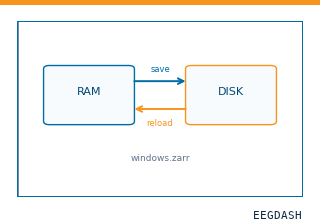 Save and reload prepared dataCache a windowed dataset to disk and reattach it without recompute.
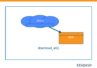 Download a dataset locallyPrefetch BIDS files to a local cache and validate the layout.
Swap any load_dataset(...) call for ds004194 to reproduce the tutorial on this dataset.
Citation
Iris Groen, Kenichi Yuasa, Amber Brands, Giovanni Piantoni, Stephanie Montenegro, … (2022). Visual ECoG dataset. 10.18112/openneuro.ds004194.v3.0.0
Provenance
¹Contributed to openneuro in BIDS format.
²Curated & ingested by the EEGDash catalog; see CITATION.cff for canonical reference.
³Persistent identifier: 10.18112/openneuro.ds004194.v3.0.0.
See Also#
eegdash.dataset.EEGDashDataseteegdash.dataset